SlideShare a Scribd company logo
1 of 122
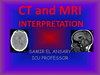
CT and MRI
INTERPRETATION
SAMIR EL ANSARY
ICU PROFESSOR
1
A midline Post-contrast Sagittal T1 Weighted MRI
2
3 4 5 6
7
8
9
10
11
12
13
14
1
6
1
5
1
7
18
19
20
21
2
2
2
3
24
Identify anatomical
structures 1 - 24
1
A midline Post-contrast Sagittal T1 Weighted MRI
2
3 4 5 6
7
8
9
10
11
12
13
14
16
15
17
18
19
20
21
2
2
2
3
24
1. Scalp fat
2. Bone
3. Inferior sagittal sinus
4. Corpus callosum
5. Internal cerebral vein
6. Vein of Galen
7. Superior sagittal sinus
8. Parietal lobe
9. Occipital lobe
10. Straight sinus
11. Vermis
12. IV ventricle
13. Cerebellar tonsil
14. Cervical cord
15. Medulla
16. Pons
17. Midbrain
18. Mass intermedia of thalamus
19. Anterior III ventricle
20. Optic chiasm
21. Pituitary gland
22. Sphenoid sinus
23. Nasopharynx
24. Frontal lobe
Coronal Section of the Brain at the level of IV Ventricle
Post Contrast Coronal T1 Weighted MRI
8
7
6
5
4
3
2
1
Identify anatomical structures
1 - 8
Coronal Section of the Brain at the level of IV Ventricle
Post Contrast Coronal T1 Weighted MRI
8
7
6
5
4
3
2
1
1. Cerebellar tonsil
2. Cerebellar hemisphere
3. IV ventricle
4. Superior vermis
5. Tentorium
6. Posterior temporal lobe
7. Choroid plexus within lateral
ventricle
8. Posterior frontal lobe
Coronal Section of the Brain at the level of Pituitary gland
Post Contrast Coronal T1 Weighted MRI
1
2
3
4
5
6
78
9
1
0
11
12 Identify anatomical structures
1 - 12
Coronal Section of the Brain at the level of Pituitary gland
Post Contrast Coronal T1 Weighted MRI
1
2
3
4
5
6
78
9
10
11
12
1. Frontal lobe
2. Corpus callosum
3. Frontal horn
4. Caudate nucleus
5. III ventricle
6. Optic nerve
7. Pituitary stalk
8. Pituitary gland
9. Internal carotid artery
10. Cavernous sinus
11. Sphenoid sinus
12. Nasopharynx
Coronal Section of the Brain at the level of the orbits.
Post Contrast Coronal T1 Weighted MRI.
1
2
3
4
5
Identify anatomical structures
1 - 5
Coronal Section of the Brain at the level of the orbits.
Post Contrast Coronal T1 Weighted MRI.
1
2
3
4
5
1. Frontal lobe
2. Orbital Fat
3. Globe
4. Nasal Cavity
5. Maxillary Sinus
Post Contrast Axial MR Image of
the brain
1
2
3
4
5
Post Contrast sagittal T1
Weighted M.R.I.
Section at the level of Foramen
Magnum
Answers
1. Cisterna Magna
2. Cervical Cord
3. Nasopharynx
4. Mandible
5. Maxillary Sinus
Post Contrast Axial MR Image of the
brain
7
6
Post Contrast sagittal T1
Wtd M.R.I.
Section at the level of
medulla
Answers
6. Medulla
7. Sigmoid Sinus
Post Contrast Axial MR Image of the brain
15
8
9
10
11
12
13
14
16
17
Post Contrast sagittal T1 Wtd M.R.I.
Section at the level of Pons
Answers
8. Cerebellar
Hemisphere
9. Vermis
10. IV Ventricle
11. Pons
12. Basilar
Artery
13. Internal Carotid
Artery
14. Cavernous Sinus
15. Middle Cerebellar
Peduncle
16. Internal Auditory
Canal
17. Temporal Lobe
Post Contrast Axial MR Image of the brain
18
19
20
21
22
Post Contrast sagittal T1 Wtd
M.R.I.
Section at the level of Mid
Brain
Answers
18. Aqueduct of Sylvius
19. Midbrain
20. Orbits
21. Posterior Cerebral
Artery
22. Middle Cerebral
Artery
Post Contrast Axial MR Image of the brain
23
24
25
26
27
Post Contrast sagittal T1 Wtd M.R.I.
Section at the level of the
III Ventricle
Answers
23. Occipital Lobe
24. III Ventricle
25. Frontal Lobe
26. Temporal Lobe
27. Sylvian Fissure
Post Contrast Axial MR Image of the brain
28
29
30
31
32
38
33
34
36
35
37
Post Contrast sagittal T1 Wtd M.R.I.
Section at the level of Thalamus
Answers
28. Superior Sagittal Sinus
29. Occipital Lobe
30. Choroid Plexus within
the occipital horn
31. Internal Cerebral Vein
32. Frontal Horn
33. Thalamus
34. Temporal
Lobe
35. Internal
Capsule
36. Putamen
37. Caudate
Nucleus
38. Frontal Lobe
Post Contrast Axial MR Image of the brain
39
40
41
Post Contrast sagittal T1 Wtd
M.R.I.
Section at the level of Corpus
Callosum
Answers
39. Splenium of corpus callosum
40. Choroid plexus within the
body of lateral ventricle
41. Genu of corpus callosum
Post Contrast Axial MR
Image of the brain
42
43
44
Post Contrast sagittal T1
Wtd M.R.I.
Section at the level of Body
of Corpus Callosum
Answers
42. Parietal Lobe
43. Body of the Corpus Callosum
44. Frontal Lobe
Post Contrast Axial MR Image of the brain
45
46
Post Contrast sagittal T1 Wtd
M.R.I.
Section above the Corpus
Callosum
Answers
45. Parietal Lobe
46. Frontal Lobe
Brain Imaging: “The Big 10”
• Infarction
• Hemorrhage
• Infection
• Tumor
• Trauma
• Dementia
• MS
• Epilepsy
• Cranial neuropathy
• Orbits / Ophtho dx
Acute Ischemic Stroke Imaging
 Confirm diagnosis
 Triage for therapy (risk / prognosis)
– Rule out hemorrhage
– Assess damage: location, pattern, extent
– Is there salvageable brain (“penumbra”)?
 Follow outcome
– Vessel patency, ultimate infarct size, hemorrhagic
transformation
CT Signs in Early MCA Ischemia
Hyperdense MCA Insular Ribbon Lentiform Nucleus
Pathophysiology of Ischemic Injury:
Duration and Degree of  CBF
Normal neuronal function
Reversible injury
(penumbra)
Infarction
25
20
15
10
5
0
CBF
ml /
100g /
min
Time (hrs)1 2
Pipes  Perfusion  Parenchyma
MRA Perfusion MR Diffusion MR
“Penumbra”
MRI in Stroke Intervention
“The 4 P’s”
MCA Infarct
MCA
PCA Infarct
PCA
ACA Infarct ACA
Brain Imaging: “The Big 10”
• Infarction
• Hemorrhage
• Infection
• Tumor
• Trauma
• Dementia
• MS
• Epilepsy
• Cranial neuropathy
• Orbits / Ophtho dx
Cerebral Hemorrhage
• Trauma
• Ruptured aneurysm
• Hypertensive
• Hemorrhagic transformation of ischemic infarction
(esp. venous)
• Venous infarction
• Tumor
• Vascular malformations
• Angioinvasive infection
• Amyloid angiopathy
Acute intraparenchymal hematoma
Cerebral Hemorrhage
Hemorrhagic melanoma metastases
Cerebral Hemorrhage
Acute subarachnoid hemorrhage
(and intraventricular)
Cerebral Hemorrhage
Subdural vs. Epidural Hematoma
Acute subdural hematoma
Cerebral Hemorrhage
Acute epidural hematoma
Cerebral Hemorrhage
Subdural: Follows inner layer of dura
“Rounds the bend” to follow falx or tentorium
Not affected by sutures of skull
Tendency for crescentic shapes
More mass effect than expected for their size
Typical source of SDH: cortical vein
Epidural: Follows outer layer of dura (periosteum)
Crosses falx or tentorium
Limited by sutures of skull (typically)
Tendency for lentiform shapes
Typical source of EDH:
skull fracture with arterial or sinus laceration
Subdural vs. Epidural Hematoma
*
Mixed acute/chronic subdural hematoma
Cerebral Hemorrhage
ACUTE
CHRONIC
Hematocrit level!
Cerebral Hemorrhage
MRI of Hemorrhage
MR appearance of hematomas depends on image type.
Magnetic properties change over time (Hgb breakdown
products), allowing approximate dating
T1 T2 T2*
Brain Imaging: “The Big 10”
• Infarction
• Hemorrhage
• Infection
• Tumor
• Trauma
• Dementia
• MS
• Epilepsy
• Cranial neuropathy
• Orbits / Ophtho dx
Infection
• Meningitis
• Encephalitis
• Cerebritis and parenchymal abscess
• Empyema (subdural/epidural)
Leptomeningitis:
pia-arachnoid
Meningitis
Pachymeningitis: dura
Most common imaging findings in meningitis: NONE !!
Herpes
Encephalitis
Cerebritis w/ Bacterial Abscess
T1 + Gd T2 Diffusion
Cerebritis w/ Subdural Empyema
T1 + Gd T2 FLAIR Diffusion
Brain Imaging: “The Big 10”
• Infarction
• Hemorrhage
• Infection
• Tumor
• Trauma
• Dementia
• MS
• Epilepsy
• Cranial neuropathy
• Orbits / Ophtho dx
Brain Tumor Imaging
Diagnosis
• Location: Intra- / Extra-axial, Supra- / Infra-
tentorial, Grey / white matter, etc.
• Single or multiple?
• Tumor or tumor-like alternatives?
• Histology: Type and grade?
Treatment Planning
• Surgery, radiation, chemo tx
• Functional MRI for eloquent brain mapping
• 3D scans to guide surgery, radiation
Follow-up
• Stable vs. recurrence / progression
• Complications
T1 + Gd T2
Intra- or Extra-axial?
Tumor vs. Other Masses
Arachnoid Cyst
Abscess
Hematoma
“Tumefactive” MS
GBM
Tumor vs. Stroke
Cytotoxic Edema Vasogenic Edema
Cellular swelling
Gray-white margin lost
Leaky capillaries
Gray matter is spared
T1 T1 + Gd
T2
T2 FLAIR
Tumor?
Stroke?
Encephalitis?
3D Imaging for XRT
or Surgical Guidance
Brain Imaging: “The Big 10”
• Infarction
• Hemorrhage
• Infection
• Tumor
• Trauma
• Dementia
• MS
• Epilepsy
• Cranial neuropathy
• Orbits / Ophtho dx
Fractures: CT not MRI !
Traumatic Brain Swelling
Cerebellopontine
angle
PontineCerebellomedullary
(Cisterna Magna)
Know your basal cisterns!
Traumatic Brain Swelling
Know your basal cisterns!
Quadrigeminal
Interpeduncular
Suprasellar
Ambient
Effacement of basal cisterns
Traumatic brain swelling with
downward herniation
Traumatic Brain Swelling
Traumatic brain swelling
Extra-axial Hemorrhage
Subdural Epidural Subarachnoid
Intra-axial Hemorrhage
Hemorrhagic contusions
Intra-axial Hemorrhage
Hemorrhagic contusions
Mechanism
Direct contact with skull
Shear-strain deformation
Lesion locations
Commonly located along inferior, lateral, and anterior
frontal and temporal lobes
Often above bony prominences (petrous pyramid,
sphenoid wing, orbital roof)
Intra-axial Hemorrhage
Hemorrhagic contusions
Appearance of cortical contusions
Overlying cortex, by definition, always involved (vs. DAI)
“Salt and pepper” appearance due to intermixed hemorrhage
and edema
Non-hemorrhagic contusions often not initially seen on CT scans
Lesions often more visible days after injury as edema and
hemorrhage increase
Acute lesions much more conspicuous on T2 or T2-FLAIR MRI
Diffuse Axonal (Shear) Injury (DAI)
Intra-axial Hemorrhage
Diffuse Axonal (Shear) Injury (DAI)
T2: Reveals non-hemorrhagic lesions occult on CT
Diffuse Axonal (Shear) Injury (DAI)
T2: Increased sensitivity to hemorrhage
Diffuse Axonal (Shear) Injury (DAI)
• Tissues w/ differing elastic properties shear against each other,
tearing axons
• Caused by rapid deceleration/rotation of head
• Locations:
• Cerebral hemispheres near gray-white junction
• Basal ganglia
• Corpus callosum, especially splenium
• Dorsal brainstem
• High morbitity & mortality – common cause of post-traumatic
vegetative state
• Initial CT often normal despite poor GCS
• Lesions often non-hemorrhagic and seen only on MRI
Brain Imaging: “The Big 10”
• Infarction
• Hemorrhage
• Infection
• Tumor
• Trauma
• Dementia
• MS
• Epilepsy
• Cranial neuropathy
• Orbits / Ophtho dx
Dementia
• Primary role of imaging is to
exclude treatable causes, e.g.:
–Hydrocephalus
–Subdural hematoma
–Neoplasm
Dementia
Irreversible dementias (imaging non-
specific):
• Alzheimer’s disease
• Multi-infarct dementia
• Dementias associated with
Parkinson’s disease and similar
disorders
• AIDS dementia complex
Alzheimer’s: Temporal-Parietal Lobe Atrophy (Late)
Brain Imaging: “The Big 10”
• Infarction
• Hemorrhage
• Infection
• Tumor
• Trauma
• Dementia
• MS
• Epilepsy
• Cranial neuropathy
• Orbits / Ophtho dx
Multiple Sclerosis (MS) Imaging
• MRI is the imaging study of choice
• Help establish “dissemination of lesions in time
and space”
• Estimate disease burden
• Identify acute (inflammatory) vs. chronic lesions
(enhancement = active inflammation)
MS
Tumefactive MS
Brain Imaging: “The Big 10”
• Infarction
• Hemorrhage
• Infection
• Tumor
• Trauma
• Dementia
• MS
• Epilepsy
• Cranial neuropathy
• Orbits / Ophtho dx
Seizure Imaging
• MRI is the imaging study of choice
• Identify and localize offending lesion
• New onset vs. chronic epilepsy
• Younger vs. older patients
• Search may be guided by EEG / clinical sx
• Preoperative planning
e.g. language lateralization before temporal
lobectomy
Congenital anomalies: Polymicrogyria
Congenital anomalies: Schizencephaly
Mesial Temporal Sclerosis
Most common pathology found in
medically refractory epilepsy patients
Rare under age 10 or with new seizures
Pathogenesis unknown
- Post ictal / kindling?
Pathology:
Hippocampal atrophy / gliosis
Mesial
Temporal
Sclerosis
FLAIR
T1
T2
• Atrophy
• Loss gray-white
•↑T2 / FLAIR
Brain Imaging: “The Big 10”
• Infarction
• Hemorrhage
• Infection
• Tumor
• Trauma
• Dementia
• MS
• Epilepsy
• Cranial neuropathy
• Orbits / Ophtho dx
Cranial Nerve Imaging
FIESTA
CN-5
CN-8
CN-7
Vestibular Schwannoma
Intracochlear Schwannoma
30 y/o F
with 6wk
h/o blurred
vision
Craniopharyngioma
CT vs. MRI
Wide doughnutOpening
10-20 minutesLength
Adjust windowTechnique
AialPlane
BrightBone
Long, narrow
30-60 min
T1, T2, Pd
3-D
Dark
Magnetic fldX-ray beamObtained
MRICT
Advantages to CT
• Costs less than MRI
• Better access
• Shows up acute bleed
• A good quick screen
• Good visualization of bony structures
and calcified lesions
Disadvantages to CT
• Resolution
• Beam-hardening artifact
• Limited views of the posterior fossa and
poor visualization of white-matter
disease
Advantages to MRI
• Good resolution—excellent view of brain
structure
• 3 dimensions
• Good gray-white differentiation
• Adjust settings based on characteristics of
the lesion
• Good view of the posterior fossa
Advantages to MRI
• No radiation exposure
• Gadolinium contrast is relatively nontoxic
• Capacity for quantitative imaging, 3-D
reconstruction, angiography, spectroscopy
Disadvantages of MRI
• Cost
• Some patients ineligible because
of pacemakers, other metal
• Claustrophobia
• Long exam
• Access
What Is Bright
on CT?
• Blood
• Contrast
• Bone
• Calcium
• Metal
What Is Dark
on CT?
•Air
•CSF/H20
Artifacts
• Beam
hardening
• Bone
• Foreign body
• Motion
Uses for SPECT and PET
• Acute stroke
• Identify a seizure focus-increased
flow during sz and decreased
interictal flow
• Dementia-frontal pattern in FTLD,
temporo-parietal pattern in AD
• Ligand imaging in PD, others
Landmarks
• Axial views
– Fourth ventricle
– Petrous bone and sphenoid ridge
– Aqueduct
– Third ventricle
– Lateral ventricles
– Frontal horns
– Calcifications in the choroid plexus, pineal,
basal ganglia and falx
– Caudate, putamen and globus pallidus
Landmarks (Cont.)
– Internal capsule—anterior and posterior limbs
– Thalami
– Sylvian fissures
• Sagittal views
– Severity of cortical atrophy
– Corpus callosum and cingulate gyrus
• Pituitary
– Coronal views
– Hippocampus and amygdala
Normal
Hippo-
Campus
Atrophic
Hippo-
campus
in AD
62 year old
woman with
rapid
progression of
memory loss
Introduction to Scan Interpretation
• Is the scan
– Contrast or noncontrast?
– Good quality?
• Describe the abnormality
– Size—small, punctuate, medium, large
– Shape—round, well circumscribed, ovoid,
irregular, patchy
Introduction to Scan Interpretation
(Cont.)
• Signal intensity
– High signal,
hyperdense
– Low signal, hypodense
– Isointense, isodense
– Mixed signal
• Location
Vascular Dementia
Three types of vascular dementia
Multiple large
Vessel infarctions
Bilateral strategic
thalamic infarcts
Binswanger’s
Disease
Normal Pressure Hydrocephalus: NPH
• Cognitive Impairment
• Gait Disturbance
• Bladder Control
• May Have:
Behavior Problems
Parkinsonism
MRI findings
• Ventricular enlargement disproportionate to the
amount of atrophy
• Bowing of the corpus callosum
• Smooth rimming of high signal around the ventricles
due to transependymal flow of CSF
NPH: pre-op NPH: post-op-130
mm H2O
Types of fMRI
• BOLD-fMRI which measures regional differences in
oxygenated blood
• Diffusion-weighted fMRI which measures random
movement of water molecules. Diffusion tensor
imaging (DTI) measures diffusion of water in different
directions and is a good test for studying white
matter tracts.
• MRI spectroscopy which can measure certain
cerebral metabolites non-invasively
DTI reconstruction of the corpus callosum
3D reconstruction
with functional
overlay
fMRI:Visual
stimulation
MR Spectroscopy
MR spectroscopy of N
acetyl aspartate
(NAA) showing
decline of NAA over
time in patients with
Alzheimer’s disease
(lower line)
compared to age-
matched controls.
GOOD LUCK
SAMIR EL ANSARY
ICU PROFESSOR
AIN SHAMS
CAIRO

More Related Content

What's hot

What's hot (20)

MRI SPINE ANATOMY
MRI SPINE ANATOMYMRI SPINE ANATOMY
MRI SPINE ANATOMY
 
MRI sequences
MRI sequencesMRI sequences
MRI sequences
 
Diagnostic Neuroradiology
Diagnostic NeuroradiologyDiagnostic Neuroradiology
Diagnostic Neuroradiology
 
Approach to head ct
Approach to head ctApproach to head ct
Approach to head ct
 
Imaging in stroke
Imaging in strokeImaging in stroke
Imaging in stroke
 
Diagnostic Imaging of Spinal Infection & Inflammation
Diagnostic Imaging of Spinal Infection & InflammationDiagnostic Imaging of Spinal Infection & Inflammation
Diagnostic Imaging of Spinal Infection & Inflammation
 
Mri brain anatomy Dr Muhammad Bin Zulfiqar
Mri brain anatomy Dr Muhammad Bin ZulfiqarMri brain anatomy Dr Muhammad Bin Zulfiqar
Mri brain anatomy Dr Muhammad Bin Zulfiqar
 
Imaging in stroke
Imaging in strokeImaging in stroke
Imaging in stroke
 
MR spectroscopy
MR spectroscopyMR spectroscopy
MR spectroscopy
 
ANATOMY OF MRI SPINE
ANATOMY OF MRI SPINEANATOMY OF MRI SPINE
ANATOMY OF MRI SPINE
 
CSF cisterns
CSF cisternsCSF cisterns
CSF cisterns
 
Presentation1, radiological imaging of pediatric leukodystrophy.
Presentation1, radiological imaging of pediatric leukodystrophy.Presentation1, radiological imaging of pediatric leukodystrophy.
Presentation1, radiological imaging of pediatric leukodystrophy.
 
Presentation1.pptx white matter disorder in pediatric
Presentation1.pptx white matter disorder in pediatricPresentation1.pptx white matter disorder in pediatric
Presentation1.pptx white matter disorder in pediatric
 
BRAIN CT SCAN
BRAIN CT SCANBRAIN CT SCAN
BRAIN CT SCAN
 
MRI of Brain: Basics
MRI of Brain: BasicsMRI of Brain: Basics
MRI of Brain: Basics
 
Basics of ct mri
Basics of ct mriBasics of ct mri
Basics of ct mri
 
Basics of neuroimaging
Basics of neuroimagingBasics of neuroimaging
Basics of neuroimaging
 
Approach to white matter disease
Approach to white matter diseaseApproach to white matter disease
Approach to white matter disease
 
Avascular necrosis Radiology
Avascular necrosis RadiologyAvascular necrosis Radiology
Avascular necrosis Radiology
 
Cvj anomalies
Cvj anomaliesCvj anomalies
Cvj anomalies
 

Viewers also liked

MRI SECTIONAL ANATOMY OF BRAIN
MRI SECTIONAL ANATOMY OF BRAIN MRI SECTIONAL ANATOMY OF BRAIN
MRI SECTIONAL ANATOMY OF BRAIN Vipin Kumar
 
Sulcal anatomy supratentorial brain, excluding the temporal lobe.
Sulcal anatomy supratentorial brain, excluding the temporal lobe.Sulcal anatomy supratentorial brain, excluding the temporal lobe.
Sulcal anatomy supratentorial brain, excluding the temporal lobe.Sarbesh Tiwari
 
Head and cervical spine imaging
Head and cervical spine imagingHead and cervical spine imaging
Head and cervical spine imagingSCGH ED CME
 
Ct & mri of central nervous system
Ct & mri of central nervous systemCt & mri of central nervous system
Ct & mri of central nervous systemghalan
 
Basics of CT & MRI
Basics of CT & MRIBasics of CT & MRI
Basics of CT & MRIVibhuti Kaul
 
MRI Procedure of Brain
MRI Procedure of BrainMRI Procedure of Brain
MRI Procedure of BrainSudil Paudyal
 
Anatomy of brain sulcus and gyrus - Dr.Sajith MD RD
Anatomy of brain sulcus and gyrus - Dr.Sajith MD RDAnatomy of brain sulcus and gyrus - Dr.Sajith MD RD
Anatomy of brain sulcus and gyrus - Dr.Sajith MD RDSajith Selvaganesan
 
Diseases of Cranial nerves 5th, 7th and 8th
Diseases of Cranial nerves 5th, 7th and 8thDiseases of Cranial nerves 5th, 7th and 8th
Diseases of Cranial nerves 5th, 7th and 8thA T M Hasibul Hasan
 
Epilepsy mri
Epilepsy mriEpilepsy mri
Epilepsy mriBhav Raj
 
Cross Sectional Anatomy Of The Abdomen Annotated
Cross Sectional Anatomy Of The Abdomen AnnotatedCross Sectional Anatomy Of The Abdomen Annotated
Cross Sectional Anatomy Of The Abdomen AnnotatedDebby Edney
 
Head Trauma Ct Evaluation
Head Trauma Ct EvaluationHead Trauma Ct Evaluation
Head Trauma Ct Evaluationsalahrad
 
Basic reading computed tomography (ct) of brain
Basic reading computed tomography (ct) of brainBasic reading computed tomography (ct) of brain
Basic reading computed tomography (ct) of brainaViVian
 
Imaging of Traumatic Brain Injury
Imaging of Traumatic Brain InjuryImaging of Traumatic Brain Injury
Imaging of Traumatic Brain InjuryRathachai Kaewlai
 
Presentation1.pptx, radiological anatomy of the abdomen and pelvis.
Presentation1.pptx, radiological anatomy of the abdomen and pelvis.Presentation1.pptx, radiological anatomy of the abdomen and pelvis.
Presentation1.pptx, radiological anatomy of the abdomen and pelvis.Abdellah Nazeer
 
Subdural And Epidural Hematomas
Subdural And Epidural HematomasSubdural And Epidural Hematomas
Subdural And Epidural HematomasJoshua Bear
 

Viewers also liked (20)

Brain herniation imaging
Brain herniation imagingBrain herniation imaging
Brain herniation imaging
 
MRI SECTIONAL ANATOMY OF BRAIN
MRI SECTIONAL ANATOMY OF BRAIN MRI SECTIONAL ANATOMY OF BRAIN
MRI SECTIONAL ANATOMY OF BRAIN
 
Sulcal anatomy supratentorial brain, excluding the temporal lobe.
Sulcal anatomy supratentorial brain, excluding the temporal lobe.Sulcal anatomy supratentorial brain, excluding the temporal lobe.
Sulcal anatomy supratentorial brain, excluding the temporal lobe.
 
Head and cervical spine imaging
Head and cervical spine imagingHead and cervical spine imaging
Head and cervical spine imaging
 
CT Scan - Basics
CT Scan - BasicsCT Scan - Basics
CT Scan - Basics
 
Ct & mri of central nervous system
Ct & mri of central nervous systemCt & mri of central nervous system
Ct & mri of central nervous system
 
Basics of CT & MRI
Basics of CT & MRIBasics of CT & MRI
Basics of CT & MRI
 
MRI Procedure of Brain
MRI Procedure of BrainMRI Procedure of Brain
MRI Procedure of Brain
 
Beta blockers in brain injuries
Beta blockers in brain injuriesBeta blockers in brain injuries
Beta blockers in brain injuries
 
Anatomy of brain sulcus and gyrus - Dr.Sajith MD RD
Anatomy of brain sulcus and gyrus - Dr.Sajith MD RDAnatomy of brain sulcus and gyrus - Dr.Sajith MD RD
Anatomy of brain sulcus and gyrus - Dr.Sajith MD RD
 
Diseases of Cranial nerves 5th, 7th and 8th
Diseases of Cranial nerves 5th, 7th and 8thDiseases of Cranial nerves 5th, 7th and 8th
Diseases of Cranial nerves 5th, 7th and 8th
 
Epilepsy mri
Epilepsy mriEpilepsy mri
Epilepsy mri
 
Cross Sectional Anatomy Of The Abdomen Annotated
Cross Sectional Anatomy Of The Abdomen AnnotatedCross Sectional Anatomy Of The Abdomen Annotated
Cross Sectional Anatomy Of The Abdomen Annotated
 
Imaging in acute stroke
Imaging in acute strokeImaging in acute stroke
Imaging in acute stroke
 
Head Trauma Ct Evaluation
Head Trauma Ct EvaluationHead Trauma Ct Evaluation
Head Trauma Ct Evaluation
 
Basic reading computed tomography (ct) of brain
Basic reading computed tomography (ct) of brainBasic reading computed tomography (ct) of brain
Basic reading computed tomography (ct) of brain
 
Imaging of Traumatic Brain Injury
Imaging of Traumatic Brain InjuryImaging of Traumatic Brain Injury
Imaging of Traumatic Brain Injury
 
Presentation1.pptx, radiological anatomy of the abdomen and pelvis.
Presentation1.pptx, radiological anatomy of the abdomen and pelvis.Presentation1.pptx, radiological anatomy of the abdomen and pelvis.
Presentation1.pptx, radiological anatomy of the abdomen and pelvis.
 
Subdural And Epidural Hematomas
Subdural And Epidural HematomasSubdural And Epidural Hematomas
Subdural And Epidural Hematomas
 
Ct Basics
Ct BasicsCt Basics
Ct Basics
 

Similar to Ct and mri interpretation

101 ct neuroimaging
101 ct neuroimaging101 ct neuroimaging
101 ct neuroimagingAhmad Shahir
 
BMS2-K13 Pemeriksaan Radiologi pada Sistem Saraf.pptx
BMS2-K13 Pemeriksaan Radiologi pada Sistem Saraf.pptxBMS2-K13 Pemeriksaan Radiologi pada Sistem Saraf.pptx
BMS2-K13 Pemeriksaan Radiologi pada Sistem Saraf.pptxssuser144901
 
Imaginginacutestroke dr anoop.k.r
Imaginginacutestroke dr anoop.k.rImaginginacutestroke dr anoop.k.r
Imaginginacutestroke dr anoop.k.ranoop k r
 
Neurosurgery 1.pptx
Neurosurgery 1.pptxNeurosurgery 1.pptx
Neurosurgery 1.pptxjoendesh
 
Imaging in head trauma
Imaging in head traumaImaging in head trauma
Imaging in head traumaSCGH ED CME
 
basicsofctmri-140310081410-phpapp01.pdf
basicsofctmri-140310081410-phpapp01.pdfbasicsofctmri-140310081410-phpapp01.pdf
basicsofctmri-140310081410-phpapp01.pdfS Gupta
 
Kuliah neoro radiologi revisi
Kuliah neoro radiologi revisiKuliah neoro radiologi revisi
Kuliah neoro radiologi revisilee25tom9
 
Acute Pediatric Neuroradiology: Pearls and Pitfalls (2020)
Acute Pediatric Neuroradiology: Pearls and Pitfalls (2020)Acute Pediatric Neuroradiology: Pearls and Pitfalls (2020)
Acute Pediatric Neuroradiology: Pearls and Pitfalls (2020)Felice D'Arco
 
Radiology of central nervous system
Radiology of central nervous systemRadiology of central nervous system
Radiology of central nervous systemWEEKLYMEDIC
 
ESSENTIALS_OF_CT_BRAIN_For_Undergraduate.pptx
ESSENTIALS_OF_CT_BRAIN_For_Undergraduate.pptxESSENTIALS_OF_CT_BRAIN_For_Undergraduate.pptx
ESSENTIALS_OF_CT_BRAIN_For_Undergraduate.pptxssuser39be96
 
skull base lesion imaging.pptx
skull base lesion imaging.pptxskull base lesion imaging.pptx
skull base lesion imaging.pptxShivanjanGoyal
 
Stroke localization by Dr. Md. firoz
Stroke localization by Dr. Md. firozStroke localization by Dr. Md. firoz
Stroke localization by Dr. Md. firozFirozMohammad8
 

Similar to Ct and mri interpretation (20)

101 ct neuroimaging
101 ct neuroimaging101 ct neuroimaging
101 ct neuroimaging
 
Acute brain
Acute brainAcute brain
Acute brain
 
BMS2-K13 Pemeriksaan Radiologi pada Sistem Saraf.pptx
BMS2-K13 Pemeriksaan Radiologi pada Sistem Saraf.pptxBMS2-K13 Pemeriksaan Radiologi pada Sistem Saraf.pptx
BMS2-K13 Pemeriksaan Radiologi pada Sistem Saraf.pptx
 
Imaginginacutestroke dr anoop.k.r
Imaginginacutestroke dr anoop.k.rImaginginacutestroke dr anoop.k.r
Imaginginacutestroke dr anoop.k.r
 
Neurosurgery 1.pptx
Neurosurgery 1.pptxNeurosurgery 1.pptx
Neurosurgery 1.pptx
 
Head Injury
Head InjuryHead Injury
Head Injury
 
Imaging in head trauma
Imaging in head traumaImaging in head trauma
Imaging in head trauma
 
Mr imaging of brain
Mr imaging of brainMr imaging of brain
Mr imaging of brain
 
basicsofctmri-140310081410-phpapp01.pdf
basicsofctmri-140310081410-phpapp01.pdfbasicsofctmri-140310081410-phpapp01.pdf
basicsofctmri-140310081410-phpapp01.pdf
 
Kuliah neoro radiologi revisi
Kuliah neoro radiologi revisiKuliah neoro radiologi revisi
Kuliah neoro radiologi revisi
 
Acute Pediatric Neuroradiology: Pearls and Pitfalls (2020)
Acute Pediatric Neuroradiology: Pearls and Pitfalls (2020)Acute Pediatric Neuroradiology: Pearls and Pitfalls (2020)
Acute Pediatric Neuroradiology: Pearls and Pitfalls (2020)
 
Basic approach to brain tumor
Basic approach to brain tumorBasic approach to brain tumor
Basic approach to brain tumor
 
Radiology of central nervous system
Radiology of central nervous systemRadiology of central nervous system
Radiology of central nervous system
 
Cerebellum
CerebellumCerebellum
Cerebellum
 
ESSENTIALS_OF_CT_BRAIN_For_Undergraduate.pptx
ESSENTIALS_OF_CT_BRAIN_For_Undergraduate.pptxESSENTIALS_OF_CT_BRAIN_For_Undergraduate.pptx
ESSENTIALS_OF_CT_BRAIN_For_Undergraduate.pptx
 
Essentials of CT brain (For Undergraduates)
Essentials of CT brain (For Undergraduates)Essentials of CT brain (For Undergraduates)
Essentials of CT brain (For Undergraduates)
 
Head injury (2)
Head injury (2)Head injury (2)
Head injury (2)
 
skull base lesion imaging.pptx
skull base lesion imaging.pptxskull base lesion imaging.pptx
skull base lesion imaging.pptx
 
Ct head
Ct headCt head
Ct head
 
Stroke localization by Dr. Md. firoz
Stroke localization by Dr. Md. firozStroke localization by Dr. Md. firoz
Stroke localization by Dr. Md. firoz
 

More from Dr. Mohamed Maged Kharabish

HTN guidelines For Elderly and whom with Renal impairement
HTN guidelines For Elderly and whom with Renal impairementHTN guidelines For Elderly and whom with Renal impairement
HTN guidelines For Elderly and whom with Renal impairementDr. Mohamed Maged Kharabish
 
NEW THERAPEUTIC OPTIONS LIFE THREATENING ARRYTHMIAS
NEW THERAPEUTIC OPTIONS LIFE THREATENING ARRYTHMIASNEW THERAPEUTIC OPTIONS LIFE THREATENING ARRYTHMIAS
NEW THERAPEUTIC OPTIONS LIFE THREATENING ARRYTHMIASDr. Mohamed Maged Kharabish
 
Atrial arrhythmia in the critically ill patients
Atrial arrhythmia in the critically ill patientsAtrial arrhythmia in the critically ill patients
Atrial arrhythmia in the critically ill patientsDr. Mohamed Maged Kharabish
 

More from Dr. Mohamed Maged Kharabish (20)

DR.HASSAN ATRIAL FIBRILLATION 2023.ppt
DR.HASSAN ATRIAL FIBRILLATION 2023.pptDR.HASSAN ATRIAL FIBRILLATION 2023.ppt
DR.HASSAN ATRIAL FIBRILLATION 2023.ppt
 
Pressure Ulcer Management dr M Gaber .pptx
Pressure Ulcer Management dr M Gaber .pptxPressure Ulcer Management dr M Gaber .pptx
Pressure Ulcer Management dr M Gaber .pptx
 
Hyponatremia Mind Maps
Hyponatremia Mind MapsHyponatremia Mind Maps
Hyponatremia Mind Maps
 
Guidelines of blood transfusion
Guidelines of blood transfusionGuidelines of blood transfusion
Guidelines of blood transfusion
 
sepsis new guidelines 2017
sepsis new guidelines 2017sepsis new guidelines 2017
sepsis new guidelines 2017
 
Tg neuralgia
Tg neuralgiaTg neuralgia
Tg neuralgia
 
HTN guidelines For Elderly and whom with Renal impairement
HTN guidelines For Elderly and whom with Renal impairementHTN guidelines For Elderly and whom with Renal impairement
HTN guidelines For Elderly and whom with Renal impairement
 
Ards management
Ards managementArds management
Ards management
 
Cerebral salt wasting and siadh
Cerebral salt wasting and siadhCerebral salt wasting and siadh
Cerebral salt wasting and siadh
 
ARDS management
ARDS managementARDS management
ARDS management
 
Hypertension
HypertensionHypertension
Hypertension
 
Haemodynamic monitoring
Haemodynamic monitoringHaemodynamic monitoring
Haemodynamic monitoring
 
Cvp
CvpCvp
Cvp
 
Copd critically ill
Copd critically illCopd critically ill
Copd critically ill
 
Bedside invasive procedures in ccu
Bedside invasive procedures in ccuBedside invasive procedures in ccu
Bedside invasive procedures in ccu
 
My shock overview
My shock overviewMy shock overview
My shock overview
 
Emergency cadiac arrhythmias
Emergency cadiac arrhythmiasEmergency cadiac arrhythmias
Emergency cadiac arrhythmias
 
Echo.basics
Echo.basicsEcho.basics
Echo.basics
 
NEW THERAPEUTIC OPTIONS LIFE THREATENING ARRYTHMIAS
NEW THERAPEUTIC OPTIONS LIFE THREATENING ARRYTHMIASNEW THERAPEUTIC OPTIONS LIFE THREATENING ARRYTHMIAS
NEW THERAPEUTIC OPTIONS LIFE THREATENING ARRYTHMIAS
 
Atrial arrhythmia in the critically ill patients
Atrial arrhythmia in the critically ill patientsAtrial arrhythmia in the critically ill patients
Atrial arrhythmia in the critically ill patients
 

Recently uploaded

Call Girls Haridwar Just Call 9907093804 Top Class Call Girl Service Available
Call Girls Haridwar Just Call 9907093804 Top Class Call Girl Service AvailableCall Girls Haridwar Just Call 9907093804 Top Class Call Girl Service Available
Call Girls Haridwar Just Call 9907093804 Top Class Call Girl Service AvailableDipal Arora
 
♛VVIP Hyderabad Call Girls Chintalkunta🖕7001035870🖕Riya Kappor Top Call Girl ...
♛VVIP Hyderabad Call Girls Chintalkunta🖕7001035870🖕Riya Kappor Top Call Girl ...♛VVIP Hyderabad Call Girls Chintalkunta🖕7001035870🖕Riya Kappor Top Call Girl ...
♛VVIP Hyderabad Call Girls Chintalkunta🖕7001035870🖕Riya Kappor Top Call Girl ...astropune
 
Best Rate (Patna ) Call Girls Patna ⟟ 8617370543 ⟟ High Class Call Girl In 5 ...
Best Rate (Patna ) Call Girls Patna ⟟ 8617370543 ⟟ High Class Call Girl In 5 ...Best Rate (Patna ) Call Girls Patna ⟟ 8617370543 ⟟ High Class Call Girl In 5 ...
Best Rate (Patna ) Call Girls Patna ⟟ 8617370543 ⟟ High Class Call Girl In 5 ...Dipal Arora
 
Bangalore Call Girls Nelamangala Number 9332606886 Meetin With Bangalore Esc...
Bangalore Call Girls Nelamangala Number 9332606886  Meetin With Bangalore Esc...Bangalore Call Girls Nelamangala Number 9332606886  Meetin With Bangalore Esc...
Bangalore Call Girls Nelamangala Number 9332606886 Meetin With Bangalore Esc...narwatsonia7
 
Call Girls Aurangabad Just Call 9907093804 Top Class Call Girl Service Available
Call Girls Aurangabad Just Call 9907093804 Top Class Call Girl Service AvailableCall Girls Aurangabad Just Call 9907093804 Top Class Call Girl Service Available
Call Girls Aurangabad Just Call 9907093804 Top Class Call Girl Service AvailableDipal Arora
 
Call Girls Siliguri Just Call 9907093804 Top Class Call Girl Service Available
Call Girls Siliguri Just Call 9907093804 Top Class Call Girl Service AvailableCall Girls Siliguri Just Call 9907093804 Top Class Call Girl Service Available
Call Girls Siliguri Just Call 9907093804 Top Class Call Girl Service AvailableDipal Arora
 
Pondicherry Call Girls Book Now 9630942363 Top Class Pondicherry Escort Servi...
Pondicherry Call Girls Book Now 9630942363 Top Class Pondicherry Escort Servi...Pondicherry Call Girls Book Now 9630942363 Top Class Pondicherry Escort Servi...
Pondicherry Call Girls Book Now 9630942363 Top Class Pondicherry Escort Servi...Genuine Call Girls
 
Call Girls Ludhiana Just Call 9907093804 Top Class Call Girl Service Available
Call Girls Ludhiana Just Call 9907093804 Top Class Call Girl Service AvailableCall Girls Ludhiana Just Call 9907093804 Top Class Call Girl Service Available
Call Girls Ludhiana Just Call 9907093804 Top Class Call Girl Service AvailableDipal Arora
 
Call Girls Visakhapatnam Just Call 9907093804 Top Class Call Girl Service Ava...
Call Girls Visakhapatnam Just Call 9907093804 Top Class Call Girl Service Ava...Call Girls Visakhapatnam Just Call 9907093804 Top Class Call Girl Service Ava...
Call Girls Visakhapatnam Just Call 9907093804 Top Class Call Girl Service Ava...Dipal Arora
 
VIP Call Girls Indore Kirti 💚😋 9256729539 🚀 Indore Escorts
VIP Call Girls Indore Kirti 💚😋  9256729539 🚀 Indore EscortsVIP Call Girls Indore Kirti 💚😋  9256729539 🚀 Indore Escorts
VIP Call Girls Indore Kirti 💚😋 9256729539 🚀 Indore Escortsaditipandeya
 
Top Quality Call Girl Service Kalyanpur 6378878445 Available Call Girls Any Time
Top Quality Call Girl Service Kalyanpur 6378878445 Available Call Girls Any TimeTop Quality Call Girl Service Kalyanpur 6378878445 Available Call Girls Any Time
Top Quality Call Girl Service Kalyanpur 6378878445 Available Call Girls Any TimeCall Girls Delhi
 
Russian Escorts Girls Nehru Place ZINATHI 🔝9711199012 ☪ 24/7 Call Girls Delhi
Russian Escorts Girls  Nehru Place ZINATHI 🔝9711199012 ☪ 24/7 Call Girls DelhiRussian Escorts Girls  Nehru Place ZINATHI 🔝9711199012 ☪ 24/7 Call Girls Delhi
Russian Escorts Girls Nehru Place ZINATHI 🔝9711199012 ☪ 24/7 Call Girls DelhiAlinaDevecerski
 
Night 7k to 12k Chennai City Center Call Girls 👉👉 7427069034⭐⭐ 100% Genuine E...
Night 7k to 12k Chennai City Center Call Girls 👉👉 7427069034⭐⭐ 100% Genuine E...Night 7k to 12k Chennai City Center Call Girls 👉👉 7427069034⭐⭐ 100% Genuine E...
Night 7k to 12k Chennai City Center Call Girls 👉👉 7427069034⭐⭐ 100% Genuine E...hotbabesbook
 
Top Rated Bangalore Call Girls Richmond Circle ⟟ 9332606886 ⟟ Call Me For Ge...
Top Rated Bangalore Call Girls Richmond Circle ⟟  9332606886 ⟟ Call Me For Ge...Top Rated Bangalore Call Girls Richmond Circle ⟟  9332606886 ⟟ Call Me For Ge...
Top Rated Bangalore Call Girls Richmond Circle ⟟ 9332606886 ⟟ Call Me For Ge...narwatsonia7
 
Call Girls Cuttack Just Call 9907093804 Top Class Call Girl Service Available
Call Girls Cuttack Just Call 9907093804 Top Class Call Girl Service AvailableCall Girls Cuttack Just Call 9907093804 Top Class Call Girl Service Available
Call Girls Cuttack Just Call 9907093804 Top Class Call Girl Service AvailableDipal Arora
 
Top Rated Bangalore Call Girls Mg Road ⟟ 9332606886 ⟟ Call Me For Genuine S...
Top Rated Bangalore Call Girls Mg Road ⟟   9332606886 ⟟ Call Me For Genuine S...Top Rated Bangalore Call Girls Mg Road ⟟   9332606886 ⟟ Call Me For Genuine S...
Top Rated Bangalore Call Girls Mg Road ⟟ 9332606886 ⟟ Call Me For Genuine S...narwatsonia7
 
Best Rate (Guwahati ) Call Girls Guwahati ⟟ 8617370543 ⟟ High Class Call Girl...
Best Rate (Guwahati ) Call Girls Guwahati ⟟ 8617370543 ⟟ High Class Call Girl...Best Rate (Guwahati ) Call Girls Guwahati ⟟ 8617370543 ⟟ High Class Call Girl...
Best Rate (Guwahati ) Call Girls Guwahati ⟟ 8617370543 ⟟ High Class Call Girl...Dipal Arora
 
Call Girls Gwalior Just Call 9907093804 Top Class Call Girl Service Available
Call Girls Gwalior Just Call 9907093804 Top Class Call Girl Service AvailableCall Girls Gwalior Just Call 9907093804 Top Class Call Girl Service Available
Call Girls Gwalior Just Call 9907093804 Top Class Call Girl Service AvailableDipal Arora
 
(Rocky) Jaipur Call Girl - 09521753030 Escorts Service 50% Off with Cash ON D...
(Rocky) Jaipur Call Girl - 09521753030 Escorts Service 50% Off with Cash ON D...(Rocky) Jaipur Call Girl - 09521753030 Escorts Service 50% Off with Cash ON D...
(Rocky) Jaipur Call Girl - 09521753030 Escorts Service 50% Off with Cash ON D...indiancallgirl4rent
 
The Most Attractive Hyderabad Call Girls Kothapet 𖠋 6297143586 𖠋 Will You Mis...
The Most Attractive Hyderabad Call Girls Kothapet 𖠋 6297143586 𖠋 Will You Mis...The Most Attractive Hyderabad Call Girls Kothapet 𖠋 6297143586 𖠋 Will You Mis...
The Most Attractive Hyderabad Call Girls Kothapet 𖠋 6297143586 𖠋 Will You Mis...chandars293
 

Recently uploaded (20)

Call Girls Haridwar Just Call 9907093804 Top Class Call Girl Service Available
Call Girls Haridwar Just Call 9907093804 Top Class Call Girl Service AvailableCall Girls Haridwar Just Call 9907093804 Top Class Call Girl Service Available
Call Girls Haridwar Just Call 9907093804 Top Class Call Girl Service Available
 
♛VVIP Hyderabad Call Girls Chintalkunta🖕7001035870🖕Riya Kappor Top Call Girl ...
♛VVIP Hyderabad Call Girls Chintalkunta🖕7001035870🖕Riya Kappor Top Call Girl ...♛VVIP Hyderabad Call Girls Chintalkunta🖕7001035870🖕Riya Kappor Top Call Girl ...
♛VVIP Hyderabad Call Girls Chintalkunta🖕7001035870🖕Riya Kappor Top Call Girl ...
 
Best Rate (Patna ) Call Girls Patna ⟟ 8617370543 ⟟ High Class Call Girl In 5 ...
Best Rate (Patna ) Call Girls Patna ⟟ 8617370543 ⟟ High Class Call Girl In 5 ...Best Rate (Patna ) Call Girls Patna ⟟ 8617370543 ⟟ High Class Call Girl In 5 ...
Best Rate (Patna ) Call Girls Patna ⟟ 8617370543 ⟟ High Class Call Girl In 5 ...
 
Bangalore Call Girls Nelamangala Number 9332606886 Meetin With Bangalore Esc...
Bangalore Call Girls Nelamangala Number 9332606886  Meetin With Bangalore Esc...Bangalore Call Girls Nelamangala Number 9332606886  Meetin With Bangalore Esc...
Bangalore Call Girls Nelamangala Number 9332606886 Meetin With Bangalore Esc...
 
Call Girls Aurangabad Just Call 9907093804 Top Class Call Girl Service Available
Call Girls Aurangabad Just Call 9907093804 Top Class Call Girl Service AvailableCall Girls Aurangabad Just Call 9907093804 Top Class Call Girl Service Available
Call Girls Aurangabad Just Call 9907093804 Top Class Call Girl Service Available
 
Call Girls Siliguri Just Call 9907093804 Top Class Call Girl Service Available
Call Girls Siliguri Just Call 9907093804 Top Class Call Girl Service AvailableCall Girls Siliguri Just Call 9907093804 Top Class Call Girl Service Available
Call Girls Siliguri Just Call 9907093804 Top Class Call Girl Service Available
 
Pondicherry Call Girls Book Now 9630942363 Top Class Pondicherry Escort Servi...
Pondicherry Call Girls Book Now 9630942363 Top Class Pondicherry Escort Servi...Pondicherry Call Girls Book Now 9630942363 Top Class Pondicherry Escort Servi...
Pondicherry Call Girls Book Now 9630942363 Top Class Pondicherry Escort Servi...
 
Call Girls Ludhiana Just Call 9907093804 Top Class Call Girl Service Available
Call Girls Ludhiana Just Call 9907093804 Top Class Call Girl Service AvailableCall Girls Ludhiana Just Call 9907093804 Top Class Call Girl Service Available
Call Girls Ludhiana Just Call 9907093804 Top Class Call Girl Service Available
 
Call Girls Visakhapatnam Just Call 9907093804 Top Class Call Girl Service Ava...
Call Girls Visakhapatnam Just Call 9907093804 Top Class Call Girl Service Ava...Call Girls Visakhapatnam Just Call 9907093804 Top Class Call Girl Service Ava...
Call Girls Visakhapatnam Just Call 9907093804 Top Class Call Girl Service Ava...
 
VIP Call Girls Indore Kirti 💚😋 9256729539 🚀 Indore Escorts
VIP Call Girls Indore Kirti 💚😋  9256729539 🚀 Indore EscortsVIP Call Girls Indore Kirti 💚😋  9256729539 🚀 Indore Escorts
VIP Call Girls Indore Kirti 💚😋 9256729539 🚀 Indore Escorts
 
Top Quality Call Girl Service Kalyanpur 6378878445 Available Call Girls Any Time
Top Quality Call Girl Service Kalyanpur 6378878445 Available Call Girls Any TimeTop Quality Call Girl Service Kalyanpur 6378878445 Available Call Girls Any Time
Top Quality Call Girl Service Kalyanpur 6378878445 Available Call Girls Any Time
 
Russian Escorts Girls Nehru Place ZINATHI 🔝9711199012 ☪ 24/7 Call Girls Delhi
Russian Escorts Girls  Nehru Place ZINATHI 🔝9711199012 ☪ 24/7 Call Girls DelhiRussian Escorts Girls  Nehru Place ZINATHI 🔝9711199012 ☪ 24/7 Call Girls Delhi
Russian Escorts Girls Nehru Place ZINATHI 🔝9711199012 ☪ 24/7 Call Girls Delhi
 
Night 7k to 12k Chennai City Center Call Girls 👉👉 7427069034⭐⭐ 100% Genuine E...
Night 7k to 12k Chennai City Center Call Girls 👉👉 7427069034⭐⭐ 100% Genuine E...Night 7k to 12k Chennai City Center Call Girls 👉👉 7427069034⭐⭐ 100% Genuine E...
Night 7k to 12k Chennai City Center Call Girls 👉👉 7427069034⭐⭐ 100% Genuine E...
 
Top Rated Bangalore Call Girls Richmond Circle ⟟ 9332606886 ⟟ Call Me For Ge...
Top Rated Bangalore Call Girls Richmond Circle ⟟  9332606886 ⟟ Call Me For Ge...Top Rated Bangalore Call Girls Richmond Circle ⟟  9332606886 ⟟ Call Me For Ge...
Top Rated Bangalore Call Girls Richmond Circle ⟟ 9332606886 ⟟ Call Me For Ge...
 
Call Girls Cuttack Just Call 9907093804 Top Class Call Girl Service Available
Call Girls Cuttack Just Call 9907093804 Top Class Call Girl Service AvailableCall Girls Cuttack Just Call 9907093804 Top Class Call Girl Service Available
Call Girls Cuttack Just Call 9907093804 Top Class Call Girl Service Available
 
Top Rated Bangalore Call Girls Mg Road ⟟ 9332606886 ⟟ Call Me For Genuine S...
Top Rated Bangalore Call Girls Mg Road ⟟   9332606886 ⟟ Call Me For Genuine S...Top Rated Bangalore Call Girls Mg Road ⟟   9332606886 ⟟ Call Me For Genuine S...
Top Rated Bangalore Call Girls Mg Road ⟟ 9332606886 ⟟ Call Me For Genuine S...
 
Best Rate (Guwahati ) Call Girls Guwahati ⟟ 8617370543 ⟟ High Class Call Girl...
Best Rate (Guwahati ) Call Girls Guwahati ⟟ 8617370543 ⟟ High Class Call Girl...Best Rate (Guwahati ) Call Girls Guwahati ⟟ 8617370543 ⟟ High Class Call Girl...
Best Rate (Guwahati ) Call Girls Guwahati ⟟ 8617370543 ⟟ High Class Call Girl...
 
Call Girls Gwalior Just Call 9907093804 Top Class Call Girl Service Available
Call Girls Gwalior Just Call 9907093804 Top Class Call Girl Service AvailableCall Girls Gwalior Just Call 9907093804 Top Class Call Girl Service Available
Call Girls Gwalior Just Call 9907093804 Top Class Call Girl Service Available
 
(Rocky) Jaipur Call Girl - 09521753030 Escorts Service 50% Off with Cash ON D...
(Rocky) Jaipur Call Girl - 09521753030 Escorts Service 50% Off with Cash ON D...(Rocky) Jaipur Call Girl - 09521753030 Escorts Service 50% Off with Cash ON D...
(Rocky) Jaipur Call Girl - 09521753030 Escorts Service 50% Off with Cash ON D...
 
The Most Attractive Hyderabad Call Girls Kothapet 𖠋 6297143586 𖠋 Will You Mis...
The Most Attractive Hyderabad Call Girls Kothapet 𖠋 6297143586 𖠋 Will You Mis...The Most Attractive Hyderabad Call Girls Kothapet 𖠋 6297143586 𖠋 Will You Mis...
The Most Attractive Hyderabad Call Girls Kothapet 𖠋 6297143586 𖠋 Will You Mis...
 

Ct and mri interpretation

  • 1. CT and MRI INTERPRETATION SAMIR EL ANSARY ICU PROFESSOR
  • 2. 1 A midline Post-contrast Sagittal T1 Weighted MRI 2 3 4 5 6 7 8 9 10 11 12 13 14 1 6 1 5 1 7 18 19 20 21 2 2 2 3 24 Identify anatomical structures 1 - 24
  • 3. 1 A midline Post-contrast Sagittal T1 Weighted MRI 2 3 4 5 6 7 8 9 10 11 12 13 14 16 15 17 18 19 20 21 2 2 2 3 24 1. Scalp fat 2. Bone 3. Inferior sagittal sinus 4. Corpus callosum 5. Internal cerebral vein 6. Vein of Galen 7. Superior sagittal sinus 8. Parietal lobe 9. Occipital lobe 10. Straight sinus 11. Vermis 12. IV ventricle 13. Cerebellar tonsil 14. Cervical cord 15. Medulla 16. Pons 17. Midbrain 18. Mass intermedia of thalamus 19. Anterior III ventricle 20. Optic chiasm 21. Pituitary gland 22. Sphenoid sinus 23. Nasopharynx 24. Frontal lobe
  • 4. Coronal Section of the Brain at the level of IV Ventricle Post Contrast Coronal T1 Weighted MRI 8 7 6 5 4 3 2 1 Identify anatomical structures 1 - 8
  • 5. Coronal Section of the Brain at the level of IV Ventricle Post Contrast Coronal T1 Weighted MRI 8 7 6 5 4 3 2 1 1. Cerebellar tonsil 2. Cerebellar hemisphere 3. IV ventricle 4. Superior vermis 5. Tentorium 6. Posterior temporal lobe 7. Choroid plexus within lateral ventricle 8. Posterior frontal lobe
  • 6. Coronal Section of the Brain at the level of Pituitary gland Post Contrast Coronal T1 Weighted MRI 1 2 3 4 5 6 78 9 1 0 11 12 Identify anatomical structures 1 - 12
  • 7. Coronal Section of the Brain at the level of Pituitary gland Post Contrast Coronal T1 Weighted MRI 1 2 3 4 5 6 78 9 10 11 12 1. Frontal lobe 2. Corpus callosum 3. Frontal horn 4. Caudate nucleus 5. III ventricle 6. Optic nerve 7. Pituitary stalk 8. Pituitary gland 9. Internal carotid artery 10. Cavernous sinus 11. Sphenoid sinus 12. Nasopharynx
  • 8. Coronal Section of the Brain at the level of the orbits. Post Contrast Coronal T1 Weighted MRI. 1 2 3 4 5 Identify anatomical structures 1 - 5
  • 9. Coronal Section of the Brain at the level of the orbits. Post Contrast Coronal T1 Weighted MRI. 1 2 3 4 5 1. Frontal lobe 2. Orbital Fat 3. Globe 4. Nasal Cavity 5. Maxillary Sinus
  • 10. Post Contrast Axial MR Image of the brain 1 2 3 4 5 Post Contrast sagittal T1 Weighted M.R.I. Section at the level of Foramen Magnum Answers 1. Cisterna Magna 2. Cervical Cord 3. Nasopharynx 4. Mandible 5. Maxillary Sinus
  • 11. Post Contrast Axial MR Image of the brain 7 6 Post Contrast sagittal T1 Wtd M.R.I. Section at the level of medulla Answers 6. Medulla 7. Sigmoid Sinus
  • 12. Post Contrast Axial MR Image of the brain 15 8 9 10 11 12 13 14 16 17 Post Contrast sagittal T1 Wtd M.R.I. Section at the level of Pons Answers 8. Cerebellar Hemisphere 9. Vermis 10. IV Ventricle 11. Pons 12. Basilar Artery 13. Internal Carotid Artery 14. Cavernous Sinus 15. Middle Cerebellar Peduncle 16. Internal Auditory Canal 17. Temporal Lobe
  • 13. Post Contrast Axial MR Image of the brain 18 19 20 21 22 Post Contrast sagittal T1 Wtd M.R.I. Section at the level of Mid Brain Answers 18. Aqueduct of Sylvius 19. Midbrain 20. Orbits 21. Posterior Cerebral Artery 22. Middle Cerebral Artery
  • 14. Post Contrast Axial MR Image of the brain 23 24 25 26 27 Post Contrast sagittal T1 Wtd M.R.I. Section at the level of the III Ventricle Answers 23. Occipital Lobe 24. III Ventricle 25. Frontal Lobe 26. Temporal Lobe 27. Sylvian Fissure
  • 15. Post Contrast Axial MR Image of the brain 28 29 30 31 32 38 33 34 36 35 37 Post Contrast sagittal T1 Wtd M.R.I. Section at the level of Thalamus Answers 28. Superior Sagittal Sinus 29. Occipital Lobe 30. Choroid Plexus within the occipital horn 31. Internal Cerebral Vein 32. Frontal Horn 33. Thalamus 34. Temporal Lobe 35. Internal Capsule 36. Putamen 37. Caudate Nucleus 38. Frontal Lobe
  • 16. Post Contrast Axial MR Image of the brain 39 40 41 Post Contrast sagittal T1 Wtd M.R.I. Section at the level of Corpus Callosum Answers 39. Splenium of corpus callosum 40. Choroid plexus within the body of lateral ventricle 41. Genu of corpus callosum
  • 17. Post Contrast Axial MR Image of the brain 42 43 44 Post Contrast sagittal T1 Wtd M.R.I. Section at the level of Body of Corpus Callosum Answers 42. Parietal Lobe 43. Body of the Corpus Callosum 44. Frontal Lobe
  • 18. Post Contrast Axial MR Image of the brain 45 46 Post Contrast sagittal T1 Wtd M.R.I. Section above the Corpus Callosum Answers 45. Parietal Lobe 46. Frontal Lobe
  • 19. Brain Imaging: “The Big 10” • Infarction • Hemorrhage • Infection • Tumor • Trauma • Dementia • MS • Epilepsy • Cranial neuropathy • Orbits / Ophtho dx
  • 20. Acute Ischemic Stroke Imaging  Confirm diagnosis  Triage for therapy (risk / prognosis) – Rule out hemorrhage – Assess damage: location, pattern, extent – Is there salvageable brain (“penumbra”)?  Follow outcome – Vessel patency, ultimate infarct size, hemorrhagic transformation
  • 21. CT Signs in Early MCA Ischemia Hyperdense MCA Insular Ribbon Lentiform Nucleus
  • 22. Pathophysiology of Ischemic Injury: Duration and Degree of  CBF Normal neuronal function Reversible injury (penumbra) Infarction 25 20 15 10 5 0 CBF ml / 100g / min Time (hrs)1 2
  • 23. Pipes  Perfusion  Parenchyma MRA Perfusion MR Diffusion MR “Penumbra” MRI in Stroke Intervention “The 4 P’s”
  • 27. Brain Imaging: “The Big 10” • Infarction • Hemorrhage • Infection • Tumor • Trauma • Dementia • MS • Epilepsy • Cranial neuropathy • Orbits / Ophtho dx
  • 28. Cerebral Hemorrhage • Trauma • Ruptured aneurysm • Hypertensive • Hemorrhagic transformation of ischemic infarction (esp. venous) • Venous infarction • Tumor • Vascular malformations • Angioinvasive infection • Amyloid angiopathy
  • 31. Acute subarachnoid hemorrhage (and intraventricular) Cerebral Hemorrhage
  • 35. Subdural: Follows inner layer of dura “Rounds the bend” to follow falx or tentorium Not affected by sutures of skull Tendency for crescentic shapes More mass effect than expected for their size Typical source of SDH: cortical vein Epidural: Follows outer layer of dura (periosteum) Crosses falx or tentorium Limited by sutures of skull (typically) Tendency for lentiform shapes Typical source of EDH: skull fracture with arterial or sinus laceration Subdural vs. Epidural Hematoma *
  • 36. Mixed acute/chronic subdural hematoma Cerebral Hemorrhage ACUTE CHRONIC
  • 38. MRI of Hemorrhage MR appearance of hematomas depends on image type. Magnetic properties change over time (Hgb breakdown products), allowing approximate dating T1 T2 T2*
  • 39. Brain Imaging: “The Big 10” • Infarction • Hemorrhage • Infection • Tumor • Trauma • Dementia • MS • Epilepsy • Cranial neuropathy • Orbits / Ophtho dx
  • 40. Infection • Meningitis • Encephalitis • Cerebritis and parenchymal abscess • Empyema (subdural/epidural)
  • 43. Cerebritis w/ Bacterial Abscess T1 + Gd T2 Diffusion
  • 44. Cerebritis w/ Subdural Empyema T1 + Gd T2 FLAIR Diffusion
  • 45. Brain Imaging: “The Big 10” • Infarction • Hemorrhage • Infection • Tumor • Trauma • Dementia • MS • Epilepsy • Cranial neuropathy • Orbits / Ophtho dx
  • 46. Brain Tumor Imaging Diagnosis • Location: Intra- / Extra-axial, Supra- / Infra- tentorial, Grey / white matter, etc. • Single or multiple? • Tumor or tumor-like alternatives? • Histology: Type and grade? Treatment Planning • Surgery, radiation, chemo tx • Functional MRI for eloquent brain mapping • 3D scans to guide surgery, radiation Follow-up • Stable vs. recurrence / progression • Complications
  • 47. T1 + Gd T2 Intra- or Extra-axial?
  • 49. Tumor vs. Other Masses Arachnoid Cyst Abscess Hematoma “Tumefactive” MS GBM
  • 50. Tumor vs. Stroke Cytotoxic Edema Vasogenic Edema Cellular swelling Gray-white margin lost Leaky capillaries Gray matter is spared
  • 51. T1 T1 + Gd T2 T2 FLAIR Tumor? Stroke? Encephalitis?
  • 52. 3D Imaging for XRT or Surgical Guidance
  • 53. Brain Imaging: “The Big 10” • Infarction • Hemorrhage • Infection • Tumor • Trauma • Dementia • MS • Epilepsy • Cranial neuropathy • Orbits / Ophtho dx
  • 56. Traumatic Brain Swelling Know your basal cisterns! Quadrigeminal Interpeduncular Suprasellar Ambient
  • 57. Effacement of basal cisterns Traumatic brain swelling with downward herniation Traumatic Brain Swelling
  • 61. Intra-axial Hemorrhage Hemorrhagic contusions Mechanism Direct contact with skull Shear-strain deformation Lesion locations Commonly located along inferior, lateral, and anterior frontal and temporal lobes Often above bony prominences (petrous pyramid, sphenoid wing, orbital roof)
  • 62. Intra-axial Hemorrhage Hemorrhagic contusions Appearance of cortical contusions Overlying cortex, by definition, always involved (vs. DAI) “Salt and pepper” appearance due to intermixed hemorrhage and edema Non-hemorrhagic contusions often not initially seen on CT scans Lesions often more visible days after injury as edema and hemorrhage increase Acute lesions much more conspicuous on T2 or T2-FLAIR MRI
  • 63. Diffuse Axonal (Shear) Injury (DAI) Intra-axial Hemorrhage
  • 64. Diffuse Axonal (Shear) Injury (DAI) T2: Reveals non-hemorrhagic lesions occult on CT
  • 65. Diffuse Axonal (Shear) Injury (DAI) T2: Increased sensitivity to hemorrhage
  • 66. Diffuse Axonal (Shear) Injury (DAI) • Tissues w/ differing elastic properties shear against each other, tearing axons • Caused by rapid deceleration/rotation of head • Locations: • Cerebral hemispheres near gray-white junction • Basal ganglia • Corpus callosum, especially splenium • Dorsal brainstem • High morbitity & mortality – common cause of post-traumatic vegetative state • Initial CT often normal despite poor GCS • Lesions often non-hemorrhagic and seen only on MRI
  • 67. Brain Imaging: “The Big 10” • Infarction • Hemorrhage • Infection • Tumor • Trauma • Dementia • MS • Epilepsy • Cranial neuropathy • Orbits / Ophtho dx
  • 68. Dementia • Primary role of imaging is to exclude treatable causes, e.g.: –Hydrocephalus –Subdural hematoma –Neoplasm
  • 69. Dementia Irreversible dementias (imaging non- specific): • Alzheimer’s disease • Multi-infarct dementia • Dementias associated with Parkinson’s disease and similar disorders • AIDS dementia complex
  • 71. Brain Imaging: “The Big 10” • Infarction • Hemorrhage • Infection • Tumor • Trauma • Dementia • MS • Epilepsy • Cranial neuropathy • Orbits / Ophtho dx
  • 72. Multiple Sclerosis (MS) Imaging • MRI is the imaging study of choice • Help establish “dissemination of lesions in time and space” • Estimate disease burden • Identify acute (inflammatory) vs. chronic lesions (enhancement = active inflammation)
  • 73. MS
  • 75. Brain Imaging: “The Big 10” • Infarction • Hemorrhage • Infection • Tumor • Trauma • Dementia • MS • Epilepsy • Cranial neuropathy • Orbits / Ophtho dx
  • 76. Seizure Imaging • MRI is the imaging study of choice • Identify and localize offending lesion • New onset vs. chronic epilepsy • Younger vs. older patients • Search may be guided by EEG / clinical sx • Preoperative planning e.g. language lateralization before temporal lobectomy
  • 79. Mesial Temporal Sclerosis Most common pathology found in medically refractory epilepsy patients Rare under age 10 or with new seizures Pathogenesis unknown - Post ictal / kindling? Pathology: Hippocampal atrophy / gliosis
  • 81. Brain Imaging: “The Big 10” • Infarction • Hemorrhage • Infection • Tumor • Trauma • Dementia • MS • Epilepsy • Cranial neuropathy • Orbits / Ophtho dx
  • 85. 30 y/o F with 6wk h/o blurred vision Craniopharyngioma
  • 86. CT vs. MRI Wide doughnutOpening 10-20 minutesLength Adjust windowTechnique AialPlane BrightBone Long, narrow 30-60 min T1, T2, Pd 3-D Dark Magnetic fldX-ray beamObtained MRICT
  • 87. Advantages to CT • Costs less than MRI • Better access • Shows up acute bleed • A good quick screen • Good visualization of bony structures and calcified lesions
  • 88. Disadvantages to CT • Resolution • Beam-hardening artifact • Limited views of the posterior fossa and poor visualization of white-matter disease
  • 89.
  • 90. Advantages to MRI • Good resolution—excellent view of brain structure • 3 dimensions • Good gray-white differentiation • Adjust settings based on characteristics of the lesion • Good view of the posterior fossa
  • 91. Advantages to MRI • No radiation exposure • Gadolinium contrast is relatively nontoxic • Capacity for quantitative imaging, 3-D reconstruction, angiography, spectroscopy
  • 92. Disadvantages of MRI • Cost • Some patients ineligible because of pacemakers, other metal • Claustrophobia • Long exam • Access
  • 93. What Is Bright on CT? • Blood • Contrast • Bone • Calcium • Metal What Is Dark on CT? •Air •CSF/H20
  • 94. Artifacts • Beam hardening • Bone • Foreign body • Motion
  • 95.
  • 96.
  • 97.
  • 98. Uses for SPECT and PET • Acute stroke • Identify a seizure focus-increased flow during sz and decreased interictal flow • Dementia-frontal pattern in FTLD, temporo-parietal pattern in AD • Ligand imaging in PD, others
  • 99.
  • 100.
  • 101.
  • 102.
  • 103.
  • 104. Landmarks • Axial views – Fourth ventricle – Petrous bone and sphenoid ridge – Aqueduct – Third ventricle – Lateral ventricles – Frontal horns – Calcifications in the choroid plexus, pineal, basal ganglia and falx – Caudate, putamen and globus pallidus
  • 105. Landmarks (Cont.) – Internal capsule—anterior and posterior limbs – Thalami – Sylvian fissures • Sagittal views – Severity of cortical atrophy – Corpus callosum and cingulate gyrus • Pituitary – Coronal views – Hippocampus and amygdala
  • 106.
  • 107.
  • 108.
  • 110. Atrophic Hippo- campus in AD 62 year old woman with rapid progression of memory loss
  • 111. Introduction to Scan Interpretation • Is the scan – Contrast or noncontrast? – Good quality? • Describe the abnormality – Size—small, punctuate, medium, large – Shape—round, well circumscribed, ovoid, irregular, patchy
  • 112. Introduction to Scan Interpretation (Cont.) • Signal intensity – High signal, hyperdense – Low signal, hypodense – Isointense, isodense – Mixed signal • Location
  • 113. Vascular Dementia Three types of vascular dementia Multiple large Vessel infarctions Bilateral strategic thalamic infarcts Binswanger’s Disease
  • 114. Normal Pressure Hydrocephalus: NPH • Cognitive Impairment • Gait Disturbance • Bladder Control • May Have: Behavior Problems Parkinsonism
  • 115. MRI findings • Ventricular enlargement disproportionate to the amount of atrophy • Bowing of the corpus callosum • Smooth rimming of high signal around the ventricles due to transependymal flow of CSF
  • 116. NPH: pre-op NPH: post-op-130 mm H2O
  • 117. Types of fMRI • BOLD-fMRI which measures regional differences in oxygenated blood • Diffusion-weighted fMRI which measures random movement of water molecules. Diffusion tensor imaging (DTI) measures diffusion of water in different directions and is a good test for studying white matter tracts. • MRI spectroscopy which can measure certain cerebral metabolites non-invasively
  • 118.
  • 119. DTI reconstruction of the corpus callosum
  • 121. MR Spectroscopy MR spectroscopy of N acetyl aspartate (NAA) showing decline of NAA over time in patients with Alzheimer’s disease (lower line) compared to age- matched controls.
  • 122. GOOD LUCK SAMIR EL ANSARY ICU PROFESSOR AIN SHAMS CAIRO