Schwannomas of parapharyngeal space
•
3 likes•245 views

Schwannomas of parapharyngeal space - anatomy, nerve of origin, surgical approaches

Recommended
More Related Content
What's hot
What's hot (20)
Similar to Schwannomas of parapharyngeal space
Similar to Schwannomas of parapharyngeal space (20)
Recently uploaded
Recently uploaded (20)
Schwannomas of parapharyngeal space
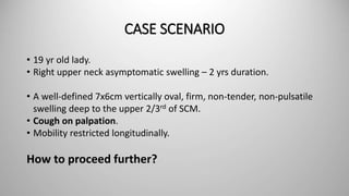
- 1. CASE SCENARIO • 19 yr old lady. • Right upper neck asymptomatic swelling – 2 yrs duration. • A well-defined 7x6cm vertically oval, firm, non-tender, non-pulsatile swelling deep to the upper 2/3rd of SCM. • Cough on palpation. • Mobility restricted longitudinally. How to proceed further?
- 3. Surgery done : Transcervical excision under GA. Mass between IJV and ICA in the right carotid space. HPE : Schwannoma. Hoarseness of voice in the post-operative period, otherwise uneventful.
- 4. SCHWANNOMAS OF PARAPHARYNGEAL SPACE Dr.Kingston.S Head & Neck Surgery CMC Vellore
- 5. OUTLINE • Anatomy of Para-pharyngeal Space (PPS) • DD for lesions in PPS • Schwannomas of the neck - Presentation - Nerve of origin (via CT/MRI) - Tips during surgical excision - Post-op neurological sequelae
- 7. • Inverted triangular pyramid on either side of pharynx. • Skull base superiorly and greater cornu of hyoid bone inferiorly. • Divided by styloid diaphragm into 2 compartments. A parapharyngeal space schwannoma arising from the vagus nerve: A case report. International Journal of Surgery Case Reports 41 (2017) 22–25 . Vagus nerve schwannoma in the parapharyngeal space: surgical, histological and immunohistochemical aspects. A case report. Rom J Morphol Embryol 2015, 56(1):273–276.
- 8. • Pre-styloid – Deep lobe of parotid, V3, IMA, APA, pharyngeal venous plexus, minor or ectopic salivary glands and fat. • Retro-styloid – ICA, IJV, cranial nerves from IX to XII, CSC, the glomus bodies and lymph nodes.
- 9. DD OF PPS LESIONS • Mostly (70-80%) benign; 20% may show malignant transformation. • Congenital – branchial cysts, teratomas. • Primary, metastatic or tumoral extensions. • Primary - salivary gland tumors or neurogenic tumors. • Others - vascular malformations. A parapharyngeal space schwannoma arising from the vagus nerve: A case report. International Journal of Surgery Case Reports 41 (2017) 22–25. Vagus nerve schwannoma in the parapharyngeal space: surgical, histological and immunohistochemical aspects. A case report. Rom J Morphol Embryol 2015, 56(1):273–276.
- 10. Parapharyngeal Space Primary Tumours. ActaOtorrinolaringol Esp. 2017;68:138---144.
- 11. SCHWANNOMAS OF THE NECK • Benign, slow growing and asymptomatic tumours. • Schwann cells of peripheral, cranial or autonomic nerves. • 45% of all extra-cranial schwannomas in H&N, with <1% in oral cavity. • Most commonly originate from the V, VII, X, XI and XII cranial nerves and CSC. (never in I & II) A parapharyngeal space schwannoma arising from the vagus nerve: A case report. International Journal of Surgery Case Reports 41 (2017) 22–25 . Schwannoma of cervical sympathetic chain: assessment and management. ACTA OTORHINOLARYNGOL ITAL 25, 191-194, 2005. Diagnosis and management of schwannomas originating from the cervical vagus nerve. Ann R Coll Surg Engl 2015; 97: 92–97.
- 12. • Incidental or asymptomatic slow growing masses (neck/throat). • Pain and paraesthesia in 50%. • Rapidly growing, trismus, otalgia, cranial palsies - ? Malignancy. • Dysphagia, difficulty breathing and hoarseness. • Occasional palpation of the mass may induce paroxysmal coughing in VNS. • Horner’s syndrome – rare. A parapharyngeal space schwannoma arising from the vagus nerve: A case report. International Journal of Surgery Case Reports 41 (2017) 22–25. Ford LC, Cruz RM, Rumore GJ, Klein J. Cervical cystic schwannoma of the vagus nerve: diagnostic and surgical challenge. J Otolaryngol. 2003 Feb; 32(1):61-63.
- 13. • Systematic neurological examination. • CT - well defined and fusiform, hypodense compared to the muscle; with contrast, shows some degree of enhancement. • MRI - low signal intensity on T1 and high signal intensity on T2- weighted images, with variable texture. • USG • Guided FNAC • IDL Primary parapharyngeal tumours: a review of 21 cases. Oral Maxillofac Surg. DOI 10.1007/s10006-014-0451-8. Schwannoma of cervical sympathetic chain: assessment and management. ACTA OTORHINOLARYNGOL ITAL 25, 191-194, 2005. Diagnosis and management of schwannomas originating from the cervical vagus nerve. Ann R Coll Surg Engl 2015; 97: 92–97. TRANSORAL FNAC/BIOPSY
- 14. NERVE OF ORIGIN
- 15. Furukawa’s rule (to identify nerve of origin) •Splaying of the CA and IJV is a sign of VNS •Absence of a gap between the CA and IJV combined with anterior displacement of both frequently occurs in CSCS. C.S. Graffeo, K.M. Van Abel, J.M. Morris, M.L. Carlson, J.J. Van Gompel, E.J.Moore, D.L. Price, J.L. Kasperbauer, J.R. Janus, K.D. Olsen, M.J. Link,Preoperative diagnosis of vagal and sympathetic cervical schwannomas basedon radiographic findings, J Neurosurg. 126 (March (3)) (2017) 690–697. 75% VNS 87% CSCS
- 16. T1 and T1 (Gd) MRI of VNS splaying the IJV and ICA Parapharyngeal Space Schwannomas. Preoperative Imaging Determination of the Nerve of Origin. Arch Otolaryngol Head Neck Surg. 2007;133(7):662-667.
- 17. T1 and T2 MRI of CSCS displacing the IJV and ICA together in a lateral direction Parapharyngeal Space Schwannomas. Preoperative Imaging Determination of the Nerve of Origin. Arch Otolaryngol Head Neck Surg. 2007;133(7):662-667.
- 18. Graffeo et al.’s additions •A medially displaced CA was more determinate of VNS and a laterally displaced CA was commonly associated with CSCS. •Peripheral enhancement of the mass on T2-MRI was significantly associated with VNS while homogenous enhancement with CSCS. C.S. Graffeo, K.M. Van Abel, J.M. Morris, M.L. Carlson, J.J. Van Gompel, E.J.Moore, D.L. Price, J.L. Kasperbauer, J.R. Janus, K.D. Olsen, M.J. Link,Preoperative diagnosis of vagal and sympathetic cervical schwannomas basedon radiographic findings, J Neurosurg. 126 (March (3)) (2017) 690–697. 97% VNS 99% CSCS Furukawa + Graffeo =
- 19. SURGICAL APPROACHES • Transcervical (retrostyloid tumours) • Transparotid (prestyloid tumours) • Combined • Transcervical-transmandibular • Infratemporal fossa Primary parapharyngeal tumours: a review of 21 cases. Oral Maxillofac Surg. DOI 10.1007/s10006-014-0451-8. COMPLETE TUMOUR EXCISION + NERVE PRESERVATION
- 20. NERVE INJURY & SURGICAL TECHNIQUE TECHNIQUE PERMANENT DEFICITS TEMPORARY Excision + nerve sacrifice and re-anastomosis 100% Excision + nerve preservation 64% 29% Tumour enucleation/emptying 29% 42% Nerve- sparing Nil <1% Nerve-sparing subcapsular resection of head and neck schwannomas: technique evaluation and literature review. The Journal of Laryngology & Otology (2013), 127, 685–690. Valentino J, Boggess MA, Ellis JL, Hester TO, Jones RO. Expected neurologic outcome for surgical treatment of cervical neurilemmomas. Laryngoscope 1998;108:1009–13
- 21. NERVE-SPARING SUBCAPSULAR RESECTION TECHNIQUE • Based on the principle that nerve fibres get expanded and stretched out on the tumour rather than getting embedded within it - herein preserving the nerve fibres without significantly compromising nerve function as nerve continuity is maintained. • 3.5 magnification surgical loupe. • Meticulous sharp dissection, parallel to the axis of the nerve. • Minimise traction to the surrounding nerve fibres. Nerve-sparing subcapsular resection of head and neck schwannomas: technique evaluation and literature review. The Journal of Laryngology & Otology (2013), 127, 685–690.
- 22. POST-OP NEUROLOGICAL SEQUELA Nerve resection is required in 87% of CSCS surgeries as compared to 52% of VNS cases. •Hoarseness, dysphonia and dysphagia in VNS. •Horner’s syndrome and first bite syndrome (FBS) in CSCS. Cervical Sympathetic Chain Schwannoma Masquerading as a Vagus Nerve Schwannoma Complicated by Postoperative Horner’s Syndrome and Facial Pain: A Case Report. International Journal of Surgery Case Reports 49 (2018) 4–7 Cervical Vagal Schwannoma. Philippine Journal Of Otolaryngology-Head And Neck Surgery Vol. 25 No. 2 Jul – Dec 2010 Parapharyngeal Space Primary Tumours. ActaOtorrinolaringol Esp. 2017;68:138---144.
- 23. A parapharyngeal space schwannoma arising from the vagus nerve: A case report International Journal of Surgery Case Reports 41 (2017) 22–25 Cervical Vagal Schwannoma. Philippine Journal Of Otolaryngology-Head And Neck Surgery Vol. 25 No. 2 Jul – Dec 2010 Vagus nerve schwannoma in the parapharyngeal space: surgical, histological and immunohistochemical aspects. A case report Rom J Morphol Embryol 2015, 56(1):273–276 S100 +ve CD34 –ve Antoni Cells A (Black Circle) Antoni Cells B (White Circle) Verocay body Palisading typical, elongated nuclei disposed in concentric rows around eosinophilic material.
- 24. TO CONCLUDE… • Common tumor in PPS – benign. • Pre- and Post-styloid compartments in PPS. • Imaging to confirm schwannoma. • If schwannoma, find nerve of origin – remember Furukawa and Graffeo. • Nerve-sparing resection technique using loupe magnification. • Postop rehabilitation.
Editor's Notes
- GM everyone. I welcome you all to the Head & Neck Surgery’s academic session. Let me start my presentation with a case scenario.
- Divided by the styloid diaphragm
- Divided by the styloid diaphragm External wall formed by an aponeurotic muscle bundle comprising the SCM, the superficial cervical aponeurosis lining the parotid, and the ascending ramus of the mandible. Posterior wall is formed by the prevertebral muscles and the cervical transverse apophyses. As we see from the contents….a wide variety of tumours can arise from this compact space
- Pleomorphic adenomas from minor salivary glands. Neurogenic tumors are second most common in prestyloid PPS, and most common in retrostyloid PPS. Shwannomas and paragangliomas.
- The relative inaccessibility of the lesions, their heterogeneous pathology, frequent lack of any tissue diagnosis prior to surgery are major challenges in preoperative assessment and surgical treatment.
- Pertaining to PPS, they commonly arise from Vagus and CSC.
- PPSS present either as an incindental finding on imaging for other issues or as asymptomatic slow-growing neck/throat mass These are the common modes of presentation
- As usual, the workup starts with the history taking, general and systemic examination. A complete neurological exam should be done to pickup any cranial nerve palsies especially the lower ones. Then comes the imaging procedures Not only for diagnosis but also to obtain topographical info to help us choose the surgical treatment. It tells about pre-styloid or retro-styloid location, its relationship with the parotid gland, the large vessels and the radiological characteristics of the tumour. On CT, internal cystic changes becoming more prominent as the tumour enlarges. Cystic changes - mucinous degeneration, haemorrhage, necrosis and the formation of microcysts. Ultrasonography has greater diagnostic utility if the diameter of the nerve of origin is large and often the tumour is connected to a clearly delineated nerve. When carotid artery involvement is suspected, surgery can be planned via preoperative angiography and executed with the help of a vascular surgeon. Although CTA and MRA now seem to have replaced catheter angiography, DSA still has a role to play in preop embolization is and balloon occlusion procedures to measure cerebral blood flow and the adequacy of the contralateral circulation Blind FNAC is not advisable in PPS tumours. If required, guided FNAC can be done and that too can be avoided in the poststyloid lesions, as it presents major risks and is often inconclusive, at least for neurogenic tumours the transoral biopsy/FNAC route is currently contraindicated – it presents greater risk of haemorrhage, tumour dissemination and, lastly, there is fibrosis increases the risk of oropharyngeal fistula in later surgery
- Paresis or paralysis of one of few of the lower cranial nerves is the mcc in the postop period. Hence finding out the nerve of origin is essential for the pre-operative counseling and to plan postop rehabilitation.
- The vagus nerve runs between the IJV and ICA in the entire carotid sheath; hence vagal tumors tend to separate these vessels. The sympathetic chain is located posteromedial to both of these vessels; hence such a separation is not usually seen with SCSCs.
- Apart from splaying…
- The approach depends on location, the relationship with the large blood vessels and suspicion of malignancy. transcervical or a transcervical/transparotid approach, neoplasms were more frequently located in the inferior or middle parts of PPS and had not extended to the skull base A combined cervical-transparotid approach, by extending the submandibular incision to the parotid region, tumours arising from the deep parotid lobe. A transcervical-transmandibular approach for large and/or malignant or vascular tumours, especially if extending into the superior part of the PPS An infratemporal fossa approach is indicated for benign or malignant tumours involving the skull base and cranial cavities As schwannomas are benign, all efforts should be made to preserve nerve function so that quality of life is unaffected. Tumor arise outside the involved nerve fascicle Tends to compress the nerve fascicles to the periphery Can be dissected free except the parent fascicle
- A number of techniques have been defined by many authors…
- The nerve-sparing subcapsular resection technique preserves the nerve fibres, without significantly compromising the nerve function as nerve continuity is maintained. the mass is delineated on all sides. The proximal and distal ends of the nerve abutting the tumour are identified and meticulous sharp dissection is carried out, parallel to the axis of the nerve
- Despite this, most complications and sequelae are well tolerated. If not, to start aggressive rehabilitation for voice and speech. Isolated injury to the vagus nerve causes dysphonia, but rarely aspirations. However, when this nerve is simultaneously involved with cranial nerves IX and XII, swal-lowing problems arise, which can even require insertion ofa nasogastric tube and, in very rare cases, performing a gas-trostomy or percutaneous endoscopic gastrostomy (PEG) toachieve contralateral compensation. Techniques for medial-isation of the paretic vocal fold, such as type I thyroplasty,can be used (3 patients in our series). We have noted that inolder patients having prior nerve palsy (X, XII, IX) after theremoval of a neurinoma or parapharyngeal paraganglioma,without additional nerve lesions after surgery, aspiration andpoor swallowing arose and worsened. Compensation of swal-lowing and phonation are closely related to patient age and,the older the patient, the worse the compensation. The classic triad of miosis, anhidrosis, and ptosis generally goes unnoticed by patients. Postoperative Horner’s syndrome rarely requires treatment and is ordinarily asymptomatic, but symptomatic patients commonly show gradual improvement. The ptosis due to paralysis of Müller’s muscle can be repaired through slight advancement of the levator aponeurosis, or resection of the conjunctiva and Müller’s muscle FBS presents with pain radiating throughout the jaw elicited on the initial bite of each meal that resolves with ensuing mastication. The pathophysiology of FBS is likely due to sympathetic nerve damage causing denervation supersensitivity of the intraparotid myoepithelial cells (treatment - botox). Although the pain appears to be aligned with the distribution of the trigeminal nerve, the duration of this patient’s pain and the lack of contact with the trigeminal nerve during surgery makes trigeminal neuralgia an unlikely diagnosis. PIFP is described as persistent facial pain recurring daily for more than 2 hours per day for greater than 3 months, in the absence of clinical neurological deficit. PIFP is often associated with post-traumatic or post-surgical onset. Post surgery atelectasis of the lung is usually the main cause of pneumonia and aggressive chest physiotherapy. Pneumonia may also occur as a result of aspiration secondary to vocal cord palsy possibly due to direct trauma to vagal trunk, traction injury or even compression from hematoma
- Histologically, schwannomas are well circumscribed and encapsulated High power view of HE stained section, highlighting hyperchromatic spindle cells with ancient change typical clusters of alternating areas of compact hypercellular with spindle shape cells (Antoni A) with areas of loose hypocellular patterns. S100 protein (neurofibroma) and GFAP were positive indicating a neural tumor CD34 (excluding a vascular tumor and a solitary fibrous tumor).