Managment of common neonatal problems
•Download as PPTX, PDF•
105 likes•38,957 views

Neonatology

Recommended
More Related Content
What's hot
What's hot (20)
Viewers also liked
Similar to Managment of common neonatal problems
Similar to Managment of common neonatal problems (20)
Recently uploaded
Recently uploaded (20)
Managment of common neonatal problems
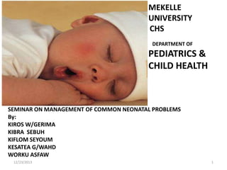
- 1. MEKELLE UNIVERSITY CHS DEPARTMENT OF PEDIATRICS & CHILD HEALTH SEMINAR ON MANAGEMENT OF COMMON NEONATAL PROBLEMS By: KIROS W/GERIMA KIBRA SEBUH KIFLOM SEYOUM KESATEA G/WAHD WORKU ASFAW 12/23/2013 1
- 2. Seminar outline Neonatal Sepsis Hypothermia Respiratory distress syndrome Perinatal Asphyxia Neonatal Seizure Neonatal Jaundice Rh and ABO incompatibility New born Anemia Hypoglycemia References and Sources 12/23/2013 2
- 3. NEONATAL SEPSIS by Kiros Weldegerima 12/23/2013 3
- 4. Sepsis Outline of presentation • • • • • • • • Classifications Risk factors Clinical Manifestations Meningitis and Pneumonia Diagnostic work up Management principles Prevention Strategies of Sepsis Hypothermia and its management 12/23/2013 4
- 5. Neonatal Sepsis Infection or sepsis is a problem faced to all new borns But the risk and severity is high in small and premature infants It is the cause of 30-50 % of Neonatal mortality in developing countries Sepsis in the Neonate includes meningitis, septicemias, pneumonia, Arthritis, o steomyelitis, and UTI or combinations of those. 12/23/2013 5
- 6. • Neonatal Sepsis can be divided as early and late onset depending on the time of occurrence • Early-onset neonatal sepsis occurs with in the first 72 hrs of life in 90% of cases – Is caused by organisms prevalent in the maternal genital tract – Or in the labor room or operation theatre where the neonate exposes initially to the Environment 12/23/2013 6
- 7. Early onset Causative Agents – Majority are caused by Group B streprococcus, E-coli, klebsiela and enterobacter – Majority manifest with respiratory distress due to an early intrauterine pneumonia – The manifestation of illness is earlier than the time limit of 1 week (24hr=85%, 2448hr=5%, 2-6 days=10) 12/23/2013 7
- 8. RISK FACTORS Neonatal sepsis is associated with certain high risk obstetric factors; -Birth asphyxia -Unclean vaginal examination -foul smelling liquor -prolonged labor(>24 hours) -preterm and low birth weight Neonates -prolonged rupture of membranes(>18 hours) -maternal pyrexia 12/23/2013 8
- 9. Late onset causative Agents • Late onset neonatal sepsis occurs after 7 days most of which is after the first week of life up to 90 days. • Most are caused by gram negative bacteria – Klebsiela, enterobacter, E-coli, pseudomonas and salmonella – Gram positives like staphylococcus aureus also contribute as causes of late onset sepsis. 12/23/2013 9
- 10. Infection Acquiring areas Late onset infections are acquired as nosocomial after delivery in: -the normal newborn nursery -Neonatal Intensive care Unit or -the Community. 12/23/2013 10
- 11. Sources of infection in late onset The usual sources of late onset neonatal sepsis: -Incubators -Resustation Equipment -Feeding bottles -Catheters -Infusion sets and sites 12/23/2013 11
- 12. Clinical features • It may be subtle especially in those who are very small and premature • This is mostly due to depressed immunity of the premature neonates • Early manifestations could be change in behavior or feeding patterns • But Gradually/sometimes suddenly they develop signs and symptoms 12/23/2013 12
- 13. Clinical features of sepsis Common clinical Features are -Lethargic/Unconscious -Inactive/Unresponsive -failure to suck -Hyperthermia/Hypothermia -Respiratory Distress/Apnea -failure to gain weight/weight loss -Anemic/pale conjunctiva, palms 12/23/2013 13
- 14. Bacterial meningitis • About a third of babies with neonatal sepsis can have coexisting meningitis • It is more common with late onset neonatal sepsis • Clinical evidence of meningial irritation are usually absent in the new born period • So to diagnose meningitis in New born we should see other Clinical clues 12/23/2013 14
- 15. Meningitis cont.… In a baby with sepsis the findings of -convulsion/twitching -staring look/fixed eye -Bulged anterior fontanel -abnormal excessive high pitched cry Should arouse the suspicion of meningitis and proceed with lumbar puncture. 12/23/2013 15
- 16. Lumbar puncture result that Indicates meningitis 12/23/2013 > 30 cells/ml in Neonates , other than Neonate >5 cells/ml of CSF >60% polymorphs cells –CSF glucose /blood glucose ratio <50% –Protein>150 mg/dl in Terms and >175 in preterm –Presence of microorganisms –If the CSF not clear may also suggest abnormality. 16
- 17. Pneumonia in the newborn • Is more common in early onset neonatal sepsis • Some peculiarities of pneumonia in the NB – Minimal clinical signs – Generalized signs of sepsis predominate esp in premature NBs – Some neonates can have apnea rather than tachypnea. – Clinical signs of pneumonic consolidation may not be evident in the neonatal period 12/23/2013 17
- 18. Diagnostic work up of sepsis • Clinical features + presence of high risk obstetric factors • Blood culture and sensitivity • CSF analysis when indicated • Chest X-ray • Urine analysis and /or urine culture • Head to Toe physical examination to identify focus of infection (bone, joint, skin, GI) 12/23/2013 18
- 19. Management • Depending on the etiologic agent but Till etiologic agent is identified:– Good broad spectrum coverage i.e. for gram +ve and gram –ve organisms is essential • First line drugs – Crystalline penicillin 100,000iu/kg /day in two divided doses and – Gentamicin 5 mg/kg/day in two divided doses – Change crystalline penicillin with Ampicillin if Listeria monocytogens is incriminated as the causative agent 12/23/2013 19
- 20. Management In meningitis the dose of Ampicillin and Crystalline penciline are doubled. Second line drugs – Ceftriaxone 50mg/kg/dose BID + – Aminoglycoside dose which is also 50mg/kg BID. • Dose of ceftriaxone is doubled in cases of meningitis • Supportive therapy to correct metabolic complications 12/23/2013 20
- 21. Management cont. Duration of therapy -suspected sepsis( blood culture –ve)= 7 days -proven sepsis(blood culture +ve)=14 days -Gram positive meningitis=14 days -Gram negative meningitis=21 days -Septic arthritis/osteomyelitis=4-6 weeks 12/23/2013 21
- 22. Prevention strategies of Neonatal infections Universal precautions -Hand washing before entering labor ward and before and after examining each infant -wear Gowns and slippers when entering NICUs -Health care providers with acute infections( fever,ARI,skin lesions and Viral exanthemas) should be restricted from providing care to the Neonates 12/23/2013 22
- 23. Prevention Cotd… -keep clean both the NICU and labor ward by Cleaning and Fumigating at regular interval -Proper skin and cord care -Keep all Equipments used in NICU and labor ward clean so there will be no infection source. 12/23/2013 23
- 24. Hypothermia • Skin temperature of <36.5 and core temperature of <35.5 • Neonates have high surface area to volume ratio ,so heat loss is so much higher. • After birth , the skin and core temperature of the baby fall by 0.1 and 0.3/min respectively .Which is equivalent to heat loss of 200kcal/kg body weight/minute.
- 25. Mechanisms of heat loss/production Mechanisms of heat loss: 1 Convection 2 Conduction 3 Radiation 4 Evaporation(common source of heat loss) Mechanism of heat production 1 muscular activity 2 metabolic thermogenesis(most important source of heat production in the new born)
- 26. Thermo Regulation Thermo neutral environment :This is the ideal temperature at which the baby can maintain normal body temperature. The optimal function of heat generating system is dependent up on the integrity of -CNS thermo regulation system -adequacy of brown fat -availability of glucose and oxygen -NBW and term gestational age.
- 27. Stages of hypothermia 36-36.4 c (96.8-97.5f) -mild hypothermia (cold stress) 32-35.9 c (89.6-96.6f) -moderate hypothermia <32 c (89.6f) -severe hypothermia (neonatal cold injury)
- 28. Warm chain system • System of keeping the baby warm immediately after birth,in delivery room,post partum ward ,transportation and while nursing the baby at home. components: -immediate drying -warm resuscitation -skin to skin contact with the mother -immediate initiation of breast feeding -bathing and weighing post pond -appropriate clothing and bedding -warm transportation.
- 29. Causes of hypothermia External factors cold environment,wet or naked baby ,blood sampling,IV sampling Poor ability to conserve heat large surface area,poor insulation,paucity of fat, Poor metabolic heat production -deficiency of brown fat(preterm ,SFD) -CNS problems -hypoxia -hypoglycemia
- 30. Sign and symptoms of hypothermia 1 peripheral vasoconstriction Acrocyanosis cold extrimity decreased peripheral perfusion 2 CNS depression Lethargy Poor feeding Apnea and bradycardia 3 Increased metabolism hypoglycemia hypoxia metabolic acidosis 4 Increased pulmonary arterial pressure tachypenia respiratory distress
- 31. Management of hypothermia There are 3 methods: 1.Kangaroo mother care 2.Warming in an open care using a radiant heater 3.Warming in an incubator Hazard’s of temperature control: hyperthermia undetected infection volume depletion.
- 32. RESPIRATORY DISTRESS and APNEA IN THE NEWBORN PREPARED BY KIBRA.S 12/23/2013 32
- 33. RESPIRATORY DISTRESS Definition • The presence of any one of the following four clinical features : – RR > 60/min ( counted two times for full one minute) – Significant lower chest indrawing – Grunting – Central cyanosis 12/23/2013 33
- 34. ETIOLOGY 1.Pulmonary cause Lung parenchyma disease Congenital airway obstruction Intrathoracic malformation Hyaline membrane disease Choanal atresia, nasal edema Pulmonary hypoplasia or agenesis Meconium aspiration syndrome Maroglossia, micrognathia, retrognathia Diaphragmatic hernia Congenital pneumonia Congenital goiter, cystic hygroma Intra thoracic cyst Transient tachypnea of the Subglottic stenosis , newborn laryngomalacia Bronchopulmonary dysplasia Congenital lobar emphysema Tracheomalacia, congenital tracheal stenosis Pulmonary hemorrhage, air leak 12/23/2013 34
- 35. 2.Extrapulmonary cause Cardiac Metabolic Neurological Hematological Congenital heart disease Hypoglycemia Neonatal meningitis Anemia Congestive heart failure Hypocalcaemia Neonatal seizure Cardiac arrhythmia Hypothermia Hypoxic ischemic encephalopathy Metabolic acidosis Extreme immaturity Polycythemia Intracranial bieed 12/23/2013 35
- 36. APPROACH History • • • • • • • 12/23/2013 Gestational age Previous preterm baby Antenatal steroid prophylaxis Maternal DM Ante partum hemorrhage PROM Prolonged duration of labor 36
- 37. CONT’D • • • • • • • 12/23/2013 Maternal fever Unclean vaginal examination Foul smelling, meconium stained liquor Hx of intrapartum or post partum suctioning Excessive salivation Difficulty of feeding Hx of traumatic delivery 37
- 38. CONT’D Physical Examination Vital sign, capillary refill time and SO2 Meconium staining of the cord or nail Hyperinflated chest or with bowel sound Cyanosis, tachycardia, murmur Scaphoid abdomen, hepatomegally Evidences of intracranial bleed Unable to pass nasal catheter 12/23/2013 38
- 39. CONT’D Investigation • • • • • • 12/23/2013 Blood group of mother and baby CBC , CXR Septic screening & complete septic work up Serum electrolytes and blood sugar gastric aspirate (shake test, PMN cells) Cord pH, arterial blood gas 39
- 40. MANAGMENT General MX – Give vit.k if not given – Basic support care • Keep in thermo neutral zone • Breast feeding or assist feeding • Maintain adequate oxygenation & circulation Specific TX for specific problems – Chest tube, decongestive measures, volume expanders, antibiotics, surgical correction 12/23/2013 40
- 41. Hyaline Membrane Disease Incidence – Primarily in premature infants; inversely proportional to GA & birth weight Risk factor – – – – – – Prematurity Maternal DM Male sex 2nd born twins C/S delivery Perinatal asphyxia Protective factor 12/23/2013 41
- 43. CONT’D Clinical Feature -Signs occur with in minutes or hours of birth, reach peak with in 3 days -Characteristically • • • • • 12/23/2013 Tachypnea Nasal flaring SC or IC retraction Cyanosis Expiratory grunting 43
- 44. CONT’D Diagnosis ─based on clinical picture in conjunction with characteristic chest radiograph ─The CXR abnormalities: Low lung volume Diffuse reticulogranular ground glass appearance with air bronchogram 12/23/2013 44
- 45. CXR findings in Classic RDS * Bell-shaped thorax * ↓lung volume * Air bronchogram extended beyond cardiac border * Absolutely opaque lung (whiteout lung) 12/23/2013 45
- 46. CONT’D Management Basic supportive care Specific measures Prevent hypoxia & acidosis Warm humified o2 administration CPAP(indication, procedure & complication) Assisted ventilation Surfactant replacement therapy (poractant,calfactant,beractant) Prevention Prenatal testing & appropriate prophylaxis 12/23/2013 46
- 47. Congenital pneumonia • Is acquired transplacentally or perinatally • Accounts for >50% of cases of RD in the new born Risk factor –PROM –Prolonged labour (> 24 hrs) –Unclean vaginal examinations. –Foul smelling liquor –Maternal fever 12/23/2013 47
- 48. CONT’D Causative organisms: -Group B streptococcus -Gram negative organisms : E.coli, klebsiela, pseudomonas -Staph aureus and Listeria monocytogens Clinical manifestations Respiratory distress soon after birth Recurrent apneic attack They are often asphyxiated and sick at birth Prolonged capillary filling time Hypothermia Cough is rare 12/23/2013 48
- 49. Investigations – CBC, esp. ANC – Gastric aspirate for PMN – Blood culture – CXR(infiltrates, lobar consolidation, interstitial reticular opacities) 12/23/2013 49
- 50. Management – Basic supportive therapy – Specific • Emperical broad spectrum antibiotics – Penicillin/Ampicillin 100mg/kg in 2 divided doses + Gentamicin 4mg/kg q24hr or 2.5 mg/kg q12hr 12/23/2013 50
- 51. Meconium Aspiration Syndrome Definition • Respiratory distress in newborn infants born through meconium stained amniotic fluid whose symptom cannot be otherwise explained Incidence ─10-15% of births-meconium stained amniotic fluid – 5% -meconium aspiration pneumonia • 30% require mechanical ventilation • 3-5% expire 12/23/2013 51
- 52. CONT’D Risk factors • Placental dysfunction • Fetal hypoxia • Ante partum haemorrhage • Post maturity or SGA • Listeriosis • Breech delivery 12/23/2013 52
- 53. CONT’D Pathophysiology ─Fetal hypoxia – Peristalsis & IU passage of meconium – Meconium stained amniotic fluid – Meconium aspiration – Peripheral & proximal air way obstruction, inflammatory &chemical pneumonitis 12/23/2013 53
- 54. CONT’D Clinical feature – Respiratory distress- onset soon after birth in a baby born to mother with meconium stained liquor . – Hyper inflated chest – Meconium stained skin and cord – Urine may appear dark and brown 12/23/2013 54
- 55. CONT’D Investigation – CXR • Hyperinflation • Bilateral fluffy shadows • Evidence of air leak 12/23/2013 55
- 56. 12/23/2013 56
- 57. CONT’D Management ─Prevention of meconium aspiration • Prevention of IU hypoxia • Intrapartum suctioning – Postnatal suctioning • Thick meconium – – – – – Direct laryngoscopy & tracheal suction Stabilize the baby Stomach wash Work up for sepsis Antibiotics can be started • Thin meconium – Depressed baby-do all like thick meconium – Active baby-no tracheal suctioning and needs close observation 12/23/2013 57
- 58. APNEA Definition – Cessation of respiratory airflow – Absence of respiratory movement • Apnea vs periodic breathing Incidence – Increases with decreasing GA 12/23/2013 58
- 59. Classification • Based on the cause – Primary(apnea of prematurity) – Secondary • Based on presence of continued inspiratory effort and upper airway obstruction – Central – Obstructive – Mixed 12/23/2013 59
- 60. CONT’D Pathogenesis unable to react to hypercapnia • • • • • • • 12/23/2013 Impaired respiratory response(hypercapnia) Apnea ↓HR, Pao2 Hypoperfusion and hypoxia Hypercapnia Recurrent apnea(≥3 attacks in one hour) Brain damage & multiorgan damage 60
- 61. CONT’D Management General management – – – – – ABC of resuscitation Avoid vigorous suctioning Keep NPO for 24 hrs put them on maintenance IVF Keep in thermoneutral environment Treat the underlying cause of apnea Specific therapy • Drug─theophylline ─aminophylline ─ caffeine • Positive pressure ventilation 12/23/2013 61
- 62. prepared by kiflom seyoum 12/23/2013 62
- 63. Layout • • • • • • • • Definition epidemiology Pathophysiology Risk factors Clinical features Management Prevention Neonatal seizure 12/23/2013 63
- 64. Perinatal Asphyxia Definition: PNA is an insult to the fetus or newborn due to lack of oxygen ( hypoxia ) and /or a lack of perfusion ( ischemia ) to various organs. Failure to establish efficient breathing at one minute of age(APGAR score 0 - 6) with hypo/hypertonia and/or seizure. This definition using APGAR score is not applicable in Babies with birth trauma 12/23/2013 Preterm babies Congenital neurologic abnormalities 64
- 65. Epidemology • • • • • Ranges 1-1.5% in gneral 9% in babes born <36 weeks of gastion 0.3% in babies born >36 weeks of gastion Accounts 23% of perinatal death 23-30% of survivors have permanent damage like CP 12/23/2013 65
- 66. Timing of ingury • Asphyxia can occur in the, Antepartem Intrapartem Postnatal priod 12/23/2013 66
- 67. Risk factors 1 .Antepartem condition Abnormal maternal oxygenation ( sever animia, cardiopulmonary disease) Inadequate placental perfusion( sever HTN, maternal vascular disease) Congenital infection or anomalies 12/23/2013 67
- 68. Conti… 2. intrapartem events Interruption of umblical circulation (true knote, cord prolaps) inadequate placental perfusion ( abrabtio placenta, utrine rupture, abnormal utrin contraction ) Abnormal maternal oxygenation Vasa previa 12/23/2013 68
- 69. Conti… • 3, post natal disorders Persistent plumnary hypertention of the new born Sever circulatory insuficency ( acut blood loss, septic shock ) 12/23/2013 69
- 70. Eitology and pathphysiology 90% of insult occur due to placental insufficiency Decrease oxygen supply & Decrease carbon dioxide and hydrogen removal <10% are post natal insult due to pulmonary, CVS or neurologic insufficiency 12/23/2013 70
- 71. Cont… • When the new born is depraved of O2 an initial period of rapid breathing occur • This is followed by primary apnea • Which responds to O2 administration and physical stimulation 12/23/2013 71
- 72. Cont… If asphyxia contnues the new born devoleps Gasping respiration Decreased puls rate Blood pressure fail And the new born develops secondary apnea which will respond only to advanced life support including CPAP 12/23/2013 72
- 73. Con… • If asphyxia is not reversed on time there will be hypoxic damage that leads to ischemic challenge • A diving reflex will be initiated that causes shunting of blood to vital organs 12/23/2013 73
- 74. Clinical feature • Depends on the organ affected • Target organs of prenatal asphyxia Kidney in 50% of cases CNS in 28% of cases CVS in 25% of cases Pulmonary 23% of cases 12/23/2013 74
- 75. Renal dysfunction • Often accompanies prenatal asphyxia • renal damage ranges from reversible cloudy swelling & hydropic digeneration of tubles to infarction of the entire nephron. • Decrease in UO(<0.5ml/kg/hr which may last 2 day to 2 weeks • Protein & cast may present in the urin • Gross hematuria may present • Elevation of BUN& creatinine may occur • Poly urea may flow oliguric phase or they may develop anuria 12/23/2013 75
- 76. CVS dysfunction • May cause myocardial ischemia which usually is transient • Patients show tachypnea,tachycardea & hepatomegally consistent with heart fealur • Rarely causes cardiogenic shock and death 12/23/2013 76
- 77. Pulmonary dysfunction 1.Plumonary edema Due to myocardial dysfunction May have sign of respiratory distress 2.Acute respiratory distress syndrome Increased plumnary capilary permebility to plasma protien which leads to inactivation of surfactant 12/23/2013 77
- 78. Hypoxic ischemic encephalopathy • The most serous complication of perenatal asphyxia • The neuralgic squale often persists • Hypoxia impairs cerebral oxidative mechanism and leads to myocardial depression this leads to fail in cerebral blood flaw • And then ischemia of the brain tissue occur • Persistance of hypoxia increase anarobic glycolysis which leads to lactic acidosis • Worsening of lactic acidosis if not corrected on time cuases loss of cerebral vascular auto regulation 12/23/2013 78
- 79. Conti… • Most of the time prolonged partial episode of aspheyxia is because of abruptio placenta • And usually it causes diffuse cerebral necrosis • Clinically this may be present in the form of seizure and paresis. • Acute total asphysia which is mainly due to cord plolapse primerly affects brain stem,thalamus, & basal ganglia. • This may manifest in the form disturbance of respiration,heart rate & blood pressure 12/23/2013 79
- 80. Investstigations • EEG-to determine severity presence of seizure • Cranial u/s- to determine presence ICH & brain edeama • CT scan –early (2-4 days) brain edema -late(2-4weeks) for encephalomalesia 12/23/2013 80
- 81. Sarnat staging of hypoxic-ischemic encephalopathy. Grade 1 (mild) Grade2 (moderate) Level of consciousness Irritable/hyperalert Lethargy Coma Muscle tone Normal or hypertonia Hypotonia Flaccid Tendon reflexes Increased Increased Depressed or absent Seizures Absent Frequent Frequent Complex reflexes Normal weak Absent Prognosis Good (100%) Normal 12/23/2013 Grade 3 (severe) Variable High mortality and (80% ) Normal neurological disability (50% Death 50% major sequelae) 81
- 82. Management of perinatal asphyxia • Anticipate need for neonatal resuscitation from maternal obstetric & labor history • Avoid blood pressure fluctuation • Intravenous fluid 2/3 of maintenance • Appropriate ventilation support • Correct metabolic abnormalities • Prophylactic anti convulsant (phenobarbital)..? 12/23/2013 82
- 83. Therapeutic hypothermia • It improves out come after perinatal asphyxia • The only effective neuro protective therapy currently available for Tx of neonatal encephalopathy • Safe and easy to administer • Selective head cooling & whole body cooling…? 12/23/2013 83
- 84. Prevention of prenatal asphyxia The minimum preventive measure which is provided during perinatal period is much better than a sophisticated care provided to an asphyxiated new born. Prenatal assessment of changing fetal and placental condition by clinical assessment and altrasonography Fetal BPP Monitor progress of labor Effective neonatal resuscitation 12/23/2013 84
- 85. Neonatal seizure The most important & common indicator of significant neurologic dysfunction in the neonatal period The immature brain is more likely to develop seizure Not similar to adult b/c of ariboraisation of axons dendrites & myelin sheath 12/23/2013 85
- 86. Cont… • They are five clinical types of NS 1.subtle 2.clonic 3.Tonic 4.spasem 5.Myoclonic 12/23/2013 86
- 87. Con… • Based on EEG NZ classified into three Electroclinical seizure Electrical seizure Clinical seizure 12/23/2013 87
- 88. causes 1.Age 1-4 days HIE IVH Drug toxicity,(lidocain,pencilin) Acute metabolic disorder 12/23/2013 88
- 89. Conti… Age (4-14 days) Infection Metabolic disorder Benign neonatal convelsion kernicterus 12/23/2013 89
- 90. Cont… Age (2-8wks) Infection Head injury Inherited disorder of cortical development 12/23/2013 90
- 91. Diagnosis Post natal & prenatal history Blood should be obtaine Lumbar puncture EEG(show paroxysmal activity, sharp wave ) 12/23/2013 91
- 92. Management of NS Treatment of the underlying etiology Complete control of clinical as well as electrographic seizure vs clinical seizure control…? Neonates required assisted ventilation after receiving IV or PO loading doses of AED 12/23/2013 92
- 93. CONT… 1.Phenobarbitol The drug of first choice in the neonatal seizure 20mg/kg loading dose, if not effective an additional dose of 5-10mg/kg until a maximam dose of 40mg/kg, the mentenance doses is 3-6mg/kg 2.Phenytoin If no response phenytoin loading dose of 15-40mg/kg can be given Rate must note exced 0 .5-1mg/kg/min to pereven cardiac toxicty 12/23/2013 93
- 94. Cont… 3.Lorazepam; the initial drug used to control acute seizure Can be used as first line or second line Tx in the new born who didn’t respond to phenobarbitol or phenytone 4.diazepam; It is highly lipophilic so cleared very quickly out carring the risk of recurrence of seizure 12/23/2013 94
- 95. When to discontinu…..? • At the time of discharge • two wks or three month after discharge if the neonate had sever PNA 12/23/2013 95
- 96. prognosis • Mortality from neonatal seizure has decreased from 40% to 20%. • The correlation b/n EEG & prognosis is very clear • Prolonged electrographic seizure >10min/hr, multifocal periodic electrographic discharge,& spread of electrogrophic seizure to contralateral hemospher has poor prognosis • Seizure due to HIE 50% of them have normal out come 12/23/2013 96
- 98. JAUNDICE 98 Visible form of bilirubinemia Adult sclera >2mg / dl Newborn skin >5 mg / dl Occurs in 60% of term and 80% of preterm neonates Is common problem and is mostly benign. However, significant jaundice occurs in 6 % of term babies 12/23/2013
- 99. 99 The conjugated bilirubin will go to bowel with bile. In adults E.coli and C.perfirngens present but absent in neonets. β-glucourinidase enzyme present in newborns. Conjugation and enterohepatic resorption. 12/23/2013
- 100. 100 Direct Vs Indirect? Van den Bergh reaction Indirect billirubin acts as an anti-oxidant It is only the indirect billirubin which crosses the BBB and results in billirubin encephalopathy 12/23/2013
- 101. Physiologic Jaundice 101 Jaundice becomes evident as physiologic in neonates B/c : A. Short life span of RBCs(70-90days) B. RBC mass is increased C. Immature ligandine D. Less UDPGT E. High activity β-glucuronidase (gut) F. Decreased flora in the gut 12/23/2013
- 102. Physiologic jaundice ( Icterus neonatorum) 102 Preterm Term Peak time 4th -7th days 2nd – 4th day Peak level 8 – 12 mg/dl 5 -6 mg/dl Resolution time Before 10th day 5th – 7th day 12/23/2013
- 103. Physiologic Vs pathologic 103 Signs PhysiologicJx Pathologic Jx Clinical Jx Visible in2-3day With in 24hrs TSB rise <5mg/dl/day >5mg/dl/day TSB Term<12mg/dl Preterm<15mg/dl Term>12g/dl Preterm>15mg/dl Conj BBn <1.5mg/dl >1.5(2)mg/dl Jaundice persisting Term <1 week Preterm <2weeks Term >1week Preterm >2weeks 12/23/2013
- 104. Jaundice associated with breast feeding 104 Breast milk Breast feeding Cause ?glucouronidase in ↓ intake BM Time of onset Max level After 1st week In the 1st week 10 – 30 mg/dl Any Treatment Discontinuation for Continuing 2 – 3 days breast feeding 12/23/2013
- 105. ETIOLOGY OF JAUNDICE 105 1. Jaundice appearing in the 1st 24 hr Rh incompatability(erythroblastosis fetalis) ABO incompatibility Mild BG incompatibility (Kelly, Duffy Ags) Defect (G6PD def. and spherocytosis TORCHS Criggler Najar syndrome Transient familial neonatal jaundice 12/23/2013
- 106. Cont… 106 2.Jaundice appearing with in 24-72hr of age Physiological jaundice Conditions w/c aggravate physiologic Jx. Cephalhematoma Hypothermia Prematurity Hypoglycemia Multiple bruises hypoxia 12/23/2013
- 107. Cont… 107 3. prolonged jaundice( after 72 hrs.) TORCHS Sepsis Hypothyroidism Biliary Artesia Breast milk jaundice Drugs (vit. K, oxytocin,diazepam) Metabolic disease eg. galactosemia 12/23/2013
- 108. 108 Hemolytic disease of the newborn Rh incompatibility Less common but sever than ABO incompatibility 90% due to D antigen Black vs white Severity from mild hemolysis to sever anemia Dx by blood group incompatiblity Reduce risk of sensitization with 300μg of human anti- D globulin during ANC ff up 12/23/2013
- 109. Cont… 109 ABO incompatibility Most common cause of HDN but mild 0.3-2.2% only manifest the disease. Type O mother with type A or B infant. Jaundice the only clinical manifestation. Hemoglobin level usually normal. Dx by ABO incompatibility (weak to moderate positive direct coombs test) 12/23/2013
- 110. Clinical assessment of jaundice 110 Jaundice in the newborn progresses in cephalocaudal direction Face =5-7mg/dl Chest =10mg/dl lower abdomen /thigh= 12mg/dl Sole/palms≥15mg/dl 12/23/2013
- 111. Work up neonates with Jx 111 History Age of onset Family history of Jaundice,pallor,splenectomy Previous sibling with Jaundice Maternal illness during pregnancy Maternal drug intake Delivery history e.g. PROM ,sepsis, prolonged labor 12/23/2013
- 112. Cont’d 112 P/E Proper classification of the newborn according to GA, & wgt. Pallor, petechea Bruises Dark and cephalhematoma urine and clay colored stool Examination geared to specific cause 12/23/2013
- 113. Investigations 113 TSB with conjugated fraction Hct with RBC morphology and reticulocyte count Bg of the baby with direct coomb’s test Bg of the mother with indirect coomb’s test. Specific investigations for suspected specific problems 12/23/2013
- 114. Management 114 Aim lower serum billirubin decrease neurtoxicity Principles of treatment Avoid drugs w/c interfere with BBn metabolism Treat factors w/c↑ neurotoxicity Give adequate feeding Specific therapy Decrease serum billirubin 12/23/2013
- 115. 115 Lower serum Billirubin Phototherapy Exchange transfusion Phototherapy Indicated when TSB rises more than normal but not exchange transfusion level May be therapeutic or prophylactic 12/23/2013
- 116. Prophylactic phototherapy 116 INDICATIONS RH isoimmunization with sever hemolysis Birth weight<1000gm(EVLBW) Sever multiple bruises SIDE EFFECTS Erythematous skin rash Retinal damage Increased insensible water loss Bronze baby syndrome Loose stool Low calcium 12/23/2013
- 117. Exchange transfusion( ET) 117 Most effective way of treating Jaundice and anemia Could be partial or double exchange transfusion INDICATIONS Rh isoimmunization with hydrops fetalis History of previous sibling required ET with pallor Cord blood Billirubin >5mg/dl Rise in Billirubin >0.5mg/dl/hr despite phototherapy Hemoglobin <11gm/dl TSB >20mg/dl VLBW, preterm, sepsis 12/23/2013
- 118. Choice of blood for exchange BT 118 ABO incompatibility Use O blood of same Rh type Rh isoimmunization Emergency 0 -ve blood Ideal 0 -ve suspended in AB plasma or baby's blood group but Rh –ve Other situations Baby's blood group 12/23/2013
- 119. Exchange transfusion 119 COMPLICATIONS Portal vein thrombosis Umbilical vein perforation/bleeding Necrotizing enterocolitis Cardiac arrest/arrhythmia Hypoglycemia, hypocalcemia, hypomagnisemia, hyperkalemia Increased risk of infection Respiratory and metabolic acidosis 12/23/2013
- 120. KERNICTERS (Billirubin encephalopathy) 120 Definition: neurologic syndrome resulting from deposition of unconjugated billirubin in brain cells . Sites of billirubin staining and necrosis include -Basal ganglia , Hippocampal cortex, Sub thalamic nucleus & cerebellum Cerebral cortex is spared Half of the neonates with kernicters at autopsy have extra neuronal lesions 12/23/2013
- 121. Pathophysiologic mechanism 121 Unconjugated BBn is nonpolar ,lipid soluble and can traverse BBB. Factors that ↑ billirubin toxicity Hypoxia (asphyxia) Hypothermia & hypoglycemia sepsis Prematurity Acidosis Hypoalbuminemia 12/23/2013
- 122. Clinical staging of KI 122 Stage-1 Poor Moro reflex, decreased tone, lethargy, poor feeding, vomiting, high pitched cry Stage-2 Opisthotonus, fever, seizure, rigidity, oculogyric crises, paralysis of upward gaze Stage-3 Stage-4 - Spasticity is decreased Late sequale including spasticity,athetosis, deafness,MR,paralysis of the upward gaze 12/23/2013
- 123. Clinical progression of encephalopathy 123 12/23/2013
- 124. Thank you!
Editor's Notes
- .