1. References : Mcleod’s cinical methods , Physical diagnosis – Vakil and Golwala
By Dr. Sristi Patodia
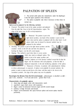
PALPATION OF SPLEEN
The normal adult spleen lies immediately under the diaphragm
in the left upper quadrant of the abdomen.
The spleen is palpable only when it increases to three times its
normal size.
Spleen can be palpated by the following methods
Classical: The patient is put in the supine position and palpated
from the right iliac fossa to the left hypochondriac region. The
edge of the spleen may be felt on deep inspiration.
Bimanual: The patient is put in the
right lateral position, one hand of the examiner is put over the
lower chest and the spleen is palpated with the other hand. A soft
spleen which may be missed by classical method may be
palpated by this method.
Hooking: The patient is put in the right lateral position and the
examiner stands on the left side and feels the spleen by
hooking his fingers over the left costal margin.
Dipping: This method is used when there is severe ascites
which may mask an enlarged spleen. The
patient is put in the supine position and
examiner palpates as in the classical method except that he dips his
fingers into the abdomen with each palpation, so that the fluid is
displaced temporarily to the side facilitating palpation of the spleen.
Manoeuvre of bockus : in patients with splanchnoptotic habitus
in whom splenomegaly is expected , if spleen is not palpable by normal methods, the
patient is made to jump up and down a few times and then made to lie down in the
recumbent position , the edge of the spleen may now be palpable.
Basedupon the distance from the left costal margin , splenomegaly is classified as mild –
upto 3 cms , moderate – 3 to 8 cms and severe – greater than 8 cms.
Characteristics of a palpable spleen:
sharp edge and angular poles with characteristic notch
superficial organ which moves with respiration
fingers cannot be insinuated between organ and left coastal margin
tendency to bulge forwards and inwards
Fingers can be insinuated into space between spleen and erector spinae muscles
posteriorly.