1. PARASTERNAL PULSATIONS :
INSPECTION :
Best performed with patientssupine and with a modest elevationof the head and chest
(not over 45º) examiner should observe by lookingdown at the chest and from the side.
Bulging of the precordium denotes long standing RV dilatation.
Pulsations visibleover the left second or third interspace. This is usuallydue to a dilated
pulmonaryartery.
The right ventricle is situated just beneath the left third, fourth and fifth intercostal spaces
close to the sternum (left parasternal area). Therefore, it is possible to assess right
ventricular activity, by palpation ofthe left parasternal area. Method of Examination
Patient should be lying supine, with the breath, held in expiration(or breathing normally).
Keep the heel (or the ulnar border) of the right hand over the left parasternalarea. Apply
sustained and gentle pressure.
PALPATION :
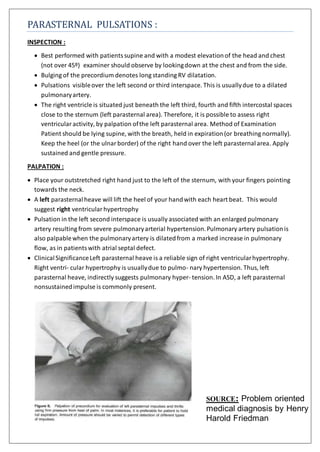
Place your outstretched right hand just to the left of the sternum, with your fingers pointing
towards the neck.
A left parasternalheave will lift the heel of your handwith each heart beat. This would
suggest right ventricular hypertrophy
Pulsation in the left second interspace is usually associated with an enlarged pulmonary
artery resulting from severe pulmonaryarterial hypertension.Pulmonary artery pulsationis
also palpablewhen the pulmonaryartery is dilatedfrom a marked increase in pulmonary
flow, as in patientswith atrial septal defect.
ClinicalSignificanceLeft parasternal heave is a reliable sign of right ventricularhypertrophy.
Right ventri- cular hypertrophy is usuallydue to pulmo- nary hypertension. Thus, left
parasternal heave, indirectly suggests pulmonary hyper- tension. In ASD, a left parasternal
nonsustainedimpulse is commonly present.
SOURCE: Problem oriented
medical diagnosis by Henry
Harold Friedman