1. PULSE
DEFINITION:
EXPANSION AND ELONGATION OF THE ARTERIAL WALL IMPARTED BY THE COLUMN OF BLOOD AND IS PASSIVELY PRODUCED BY THE
PRESSURE CHANGES DURING VENTRICULAR SYSTOLE AND DIASTOLE.
Points to note:
07 Lippincott Williams & Wilkins
CLINICAL NOTES:
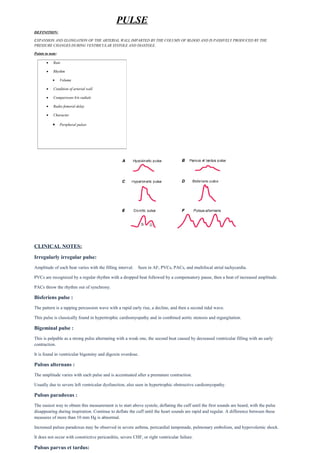
Irregularly irregular pulse:
Amplitude of each beat varies with the filling interval. Seen in AF, PVCs, PACs, and multifocal atrial tachycardia.
PVCs are recognized by a regular rhythm with a dropped beat followed by a compensatory pause, then a beat of increased amplitude.
PACs throw the rhythm out of synchrony.
Bisferiens pulse :
The pattern is a tapping percussion wave with a rapid early rise, a decline, and then a second tidal wave.
This pulse is classically found in hypertrophic cardiomyopathy and in combined aortic stenosis and regurgitation.
Bigeminal pulse :
This is palpable as a strong pulse alternating with a weak one, the second beat caused by decreased ventricular filling with an early
contraction.
It is found in ventricular bigeminy and digoxin overdose.
Pulsus alternans :
The amplitude varies with each pulse and is accentuated after a premature contraction.
Usually due to severe left ventricular dysfunction, also seen in hypertrophic obstructive cardiomyopathy.
Pulsus paradoxus :
The easiest way to obtain this measurement is to start above systole, deflating the cuff until the first sounds are heard, with the pulse
disappearing during inspiration. Continue to deflate the cuff until the heart sounds are rapid and regular. A difference between these
measures of more than 10 mm Hg is abnormal.
Increased pulsus paradoxus may be observed in severe asthma, pericardial tamponade, pulmonary embolism, and hypovolemic shock.
It does not occur with constrictive pericarditis, severe CHF, or right ventricular failure.
Pulsus parvus et tardus:
• Rate
• Rhythm
• Volume
• Condition of arterial wall
• Comparisons b/n radials
• Radio femoral delay
• Character
• Peripheral pulsesto Bedside
2. The classic finding in hemodynamically significant aortic stenosis, the carotid pulse is low in volume and has a slowly rising upstroke
with a prolonged plateau. A “shudder” may also be felt.
Asymmetric pulses: Consider subclavian artery atherosclerosis, arterial thrombosis (especially with atrial fibrillation), thoracic
outlet compression, or aortic dissection.
Thready pulse : A low-volume thready pulse is found in hypovolemic or septic shock, severe aortic stenosis, and severe left
ventricular dysfunction. Intense vasoconstriction may produce a diminished pulse with normal stroke volume.
Bounding pulses: Hyperkinetic pulse with a rapid large-amplitude upstroke and rapid collapse is associated with increased stroke
volume or decreased arterial compliance. The classic “collapsing pulse” is found in aortic regurgitation. It also occurs in thyrotoxicosis.