1. S3 and S4 Heart
Sounds
Mid-diastolic sounds are,either normal or
abnormal S3 sounds, and most late diastolic or
presystolic sounds are S4 sounds.
Low frequency sounds heard with the bell of
stethoscope
LV S3, S4 heard over the left ventricular apex
with the patient in the left lateral decubitus
position
RV S3, S4 heard over the lower left sternal edge,
occasionally subxiphoid with the patient supine
becoming more prominent during inspiration.
A left sided S3 AND S4 is augmented post-
tussively and with sustained handgrip exercise.
S3 heart sound
Ventricular gallop sound
Heard during the rapid ventricular filling phase
Physiological: Children and young adults
A normal S3 sometimes persists beyond the age of 40,
especially in women. Often associated with a thin,
asthenic body habitus
Decreased prevalence with increasing age
Triple rhythm-S1+S2+S3 OR S4
Quadruple rhythm-S1+S2+S3+S4
Pathological S3
-Ventricular dysfunction- poor systolic function,
increased end-diastolic and end-systolic volume,
decreased EF, and high filling pressures
1. Idiopathic DCM
2. IHD
3. Valvular heard disease- Chronic MR, Chronic AR
4. Congenital heart disease-VSD, PDA, ASD with
high flow across tricuspid valve
5. Systemic and pulmonary hypertension
-Excessive rapid early diastolic ventricular filling
1. HYPERKINETIC STATES- Anemia,
Thyrotoxicosis, AV fistula
2. AV valve incompetence- Left-to-right shunts
-Restrictive myocardial or pericardial disease
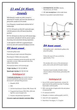
S4 heart sound
Atrial gallop sound – atrial diastolic gallop or the
presystolic gallop
Follows the onset of P wave on ECG by
approximately 70 ms
S4 coincides with a waves of the atrial pressure
pulse, atrial systole and with the apical impulse.
Sometimes heard in healthy old adults without
clinical evidence of heart disease, particularly after
exercise
Pathological S4
1. Severe LVH- systemic hypertension, valvular AS
(peak transvalvular gradient more than 70 mmHg),
hypertrophic cardiomyopathy
2. IHD- acute episode of angina and in the early phases
of transmural MI
3. LV aneurysm (with S3)
4. Idiopathic or ischemic cardiomyopathy (with S3)
5. Acute AV valve regurgitation
6. Heart block- First degree, 2:1, Complete
2. A loud S4 that is also usually palpable is a frequent
finding in acute and severe MR or AR. It is almost
always associated with an increased LV end-diastolic
pressure (>15 mmHg).
PERICARDIAL KNOCK
Ventricular filling is confined to early diastole in
constrictive pericarditis and terminates with a sharp
S3. This is termed pericardial knock. Its timing is
earlier than a normal S3 and typically occurs 0.1 to
0.12 seconds after S2.
Dr Nikita J.Mehra
II year MD General Medicine Post
Graduate