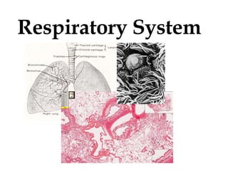
Histology of the Respiratory System
•
66 likes•31,194 views

The epithelium lining the respiratory tract from the nasal fossa through the bronchi is called the respiratory mucosa and is characterized by a pseudostratified ciliated epithelium with abundant non-ciliated cells known as goblet cells. - [Source: medcell.med.yale.edu/histology/respiratory_system_lab.php]

Recommended
More Related Content
What's hot
What's hot (20)
Similar to Histology of the Respiratory System
Similar to Histology of the Respiratory System (20)
More from Garry D. Lasaga
More from Garry D. Lasaga (20)
Recently uploaded
Recently uploaded (20)
Histology of the Respiratory System
- 2. Functions of the Respiratory System This “gas exchange” is the function of the respiratory system. All higher animals require a mechanism to: 1.Obtain O2 from the environment 2.and get rid of CO2
- 3. Epithelium in the respiratory system Olfactory epithelium Respiratory epithelium Olfactory
- 4. Epithelium in the respiratory system Olfactory Nose Skin junction Nasal cavity Air sacs Air sac Respiratory epithelium Histo 36 Conducting bronchiole
- 5. Conducting Portion Extrapulmonary • Nasal cavities • Pharynx • Larynx • Trachea • Bronchi Intrapulmonary • Intra. Bronchi, bronchioles, terminal bronchioles
- 6. Respiratory Portion Respiratory bronchiole Alveolar sacs Alveolar ducts Alveoli
- 7. Extrapulmonary region Function: Humidifies, cleanses and adjusts the temperature of inspired air. Mucosa: PSEUDOSTRATIFIED CILIATED COLUMNAR w/ GOBLET CELLS (w/ seromucous glands)
- 8. Nasal Cavity 1. Cutaneous region 2. Respiratory region 3. Olfactory region
- 9. Cutaneous region aka nasal vestibule rostral: lined by a thick SSE midvestibule: thinner & nonkeratinized caudal: stratified cuboidal (transitional zone)
- 10. Respiratory region Epithelium lining the caudal 2/3 of the nasal cavity proper Lining: respiratory epithelium • Cells: ciliated, secretory, brush, basal cells • *The respiratory mucosa is more vascular than other regions; cavernous stratum – it is the highly vascular propria- submucosa.
- 11. Olfactory region Dorsocaudal portion of the nasal cavity. Lining: ciliated pseudostratified columnar epithelium. Cell types: basal, sustentacular olfactory cells Distinguished from respiratory mucosa: (1) thicker epithelium, (2) numerous tubular glands, (3) bundles of nonmyelinated nerve fibers in lamina propria.
- 12. Olfactory glands Characteristic feature; branched tubuloalveolar serous glands – deliver secretions to olfactory surface. Function: secretion serves as a trap and solvent for odoriferous substances.
- 13. Olfactory epithelium has no goblet cells.
- 14. Conditioning Air By The Conducting Portion Nasal fossae – Superior conchae - olfactory epithelium long cilia, nervous cells – Middle conchae - respiratory epithelium – Inferior conchae - respiratory epithelium Swell bodies – large venous plexus that direct air (occludes air way) – Allergic reaction or inflammation restrict air flow counter current system warms air
- 15. Respiratory (Olfactory) mucosa and nasal septum Bowman’s glands Highly vascular lamina propria Swell bodies Olfactory epithelium Respiratory epithelium
- 16. Respiratory (Olfactory) mucosa and nasal septum Nerves Highly vascular lamina propria Olfactory epithelium Bowman’s glands Nerve
- 17. Larynx Formed by irregularly shaped plates of hyaline & elastic cartilage. Lining: respiratory epithelium; some – SSE – NK. Lumen: vestibule, ventricle, infraglottic cavity
- 18. Hyaline cartilage provides flexible support in the respiratory system to hold the air way open. Elastic cartilage in epiglottis Hyaline cartilage
- 19. Larynx Thyroid cartilage Cricoid cartilage Tracheal cartilages True vocal cords The false vocal cords Laryngeal ventricle Vocal cord muscles Air space lumen
- 20. Larynx Thyroid cartilage Vocal cord muscles Respiratory epithelium Stratified squamous epithelium
- 21. Larynx (lower portion) Cricoid cartilage Tracheal cartilage Respiratory epithelium lining Esophagus
- 22. Trachea 1. Mucosa 2. Submucosa 3. Cartilaginous layer 4. Adventitia
- 23. Trachea (Mucosa) Lining: respiratory epithelium Principal cell types: (1) ciliated, (2) secretory, (3) basal Lamina propria, elastic lamina
- 24. Goblet cells – readily seen after they have accumulated mucinogen granules in their cytoplasm (EM of human trachea).
- 25. Trachea (Submucosa) Serous & seromucous glands Tracheal glands – provide most of the secretory material that covers the ciliated surface in the trachea.
- 26. Trachea (Adventitia) Hyaline cartilage – most distinctive feature; C- or U-shaped stacked one on top of each other – support. Trachealis muscle
- 27. Trachea (rat) Principal features. 1.0 mm Cartilage of trachea Oesophagus Muscle (skeletal) Line across lumen is a fold in the section DorsalVentral Thyroid Lumen of trachea
- 28. Trachea (rat) Tracheal wall 1.0 mm •Hyaline cartilage is present. It gives support during respiration and rigidity prevents collapse. •Cartilage rings (incomplete): radial expansion possible; bolus of food can pass down the esophagus. cartilage lumen
- 29. Trachea (rat) Tracheal wall 1.0 mm Trachealis muscle at the dorsal aspect of the trachea.
- 30. Trachea (rat) Epithelial lining 25 µm Pseudostratified, ciliated, columnar epithelium. mucous on surfacecilia pseudostratified epithelium
- 31. Esophagus and trachea, monkey – glands in trachea Esophagus Trachea, whose lumen is lined with pseudostratified ciliated epithelium with goblet cells Trachea Thick hyaline cartilage bridged by smooth muscle bundle posteriorly Submucosa with glands Elastic fiber layer beneath the epithelium
- 32. Trachea epithelial lining Pseudostratified, ciliated, columnar epithelium from cat trachea. 25 µm
- 33. Trachea, monkey Pseudostratified ciliated epithelium with goblet cells Thick basement membrane Goblet cell Rich vascular supply to warm air Plasma cells to produce antibodies
- 34. Trachea, monkey Trachea, whose lumen is lined with pseudostratified ciliated epithelium with goblet cells Submucosa with glands Thick hyaline cartilage
- 35. Trachea (rat) Tracheal wall 50 µm Submucosal glands lumen epithelium submucosal glands
- 36. EM 8 trachea; 20630x 1. Mucous 2. Microvilli 3. Cilia 4. Goblet cell
- 37. Trachea (cat) Stained for elastic fibres 50 µm Elastic fibres below the epithelium. They are part of the elastic continuum of the lung. Add to the elastic recoil of the lungs after inspiration and expiration. epithelium pseudostratified columnar, ciliated. band of black stained elastic fibres in lamina propria.
- 38. Intrapulmonary bronchi Mucosa: resp. epithelium w/ goblet Muscle: SM Cartilage: hyaline cartilage plates Glands: serumocous
- 40. Lung (adult) Bronchus Low magnification view of bronchus. Plates of cartilage can be identified. 1.0 mm cartilage cut edge of section lumen
- 41. Lung (adult) Bronchus 250 µm C C : cartilage M : smooth muscle epithelium lumen G : glands M G
- 42. Lung (adult) Bronchus 25 µm Typical respiratory tract epithelium lines the bronchus. Pseudostratified, ciliated, columnar with goblet cells. Further down as the bronchus gets smaller the epithelial lining also reduces in height. cilia RB : blood cells in capillary G : goblet cells mucous on surface RB G G
- 43. SLIDE 67 Lung (adult) – Bronchus Bronchus and blood vessels 0.5 mm BR : bronchus BV : blood vessel BR BV
- 44. Lung (adult) – Bronchus Although there is little evidence of cartilage in this structure, some glands can be seen suggesting that this is a small bronchus. 250 µm glands
- 45. Bronchus (40x) - Slide 70: Mammalian Lung 1. Lumen 2. Pseudostratified ciliated epithelium 3. Mixed glands in the submucosa 4. Hyaline cartilage 5. Alveoli
- 46. 1. Lumen 2. Pseudostratified ciliated epithelium 3. Lamina propria 4. Smooth muscle 5. Mixed glands in the submucosa 6. Hyaline cartilage Bronchus (100x) - Slide 70: Mammalian Lung http://cal.vet.upenn.edu/projects/histo/Labrespiratory.htm
- 47. Lung (adult) Smooth muscle runs in a spiral around the bronchus. 100 µm epithelium cartilage glands muscle blood vessel
- 48. Bronchi vs. Trachea Various layers of the bronchi are thinner than the trachea. Mucous & basal cells decrease & bronchiolar exocrine cells increase in number. Cartilage rings replaced by cartilage plates. Location of the smooth muscle – between the mucosa & submucosa; arranged in a circular fashion.
- 49. Bronchioles Lining: ciliated simple columnar to cuboidal epithelium Clara cells (bronchiolar exocrine cells). Clara cells – interspersed among ciliated cells; nonciliated cells that have a characteristic rounded apical projection.
- 50. Diagram of a Clara cell between bronchiolar ciliated epithelial cells.
- 51. Terminal Bronchioles Most distal part of the conducting portion of the RS. Lined by simple cuboidal epithelium w/ Clara cells. CT & muscle greatly reduced.
- 53. Lung (adult) Bronchiole 250 µm Cartilage is absent from the wall of a bronchiole. To allow a change in diameter. They must be flexible in a way that minimises ‘dead space’ but does not cause too great a resistance to air flow. epithelium M : muscle M
- 54. TERMINAL BRONCHIOLE CLARA CELLS Respiratory BRONCHIOLE Ciliated cells Elastic fibers Smooth muscle Smooth muscle cells Cells in the respiratory portion
- 55. SEM of a terminal bronchiole. A longitudinal section of terminal bronchiole & surrounding alveoli. CLARA CELLS – secrete a surface-active agent that prevents luminal adhesion should the wall of the airway collapse on itself.
- 57. Lung (bronchus) Bronchus: 1) pseudostratified ciliated columnar epithelium with goblet-cells; 2) smooth muscle band between the lamina propria and the cartilage. The smooth muscle is not continuous around the bronchus as it spirals. 3) a change from cartilage rings to cartilage plates surrounding the tube; 4) glands in the submucosa. Bronchioles: 1) have a ciliated columnar epithelium; 2) do not have cartilage plates or glands; 3) have well organized muscle layers.
- 58. Bronchus vs. Bronchiole Bronchus Bronchiole Lining cells Cartilage present absentMucosal glands pseudostratified ciliated columnar epithelium ciliated columnar epithelium plates of cartilage cartilage absent
- 59. Lung (adult) At low magnification identify : bronchus bronchiole large blood vessels 1.0 mm blood vessels bronchus bronchiole
- 60. Respiratory portion Respiratory bronchiole Alveolar ducts Alveolar sacs Alveolus
- 61. Respiratory portion Gas exchange area/parenchyma ACINUS – functional unit of GEA. include respiratory bronchioles, alveolar ducts, alveolar sacs & alveoli. LOBULE – structural unit of GEA. cluster of acini separated by interlobular septa.
- 62. Species differences Lungs of cattle, sheep & pigs – complete septa (highly lobulated). Lungs of horses – incomplete septa (poorly lobulated). Carnivores – do not have interlobular septa.
- 63. Respiratory bronchiole Similar to the terminal bronchioles, except that the epithelium is interrupted by alveoli. Epithelium consists almost entirely of Clara cells.
- 64. Lung (adult) Respiratory Bronchiole Respiratory bronchioles are lined by a cuboidal epithelium. Alveolar ducts open into alveoli. 100 µm blood vessel alveoli
- 65. Alveolar ducts Elongate airways that have almost no walls, only alveoli, as its peripheral boundary. Lined by simple squamous epithelium. Terminate in clusters of alveoli – alveolar sacs.
- 66. Alveoli Terminal air spaces of the respiratory system & are the actual sites of gas exchange. Cells in alveolar lining: 1. Type I pneumocytes (lining cells) 2. Type II pneumocytes (produce surfactant)
- 67. Alveoli Terminal air spaces of the respiratory system & are the actual sites of gas exchange. Cells in alveolar lining: 1. Type I pneumocytes (lining cells) 2. Type II pneumocytes (produce surfactant)
- 68. Terminal and Respiratory Bronchioles (40x) - Slide 16: Mammalian Lung http://cal.vet.upenn.edu/projects/histo/Labrespiratory.htm 1. Terminal bronchiole 2. Respiratory bronchiole 3. Respiratory bronchiole to alveolar duct 4. End of alveolar duct 5. Alveolar sac 6. Alveoli 7. Smooth muscle
- 69. Terminal and Respiratory Bronchioles (100x) - Slide 16: Mammalian Lung 1. Terminal bronchiole 2. Respiratory bronchiole 3. Smooth muscle 4. Alveolar duct 5. End of Alveolar duct 6. Alveolar sac 7. Alveoli http://cal.vet.upenn.edu/projects/histo/Labrespiratory.htm
- 70. Aveolar Sac (100x) - Slide 72: Mammalian Lung 1. Alveolar sac 2. Alveoli 3. Elastic fiber http://cal.vet.upenn.edu/projects/histo/Labrespiratory.htm
- 71. 1. Type I pneumonocyte 2. Type II pneumonocyte • Extremely thin squamous cells that line most (97%) of the surface of the alveoli. • A cuboidal cell with a central nucleus; it covers the remainder of the interalveolar septal surface (3%) • Surfactant – decreases the alveolar surface tension and actively clears foreign materials.
- 72. Type II pneumonocyte (EM 18c). 1. Nucleus 2. Surfactant bodies
- 73. Surfactant bodies in Type II cells Type II pneumocytes
- 74. Macrophages in Air Space of Alveoli Alveolus Air space
- 75. Lung (marsupial mouse) alveolar phagocytes 25 µm P : phagocytes PP
- 76. Lung stained for elastic tissues Note the distribution of these fibres throughout the section especially in relation to the alveolar ducts and alveoli. 100 µm elastic fibres surrounding alveoli elastic fibres and smooth muscle cells below the cuboidal lining of the alveolar ducts
- 77. Lung Mesothelium and connective tissue of lung capsule Type I & Type II pneumocytes Alveolar macrophageCapillary endothelial cells and fibroblasts
- 78. Alveolus (Air) Capillary (Blood) Oxygen CO2 Respiratory Portion Respiratory bronchioles Alveolar ducts Alveolar sacs Alveoli
- 80. Scanning electron micrograph of adult sheep lung 50 µm B : cut bronchioles alveoli B B
- 81. Lung Bronchus Respiratory bronchiolesConducting bronchiole Alveolar duct Alveolar sac Alveoli The air-conducting tubes of the respiratory system can be thought of as a series of ducts which carry air to the sites of gaseous exchange - the alveoli
- 82. Lung (adult) Respiratory duct/sac The lips of the alveoli contain smooth muscle. Respiratory bronchiole → alveolar ducts → alveolar sacs → alveoli. 100 µm A AS AD A : alveoli AS alveolar sacs AD alveolar ducts A
- 83. Bronchus Conducting bronchiole Respiratory bronchiole Alveolar duct Alveolar sac Alveoli Mast cell Mesothelium Alveolar macrophage Type II pneumocyteType I pneumocyte Lung (mast cells) Terminal bronchiole
- 84. Mast cells function in the localized release of many bioactive substances with roles in the local inflammatory response, innate immunity, and tissue repair. Mast cell granules normally contain: heparin, histamine, serine proteases, eosinophil and neutrophil chemotactic factors, cytokines, etc. 48 Mast cells Type II pneumocyte
- 86. Pleura VISCERAL (PULMONARY) PLEURA – serous membrane that completely covers both lungs, except at the hilum & pulmonary lig. Lined by a squamous to cuboidal mesothelial cells.
- 87. Lung (adult) 1 mm Visceral pleura. outer surface of lung covered by visceral pleura B : blood vessels small bronchus B B
- 88. Lung (adult) Visceral pleura. 100 µm surface mesothelium connective tissue layer with elastic fibres
- 89. Avian Lung The duct system bears no similarity to that in mammals. The primary bronchi enter the lung and expand as the vestibulum. The latter continues through the lung as the mesobronchus and is connected to the abdominal air sac. Secondary bronchi give rise to tertiary bronchi (parabronchi).
- 90. Avian Lung The parabronchi are analogous to the alveolar duct of mammals. Atria project radially from the parabronchi. These atria are continuous with the air capillaries. The air capillaries are responsible for the actual exchange of gases with the closely associated vascular capillaries.
- 91. Avian Lung The air sacs associated with the avian lung aid in the movement of air through the lung. They are membranous structures, which do not contribute to the exchange of gases. Most birds have nine air sacs: single (unpaired) cervical, paired claviculars, paired cranial thoracics, paired caudal thoracics, paired abdominals.
- 92. Avian THE LUNG OF THE FOWL
- 93. Trachea (25x) - Slide C15: Avian Trachea 1. Tracheal lumen 2. Pseudostratified ciliated columnar Jeepithelium 3. Complete hyaline cartilage ring http://cal.vet.upenn.edu/projects/histo/Labrespiratory.htm
- 94. Avian Lung (25x) - Slide C17: Avian Lung 1. Parabronchus in l.s. 2. Parabronchus in x.s. 3. Atria http://cal.vet.upenn.edu/projects/histo/Labrespiratory.htm
- 95. Summary Table of Respiratory System (Nasal cavity) Region Skeleton Glands Epithelium Cilia Goblet Special features Vestibule Hyaline cartilage Sebaceous & sweat g. SSK No No Vibrissae Respiratory Bone & hyaline cart. Serumocous PCC Yes Yes Large venous plexus Olfactory Nasal conchae Bowman’s g. PCC Yes Yes Basal cells; sustentacul ar, olfactory cells; nerve fibers
- 96. Summary Table of Respiratory System (Pharynx) Region Skeleton Glands Epithelium Cilia Goblet Nasal Muscle Seromucous PCC Yes Yes Oral Muscle Seromucous SSE-NK No No
- 97. Summary Table of Respiratory System (Larynx) Skeleton Glands Epithelium Cilia Goblet Special features Hyaline & elastic cartilage Mucous & seromucous SSE-NK & PCC Yes Yes Vocal cords, epiglottis, some taste buds
- 98. Summary Table of Trachea & Primary Bronchi Skeleton Glands Epithelium Cilia Goblet Special features C-rings of hyaline cartilage Mucous & seromucous PCC Yes Yes Trachealis m.; elastic lamina
- 99. Summary Table of INTRAPULMONARY CONDUCTING Region Skeleton Glands Epithelium Cilia Goblet Features SECONDARY BRONCHI Plates of hyaline cartilage Seromucous PCC Yes Yes 2 helically oriented ribbons of smooth mucle BRONCHIOLES Smooth m. None Simple columnar to simple cuboidal Yes Only in larger bronc hioles Clara cells TERMINAL BRONCHIOLE Smooth m. none Simple cuboidal Some None Clara cells
- 100. Summary Table of RESPIRATORY REGION Region Skeleton Glands Epithelium Cilia Goblet Features RESPIRATORY BRONCHIOLE Some smooth m. None Simple cuboidal & simple squamous Some None Outpocketings of alveoli ALVEOLAR DUCT None None Simple squamous None None Outpocketings of alveoli; type I & II pneumocytes; dust cells ALVEOLUS None None Simple squamous None None Type I & II pneumocytes; dust cells