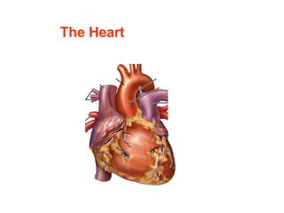
13. SA node - located in the right atrial wall, just inferior to the entrance of the superior vena cava. Original Impulses from S-A Node The electrical impulses are normally generated by a group of specialized pacemaker cells at sinoatrial (SA) node.
17. Conduction in Atria The electrical impulses from SA node spread through the entire right and left atrial muscle mass, triggering contraction of the right and left atrium.
18. Delay at A-V Node - The impulses from S-A node travel to atrioventricular (A-V) node . - A-V node is located in lower end of the interatrial septum near the tricuspid valve. A-V node
19. Delay at A-V Node - A-V node is the only normal route that impulses from SA node are transmitted into ventricles. - Conduction speed in A-V node is slow (delay). - This delay allows time for the atria to finish contraction and empty their contents into the ventricles before ventricles start to contract.
20.
21.
22. Rapid conduction in the ventricles simultaneous excitation of the ventricles functional syncytium
23. NNote : - Each electrical impulse can trigger cardiac muscle contraction normally only once. - A normal heart generates 60 to 100 impulses in 1 minute at resting state. 1 1
24. Excitation Contraction [ Ca ++ ] i (Action Potentials) (shortening) Properties of Cardiac Muscle Excitation of the heart is triggered by electrical impulse rather than neural transmitters. Contraction of the heart is triggered by elevation of intracellular calcium influx.
25. Properties of Cardiac Muscle - Myocytes depend heavily on oxygen and blood supply. - Not fatigue - Excitability Cycle The myocytes have Long refractory period during which they do not respond to any electrical impulses.
26. RRole of a Long Refractory Period – 1 prevent ventricles from contracting at too high rates so that enough time is allowed for refill of the ventricles
27. Role of Long refractory period - 2 Prevent retrograde excitation
33. Disorders of the Cardiac Conduction System ---- Arrhythmias - refers to abnormal initiation or conduction of electrical impulses in the heart. - caused by ischemia, fibrosis, inflammation, or drugs.
34.
35. - contract uncoordinatedly and extremely rapidly. - Ventricular fibrillation is lethal. Atrial or Ventricular Flutter and Fibrillation
36. is when the heart beat is triggered by ectopic pacemakers (cells other than SA node). Premature contraction
38. Artificial Pacemaker Application: sinus abnormality, complete AV or ventricular block Function: - generate electric pulses - sensing - antitachyarrhythmia
39. Heart Sounds Four heart sounds can be recorded via phonocardiography, but normally only two, the first and the second heart sounds, are audible through a stethoscope.
40.
41. - occurs when aortic and pulmonary semilunar valves close at the beginning of ventricular dilation - generated by the vibration of the blood and the aorta - Aortic valve closes slightly before pulmonary valve. Second heart sound
42. Heart Murmur - abnormal heart sound - occur in valvular diseases and septal defects
43. Two Basic Types of Valvular Diseases 1) valvular stenosis , a narrowing of the valve 2) valvular insufficiency (incompetence). A valve is unable to close fully; so there is some backflow (regurgitation) of blood.
44.
45. Heart Rate the number of heart beats in 1 minute. Normal value: 60-100/min Stroke volume the volume of blood pumped out by each ventricle per each contraction. SV
46. Cardiac Output (CO) the amount of blood pumped out by each ventricle in 1 minute. Cardiac output = stroke volume x heart rate Example: 70 ml x 75 beat/min = 5,250 ml/min 70 75 beat/min ml
47. Ejection Fraction = stroke volume end-diastolic ventricular volume 70 ml 130 ml = 54% 60 ml End of diastole 130 ml 70 ml End of systole SV =
48. End of diastole 133 ml 120 ml End of systole SV = Ejection Fraction 120 ml 133 ml = 90% increases during exercise
49.
50. Preload to ventricles = ventricular end diastolic pressure - the degree of stretch of the ventricular muscle cells just before they contract. - determined by ventricular filling.
51. Afterload to left ventricle: aortic arterial pressure Afterload to right ventricle: pulmonary arterial pressure Afterload to the left ventricle is greater than that to the right ventricle. Aortic arterial pressure
52. Contractility - the intrinsic strength of cardiac muscles.
66. Centers in Medulla Oblongata Sympathetic center: distinct accelerator and augmentor Parasympathetic center: Nucleus vagus and nucleus ambiguus
67. Hypothalamus, Thalamus, Cerebral cortex Involved in the cardiac response to environmental temperature changes, exercise , or during excitement , anxiety , and other emotional states
70. 1) Baroreceptor Reflex - stimulated by increase in arterial pressure (stretch) - Effect: negative chronotropic and inotropic - regulate the heart when BP increases or drops - involved in short term regulation of BP
73. 2) Chemoreceptor Reflex - stimulated by oxygen , pH , or CO 2 - overall effect: positive choronotropic and inotropic. - less important in regulating cardiac function
74. 3) Proprioceptor Reflex - Stimulated by muscle and joint movement - Effects: increase heart rate during exercise
75.
76. Autoregulation of the Heart Stroke volume is autoregulated by ventricular filling ( Frank-Starling law ). SV More in More out
85. dull white and slightly elevated fibrous plaque ( atheroma ) on coronary arterial lumen. Typical lesion of Coronary Atherosclerosis
86. composed of lipid, smooth muscle, macrophages, and connective tissues. cause stenosis of coronary arteries Histology of the plaque occlude arterial lumen when combined with internal hemorrhage, thrombosis, and arterial spasm