

Recommended
Recommended
More Related Content
What's hot
What's hot (20)
Viewers also liked
Viewers also liked (20)
Similar to Rib notching
Similar to Rib notching (20)
More from Madhu Reddy
Recently uploaded
Recently uploaded (20)
Rib notching
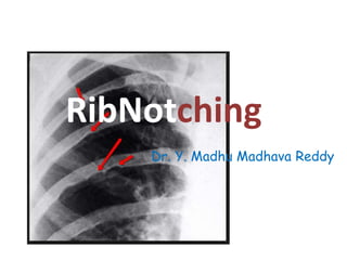
- 1. RibNotching Dr. Y. Madhu Madhava Reddy
- 2. Introduction • Rib notching refers to deformation of the superior or inferior surface of the rib. • It can affect single ribs (from trauma or solitary masses e.g. schwannoma) or can affect multiple ribs. • The differential differs according to whether it is the superior or inferior surface that is notched.
- 3. Differentials • Superior rib notching: • The superior rib notching can be caused by 1. Abnormal osteoblastic activity – osteogenesis imperfecta – connective tissue diseases • rheumatoid arthritis • systemic lupus erythematosus (SLE)
- 4. Superior rib notching 2. Abnormal osteoclastic activity – hyperparathyroidism 3. Miscellaneous – neurofibromatosis type 1 – restrictive lung disease – Poliomyelitis – Marfan syndrome
- 5. Inferior rib notching • Aka Roesler sign 1. Enlarged collateral vessels – coarctation of the aorta – interrupted aortic arch – subclavian artery obstruction • Takayasu disease • Blalock-Taussig shunt : involves only upper 2 rib spaces – AVM of the chest wall – SVC obstruction with enlarged venous collaterals – pulmonary AVM
- 6. Inferior rib notching 2. Neurogenic tumours – schwannoma (usually single) – neurofibromatosis type 1 (rarely can be superior if neurofibroma is very large)
- 7. Superior and Inferior Rib notching • 3. Superior and Inferior Rib notching - hyperparathyroidism
- 8. Superior rib notching • Osteogenesis imperfecta: • The hallmark feature of osteogenesis imperfecta is that fragile bones that fracture easily. It affects both bone quality and quantity. • It is due to abnormality of type 1 collagen, so sclera, cornea, joints, and skin are abnormal. • Due to the abnormal osteoblastic activity the superior part of ribs is deformed.
- 9. Types of OI • Type 1: Most common. • Sclera are blue, Bony fragility is mild, Stature is reduced, Deafness can occur in adult life. • Osteoporosis occur with cortical thinning. Long Bones are bowed and gracile.
- 10. OI
- 11. Types of OI • Type 2: Gross demineralisation of long bones with thin cortices. Numerous healing or healed rib fractures are seen. • Type 2A: Long bones are bowed, short and broad. Numerous fractures are seen. Ribs are broad with continuous beading. • Type 2B: Long bones as in case of type 2A, but ribs show less or no beading.
- 12. Types of OI • Type 2C: the long bones are thinned, show numerous fractures and ribs are too thin and beaded. • Type 3: over all bones are demineralised, Vertebral compression is seen, kyphoscoliosis may present. • Sutures may be wide and wormian bones persists. Associated with dentinogenesis imperfecta.
- 13. Types of OI • Type 4: the ribs are so soft and thin that the down ward pull of the intercostal muscles make their posterior portion convex downward.
- 14. OI
- 15. OI
- 16. ZEBRA LINES IN BISPHOSPHONATE THERAPHY
- 17. Rheumatoid Arthritis • It is a chronic systemic inflammatory disease which affects many organs, but predominantly attacks the synovial tissues and joints. • Proximal joints are affected first, with symmetrical involvement of joints.
- 18. Rheumatoid Arthritis • Diagnosis is based on a combination of clinical, radiographic and serological criteria. 1. Morning stiffness lasting at least 1 hour before maximal improvement 2. Soft tissue swelling of 3 or more joints observed by a physician 3. Swelling of the proximal interphalangeal, metacarpophalangeal, or wrist joints 4. Symmetric swelling 5. Rheumatoid nodules 6. The presence of rheumatoid factor; and 7. Radiographic erosions and/or periarticular osteopenia in hand and/or wrist joints.
- 19. Rheumatoid Arthritis • The erosions are typical being localized defects in the superior aspect of the upper ribs towards their posterior ends. These erosions occur symmetrically and involve each particular rib at a constant distance from the costo verterbral joint.
- 20. RA
- 22. SLE • It is a complex autoimmune disease with multisystem involvement. • Diagnosis is made using the following criteria: • Malar rash, discoid rash, photo sensitivity, pleuritis / pericarditis etc. • SLE affects bone formation and bone strength there by causing the rib notching.
- 23. Marfan syndrome • Marfan syndrome is a multi system hereditary connective tissue disease with a high penetrance and variable expression. Pathology:- • Results from a defect in fibrillin 1 (FBN1) gene located in chromosome 15 which is responsible for cross linking collagen.
- 24. Disease spectrum and associated features Skeletal:- • General – tall stature – osteopaenia – joint laxity • recurrent dislocations
- 25. Marfan syndrome • Spine / skull – High arched palate – Atlanto-axial subluxation – Dural ectasia – Increased interpedicular distance – kyphoscoliosis – Enlarged sacral foramina – Meningocoele • pre sacral meningocoele • lateral sacral meningocoele – Vertebral scalloping
- 26. Marfan syndrome Cardiovascular:- • Arterial dissection • Aortic aneurysm • Aortic valve regurgitation (AR) • Aortic coarctation • Aortic sinus dilatation • Myxomatous degeneration of the mitral valve • Pulmonary arterial dilatation
- 27. Marfan syndrome Ocular:- • Ectopia lentis lens usually displaced upwards and out • Myopia • Retinal detachment • Megalocornea
- 28. Hyperparathyroidism • It is the effect of excess parathyroid hormone in the body. • Increased levels of parathyroid hormone (PTH) lead to increased osteoclastic activity. The resultant bone resortpion produces cortical thinning (subperiosteal resorption) and osteopaenia. This leads to the rib notching on Chest X ray.
- 29. Hyperparathyroidism • Hyperparathyroidism causes both superior and inferior rib notching due to the increased levels of PTH in blood initiating the osteoclastic reaction.
- 30. Inferior rib notching (Roesler sign) • Enlarged collateral vessels:- – Coarctation of the aorta – Interrupted aortic arch – Subclavian artery obstruction • Takayasu disease • Blalock-Taussig shunt: involves only upper 2 rib spaces – AVM of the chest wall – SVC obstruction with enlarged venous collaterals • Neurogenic tumours:- – Schwannoma (usually single) – Neurofibromatosis type 1 (rarely can be superior if neurofibroma is very large)
- 31. Coarctation of the aorta • Coarctation of the aorta refers to a narrowing of the aortic lumen. It can be primarily divided into two types • Infantile (pre-ductal) form • Adult (juxta-ductal, post-ductal or middle aortic) form
- 32. Coarctation of the aorta • Figure of 3 sign - contour abnormality of the aorta • Inferior rib notching - Roesler sign – Secondary to dilated intercostal collateral vessels – Seen only in long standing cases, and therefore not seen in infancy – Seen in 70% of cases presenting in older children or adults – If unilaterally seen on the left, then this suggests an associated aberrant right subclavian artery arising after the coarctation.
- 33. Coarctation of the aorta – If unilaterally seen on the right, then the origin of the left subclavian artery is distal to the coarctation. – Most often involves 4th-8th ribs – Occasionally involves 3rd and 9th ribs – Does not involve 1st and 2nd ribs (the associated arteries are branches of the costocervical trunk, and thus proximal to coarctation)
- 34. Coarctation of the aorta • Coarctation of the aorta with an aberrant right subclavian artery: Left-sided rib notching is seen. Occurs when the aberrant right subclavian artery arises after the coarctation. • Coarctation of the aorta proximal to the left subclavian artery: Right-sided rib notching occurs.
- 35. Coarctation of the aorta • Rib notching in Coarctation of aorta occurs due to dilatation of the posterior intercostal arteries, which act as collateral vessels. • Since 1st and 2nd posterior intercostal arteries arise from the costocervical trunk of sub clavian artery rather than descending aorta, they do no form a collateral path and hence do no cause rib erosion.
- 36. COA
- 37. FIGURE OF 3
- 38. COA
- 39. COA
- 40. Interrupted aortic arch • It is an uncommon congenital cardiovascular anomaly where there is a separation between the ascending and descending aorta . • It can either be complete or connected by a remnant fibrous band . • An accompanying large ventricular septal defect (VSD) and or patent ductus arteriosus (PDA) is frequently present. Pathology • Faulty embryological development of the aortic arch (thought to occur during the 5th to 7th week of intra uterine life).
- 42. Classification:- • Type A : second commonest : interruption occurs distal to the left subclavian arterial origin • Type B : commonest (> 50 %) : interruption occurs between left common carotid arterial and left subclavian origins • Type C : rare : interruption occurs proximal to left common carotid arterial origin Each type is divided into 3 subtypes : • sub-type 1 : normal subclavian artery • sub-type 2 : aberrant subclavian artery • sub-type 3 : isolated subclavian artery that arises from the ductus arteriosus.
- 43. Type A : second commonest : interruption occurs distal to the left subclavian arterial origin Type B : commonest (> 50 %) : interruption occurs between left common carotid arterial and left subclavian origins Type C : rare : interruption occurs proximal to left common carotid arterial origin
- 44. Abberant Right Subclavian Art.
- 45. Radiographic features Plain film - CXR • Plain film features are often non specific • The aortic knuckle may be absent • May show cardiomegaly Antenatal ultrasound • The right ventricle may appear a lot larger than the left The ascending aorta may also appear more vertical than usual. MRI / MRA • Non visualisation of portion of interruption • Great vessels may show a "V" configuration on coronal imaging 2
- 46. IAA
- 47. Subclavian artery obstruction – Takayasu disease – Blalock-Taussig shunt : involves only upper 2 rib spaces
- 48. Takayasu arteritis • Takayasu arteritis (TA) (also known as idiopathic medial aortopathy or pulseless disease) is a granulomatous large vessel vasculitis predominantly affecting the aorta and its major branches. • It may also affect the pulmonary arteries.
- 49. Takayasu arteritis Pathology • There is segmental and patch granulomatous inflammation of the aorta which results in stenosis, thrombosis and aneurysm formation. Half of patients present with an initial systemic illness whereas the other 50% present with late-phase complications.
- 50. Takayasu arteritis Two phases of the disease are classically described :- • Pre pulseless phase : characterised by nonspecific systemic symptoms • Pulseless phase : presents with limb ischaemia or renovascular hypertension.
- 51. Takayasu arteritis • The initial systemic illness may include symptoms of malaise, fever, night sweats, weight loss and arthralgia. There is often anaemia with raised inflammatory markers. • This phase gradually resolves with initiation of the chronic phase which is characterised by inflammatory and obliterative changes in the aorta and its branches. • There are often reduced or absent peripheral pulses, giving rise to it's alternative name of "pulseless disease
- 52. Takayasu arteritis • Subtle notching of the first three ribs may be an expression of sub clavian artery occlusion. • Nothing of the lower ribs is usually associated with severe narrowing or occlusion of the upper abdominal aorta with intercostal arteries serving as prominent collateral channels.
- 53. Location and classification • Type I - classic type involving the solely the aortic arch branches : brachiocephalic trunk, carotid and subclavian arteries • Type II - – IIa - involvement of the aorta solely at its ascending portion and/or at the aortic arch +/- branches of the aortic arch – IIb - involvement of the descending thoracic aorta +/ - ascending or aortic arch + branches • Type III - involvement of the thoracic and abdominal aorta distal to the arch and its major branches (i.e. descending thoracic aorta + abdominal aorta +/ - renal arteries) • Type IV - sole involvement of the abdominal aorta and/or the renal arteries • Type V - generalised involvement (all aortic segments)
- 54. Radiographic features Ultrasound :- • Homogeneous circumferential thickening of affected vessels (indistinguishable from atherosclerotic plaque) • Vascular occlusion +/- dilation • Flow velocity elevations beyond stenotic segments
- 55. CT/MRI:- • Wall thickening - acute active phase • Wall enhancement - acute active phase • Aortic valve disease - stenosis, regurgitation • Occlusion of major aortic branches • Aneurysmal dilatation of the aorta or its branches. • Pseudoaneurysm formation • Diffuse narrowing distally (i.e. descending and abdominal aorta ) - in late phase
- 56. Blalock-Taussig shunt • Blalock-Taussig shunt: involves only upper 2 rib spaces. • A Blalock-Taussig (BT) shunt is a palliative procedure performed in patients with tetralogy of Fallot (prior to the ability to repair the defect) to increase the pulmonary blood-flow. • The shunt sacrificed the subclavian artery (with a distal ligation) and the proximal portion is routed downwards to an end to side anastomosis with the ipsilateral branch of the pulmonary artery.
- 58. Blalock-Taussig shunt • Rib notching is Ipsilateral to the side of shunt. Due to division of all the branches of the first part of the subclavian artery performed during shunt creation.
- 60. Points to remember • Coarctation of aorta proximal to left subclavian artery : Right sided rib notching • Coarctation of aorta with an abberant right subclavian artery: left sided rib notching • Sub clavian stenosis: ipsilateral to the side of stenosis. • Blalock taussig shunt: ipsilateral to the side of shunt.
- 61. Superior vena caval obstruction • Superior vena caval obstruction can occur from of a wide variety of pathologies. • Superior vena caval syndrome refers to the clinical syndrome with symptoms that results from obstruction of the superior vena cava.
- 62. Causes include: - • Malignancy, e.g. lung cancer (the most common cause) • Central venous catheters • Pacemaker wires • Fibrosing mediastinitis • Due to development of collateral channels to the inferior vena cava via the intercostal veins rib notching occurs in cases of SVC obstruction
- 63. • Neurogenic tumours:- – Schwannoma (usually single) – Neurofibromatosis type 1 (rarely can be superior if neurofibroma is very large) – They arise from the intercostal neurovascular bundles. They grow large in size causing rib notching.
- 65. Neurogenic tumor
- 66. NF 1
- 67. RIBBON RIBS
- 69. PROGERIA
- 71. Thank you
Editor's Notes
- cyclic bisphosphonate treatment produces sclerotic growth recovery lines in the long bones
- S: scleroderma H: hyperparathyroidism I: infection (osteomyelitis) R: rheumatoid arthritis T: trauma P: progeria Mnemonic : SHIRT Pocket