ACUTE and CHRONIC AORTIC INSUFFICIENCY-DR MAGDI SASI 2016
1. ACUTE AORTIC REGURGITATION DR MAGI AWAD SASI 2016
ACUTE and CHRONIC AORTIC INSUFFICIENCY
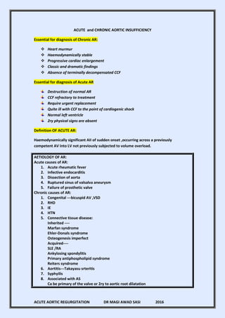
Essential for diagnosis of Chronic AR:
Heart murmur
Haemodynamically stable
Progressive cardiac enlargement
Classic and dramatic findings
Absence of terminally decompensated CCF
Essential for diagnosis of Acute AR
Destruction of normal AR
CCF refractory to treatment
Require urgent replacement
Quite ill with CCF to the point of cardiogenic shock
Normal left ventricle
2ry physical signs are absent
Definition OF ACUTE AR:
Haemodynamically significant AII of sudden onset ,occurring across a previously
competent AV into LV not previously subjected to volume overload.
AETIOLOGY OF AR:
Acute causes of AR:
1. Acute rheumatic fever
2. Infective endocarditis
3. Dissection of aorta
4. Ruptured sinus of valsalva aneurysm
5. Failure of prosthetic valve
Chronic causes of AR:
1. Congenital ---bicuspid AV ,VSD
2. RHD
3. IE
4. HTN
5. Connective tissue disease:
Inherited ----
Marfan syndrome
Ehler-Donals syndrome
Osteogenesis imperfect
Acquired----
SLE /RA
Ankylosing spondylitis
Primary antiphospholipid syndrome
Reiters syndrome
6. Aortitis---Takayasu srteritis
7. Syphyilis
8. Associated with AS
Ca be primary of the valve or 2ry to aortic root dilatation
2. ACUTE AORTIC REGURGITATION DR MAGI AWAD SASI 2016
Aetiology :
1. Infective endocarditis:
A. Staphylococcus Aureus----acute AR
It causes necrosis ,perforation ,and detachment of valve leaflets.
Infection of aortic annulus with necrosis and abscess formation may cause
weakness and progressive dilatation.
An annular abscess may distort aortic valve.
One or two large valvular vegetation occurs.
B. Fungal endocarditis:
Bulky vegetation
Asperigillus
Candida albicans
Histoplasma
C. Acute endocarditis in drug addicts ,in S/C drug users –skin popping
D. Patient with congenitally bicuspid AV at increased risk for bacterial endocarditis
even in absence of AS/AR because:
i. Bicuspid valve is the commonest congenital valvular malformation
associated with turbulent flow
ii. The valve lesion is commonly asymptomatic and escape detection
iii. Antibiotic has not be prescribed prophylatically.
COMPLICATIONS:
A. Annular abscesses ,nearly always associated with severe AR
Gradual or sudden
Necrosis ,inflammation encroach AV node and proximal his prukinje region
First /second degree heart block
LBBB
CHB
B. Abscess may erode into pericardium
Purulent pericarditis
Hemopericardium
Cardiac temponade
C. Annular abscess extending into the membranous interventricular septum
Septal rupture with left to right shunt.
D. Extension of the infection to the muscular part
This leads to ventricular irritability and infranodal block
E. Infection may extend into the contagious right ventricle or right atrium
This leads to development of an aorto ventricular /right atrial fistula
C/F:
Machinery murmur related to left to right shunt & worsening CCF
F. Superior extension of infection may cause a mycotic aneurysm
This involves sinus valsalva and proximal ascending aorta.
3. ACUTE AORTIC REGURGITATION DR MAGI AWAD SASI 2016
THE TREATMENT FOR ALL IS AORTIC VALVE REPLACEMENT.
Diagnosis of infective endocarditis is suggested by:
1) One /more large or small vessel embolic events
2) Systemic signs of toxicity
2. Dissecting of ascending aorta:
Ascending aorta dissection with consequent hematoma may extend to involve the
aortic valve.
Displace the cusps downward &medially
One/more prolapsed or Evert into the outflow tract of the LV during diastole
AR
AR occurs in 65% of patients with dissection of ascending aorta.
Another complication ----acute myocardial infarction that usually involves the inferior wall
due to compromise of right coronary ostium by hematoma ---dissecting medial.
Sudden death may occur due to cardiac temponade if the process is protracted by
extension through aortic adventia.
C/F:
Severe chest pain
Evidence of vascular compromise to:
1. Head 3. Upper extremities
2. Lower extremities 4. Gut 5. Kidney
Treatment:
a. Urgent CT scan chest , aorto-graphy
b. Operative intervention
3. Connective tissue disease:
SLE---sterile perforation of one/more aortic valve cups by fibrinoid necrosis
involving valve parenchyma.
Other :
i. Aortitis
ii. Ankylosing spondylitis
iii. Whipples disease
iv. Giant cell aortitis
v. Takayusu s arteritis
4. ACUTE AORTIC REGURGITATION DR MAGI AWAD SASI 2016
4. Trauma :
Pathogenesis:
Closed chest or abdominal trauma ----falls ,crush injuries ,automobile accident
Water hammer effect
Sudden compression of the thoracoabdominal aorta diastole with the aortic valve
closed may acutely increase intravascular pressure and cause aortic cuspal tearing
,perforation or detachement.
Trauma cause damage to normal valve and myxematous degenerated valves are
more susceptible.
Traumatic AR may occur as a consequence of severe muscle strain or strenuous
physical activity.
TO DIAGNOSE:
Require careful and thorough careful CVS examination
1) Initially when the victims presents to the hospital
2) After resuscitation and interventions complete
3) After several weeks of recovery
5. Spontaneous AR:
AV may be normal or myxematous.
Sudden eversion of an aortic cusp may result in spontaneous acute AR .
Bicuspid valve have an asymmetric nature ,the larger valve leaflet must oppose an
abnormally large total force in diastole ;resulting in an asymmetric + deformed
raphe.
Larger valve leaflet may not be sufficiently buttressed by smaller leaflet along
the line of coaptation leading to AR.
6. Prosthetic aortic valve insufficiency:
Can be caused by:
i. Sudden dehiscence of the sewing ring of prosthetic valve from the aortic
annulus .
It is an occasional complication of emergency AVR for acute fulminant ABE
when the valve must be implanted into an infected annulus.
ii. Pannus ingrowth ,thrombus formation ,vegetation may impede proper
seating of the ball or poppet in diastole.
iii. Poppet wear /failure
iv. Homograft valves and porcine bio-prostheses previously prepared undergo
progressive degeneration
Pathophysiology :
Chronic AR
Volume overload regurgitating into the LV during diastole increased overtime
LV wall undergoing minimal thickening ((septal +posterior wall thickening))
Eccentric hypertrophy((LVEDP+LVEDV))
5. ACUTE AORTIC REGURGITATION DR MAGI AWAD SASI 2016
Allows LV to operated at a larger end diastolic volume
Increased LV size to maintain CO ((starling law))
((LV dilatation ---apex beat deviated line))
As long as systolic function is preserved ,the ventricle is capable to ejecting this
abnormal large total volume + LV diastolic pressure dose not raise and symptoms
of decompensation does not ensure.
Ejection of this large stroke volume results in widened arterial pulse pressure.
This widened arterial pulse pressure of brisk upstroke and fall-off with its myriad of
associated peripheral signs is used to assess the severity of AR.
These signs also indicate reasonable preservation of ventricular systolic function in
chronic AR.
At later stages ,due to volume overload or other intercurrent myocardial insult
Congestive cardiomyopathy
Systolic function deteriorates
+
Left ventricle diastolic pressure rises
This results in :
1. Decrease organ perfusion
2. Pulmonary congestion
CARDIAC DECOMPENSATION
LV overload LV size ((eccentric hypertrophy))
to maintain COP
Run off phenomena
Large amount of blood escape Large LV needs more O2
From cardiac cycle
Angina pectoris
Diastolic BP/ LVEDP
Takes decades to develop CCF if chronic
Coronary perfusion
CCF
Decompensation occurs when:
1. LV systolic pressure begins to fail
2. Progressive LV dilatation occurs
3. Spherical geometry develops
6. ACUTE AORTIC REGURGITATION DR MAGI AWAD SASI 2016
C/F: chronic AR
Fatigue
Decreased exercise capacity
Dyspnea
As forward COP declines ,arterial resistance rises to preserve blood pressure.
The peripheral manifestation of chronic AR are no longer manifest ,although the
patient is now severely ill from cardiac decompensation.
The dramatic clinical ,radiographic ,echocardiographic of ventricular enlargement
is helpful to testify the chronicity of volume overload state.
LV systolic function and ESD are the most predictors of post operative survival and
LV function.
Symptoms of LV dysfunction---Dyspnea ,PND ,orthopnea
Symptoms of coronary insufficiency—angina ,nocturnal > exertional
With extreme reduction of diastolic pressure <40mmHg ,angina may be seen.
Symptoms of high COP----------pounding of the heart
The peripheral signs of chronic AR "signs of high COP in CHRONIC AR":
Presence of large bounding collapsing pulse is the first clue to chronic AR.
1. De Mussets sign:
The head frequently bobs with each heart beat ,caused by large stroke volume.
2. Müller sign is systolic pulsations of the uvula
3. Corrigans sign:
the pulse is of the water hammer or collapsing with abrupt distention and
quick collapse" sharp & full upstroke of the carotid pulse and a precipitous
decrease in diastole" .
4. Dancing carotid
5. Bisferiens pulse may be present & recognized in the brachial and femoral
It is bounding ,visible and forceful peripheral pulses.
6. Signs in femoral artery:
a. Traubes sign or pistol shot sound:
Booming sharp systolic sound heard over femoral artery((like gunshot)).
b. Duroziezs sign:
Systolic murmur ((bruit))heard over the femoral artery when it is compressed
proximally and diastolic murmur when it is compressed distally.
7. Quinckes sign:
By pressing a glass slide on the patients lip or transmitting a light through the
patients finger tips. It is a systolic blushing and diastolic blanching of the nail
skin when pressure is applied gently to the tip of nail.
8. Hills sign:
Disproportionate increase of systolic BP> 2ommHg when measured in the leg
as compared with systolic BP in the arm.
Mild > 20 mmHg , Modearte>40mmHg , Sever >60 mmHg
Popliteal cuff systolic pressure >brachial cuff pressure by > 60mmHg
The presence of such sign suggests sever chronic AR.
9. Final Jeopardy:
Landolfi’s sign = alternating constriction/dilatation of pupils
7. ACUTE AORTIC REGURGITATION DR MAGI AWAD SASI 2016
10. Mayne’s sign - moderate or mild degrees of of AR may be detected by
demonstration of a diminution of >15 mmHg in the diastolic BP in the arm
when it is elevated over the head compared with values taken when the arm is
at heart level
11. Blood pressure:
Systolic blood pressure is elevated
Diastolic pressure is abnormally low.
Korotkoff sound often persistent to zero.
As heart failure develops ,peripheral vasoconstriction occurs ;arterial diastolic
pressure rise------------------ increase severity of AR
The consequences are:
1. Increased LVEP---increased left atrium P---increased PCP ---PULMONARY E.
2. Early in the diastole ;LVP rises rapidly causing mitral leaflets to drift
toward closed position -----------------AUSTIN FLINT MURMUR.
3. Premature closure of mitral valve before onset of systole
4. The mitral valve may open late because the ejections period of acutely
volume---overloaded ventricle is prolonged.
5. Compensatory tachycardia shortens diastole
All factors reduce the time during which MV is open in diastole.
Premature closure of mitral valve causes:
A. Limits the degree of transmission of high pressure from LVEDP to left atrium and
pulmonary capillary bed.
B. Absence of the first heart sound
C. Limitation of diastolic interval compromises LV inflow and effective COP
IN ACUTE AR:
Acute damage to AV :
A. Lacking eccentric hypertrophy
Ventricle compliance is not increased
B. LV remains normal and poorly tolerated
If AR is severe ,end diastolic LV pressure may approaches aortic diastolic P.
The normal ventricle (( neither hypertrophied nor dilated)) cant acutely increased LV
stroke volume sufficiently to maintain stoke volume.
In normal ventricle ,there is little increase in stroke volume when EDP exceeds 12—
15mmHg .The starling curve relating ventricle stroke volume to EDV is rather flat at
pressures>15mmHg.So; increased EDP doesn't improve COP.
Pericardium may be a rule as it is suddenly stretched to the limit of its distensibility.
This leads to forward COP to declines.
This results in reflex increased in peripheral vascular resistance.
Acute AR:
Pallor and coolness of the skin
Decreased cutaneous blood flow
Impaired regional arterial flow
8. ACUTE AORTIC REGURGITATION DR MAGI AWAD SASI 2016
Reduced COP
Oliguria due to decreased blood flow
Deranged temperature regulation
GIT dysfunction
Hepatic dysfunction
Decreased tissue perfusion : a. Cardiogenic shock b. Lactic acidosis
Clinical presentation:
Acute AR---
The symptoms are of rapid onset due to increased left atrial pressure—pulmonary
congestion and decreased forward cardiac output.
Symptoms include: ACUTE PULMONAR EDEMA2
1. Exertional dyspnea
2. Orthopnea +dry cough increased by recumbency
3. PND
4. Dyspnea at rest even while sitting upright
Symptoms reflecting decreased COP are more subtle and overshadowed by those of
capillary congestion.
1. Fatigue on exertion
2. Apathy
3. Agitation
4. Deterioration in intellectual function
This reflects impaired skeletal muscle and cerebral perfusion.
An etiological diagnosis;
Sever chest/back pain of abrupt onset--------Dissection
Fever, Chills ,Malaise ,evidence of peripheral arterial emboli---infective endocarditis
H/O chest /abdominal trauma------trauma induced
Absence of symptoms or H/O heart disease suggest sudden perforation of AV
Chronic AR
There is a long period during which LV under goes enlargement while the
patient remains asymptomatic.
4th
;5th
decade -----decreased cardiac preserve or myocardial ischemia develop
after cardiomegally and myocardial dysfunction have occurred.
Principal symptoms -----exertional dyspnea ,orthopnea and PND.
In comparison to AS, ((where syncope and angina pectoris are common));
nocturnal angina often accompanied by diaphoresis which occurs when heart
rate slows and diastolic pressure falls to low levels may be trouble.
These episodes may be accompanied by abdominal discomfort.
Patient with severe AR complain of :
Uncomfortable awareness of the heart beat---on laying down
9. ACUTE AORTIC REGURGITATION DR MAGI AWAD SASI 2016
Thoracic pain due to bounding of heart against chest wall
Tachycardia ,stress or exertion may produce palpitation and head
pounding.
VPC are distressing due to great heave of volume loaded LV.
NOTE:
These complaints may be present for many years before symptoms of LV
dysfunction develops.
Acute AR:
Gravely ill patients
Sever dyspnea
weakness
hypotension
Sever peripheral vasoconstriction
Tachycardia
Cyanosis
Angina is uncommon
Acute AR--Physically :
Acute AR----ill patient ,dyspnic
The finding are related to:
1. Severity of pulmonary congestion
2. Impairment of forward COP + tissue perfusion
Thorough examination is mandatory.
3 major category:
a. Pulmonary congestion
b. Decreased COP
c. No signs of LV volume overload +stroke volume decreased
Tachycardia is the rule.
LV stroke volume is reduced(systolic BP is normal/increased;diastolic BP slightly
increased)
Precordium is relatively quiet to inspection and palpation.
There is lacking lateral displacement and heaving.
Auscultation in acute AR :
S1—first heart sound is usually soft as regurgitant volume rapidly fills ventricle and drives
the mitral leaflets toward the closed position at time of systole.
Absent S1---LVDP exceeds left atrial pressure prior to ventricular activation ,MV closure
occurs late in diastole ,preceding ventricular and atrial systole.
Premature MV closure have a protective rule over pulmonary circulation +pul.HTN
S2--- soft + may be absent if AV leaflets destroyed as no diastolic coaptation.
The murmur ---an ejection of variable intensity is heard to the base.
The diastolic murmur of AR is low pitched +shorter because as LVEDP raises ,the pressure
gradient between aorta and left ventricle is rapidly decreased.
10. ACUTE AORTIC REGURGITATION DR MAGI AWAD SASI 2016
A flail cusp may evoke a musical ((narrow frequency band)) and very intense diastolic M.
The Austin flint murmur if present is brief.
S3 is often present ,reflecting rapid early diastolic ventricular filling from LT atrium& aorta.
S3 S4 may be heard.
Loud P2 indicate pulmonary HTN.
The cacophony of sounds and murmurs may mimic pericarditis.
SUMMARY POINTS:
A. Fever, petechia ,purpura, arterial emboli (small or large)---INFECTIVE ENDOCARDITIS
B. Sever chest or back discomfort or unequal pulse in upper limbs—DAA
C. H/O of trauma------------------------------------------------------------------TRAUMATIC AR
D. Tall ,thin ,long arm span ,hyper-extensile joints ,ectopia lentis ----MARFANS
E. No finding---------------------------------------------------------------------------SPONTANEOUS
COMPARISON OF CLINICAL FINDING
CLINCAL FAETURES ACUTE AR CHRONIC AR
CCF Early & sudden Late &insidious
Arterial pulse
1. Rate/min Increased N
2. Rate of rise Not increased INCRAESED
Blood pressure
1. Systolic BP N/ low INCREASED
2. Diastolic BP N/low DECREASED
3. Pulse pressure N INCREASED
Contour of peak single bisferians
Left ventricle impulse Near normal Laterally displaced
Pulsus alternus common uncommon
Ausculatation
S1 Soft to absent Normal
A2 Soft N/decraesed
P2 N/decraesed N
S3 COMMON Absent
S4 Consistently absent Usually absent
Aortic systolic murmur Grade 3/less Grade 3/ more
Aortic regurgitant murmur Short .medium pitched Long ,high pitched
Austin flint murmur Mid diastolic Presystolic ;mid diastolic
Peripheral signs Absent Present
11. ACUTE AORTIC REGURGITATION DR MAGI AWAD SASI 2016
From such comparisons ,vital signs are helpful to clarify the cause of aortic regurgitation
weather it is acute or chronic .
It is mandatory to check the pulse and blood pressure and CVS examination.
WHAT ARE THE DIGANOSTIC DIFFERENCES BETWEEN ACUTE AR& CHRONIC AR?
Acute AR Chronic AR
ECG NORMAL LVH
CXR LV NORMAL PROMINANT
Aortic root and arch NORMAL PROMINENT
Pulmonary venous pattern Redistributed to upper lobes NORMAL
Interstitial and alveolar fluid Present absent
Chronic AR:
Apical impulse -----diffuse, displaced laterally & inferiorly ,forceful &unsustained.
There may be systolic retraction over the parasternal region.
A rapid ventricular filling wave is palpable at the apex.
A systolic thrill at the base of the heart ,suprasternal notch ,carotid arteries is palpable.
Auscultation :
S1—soft and prolongation of PR
A2—soft /absent
P2—obscured by murmur
S2—variable –absent ,single ,narrow or paradoxical splitting
An ejection systolic murmur is audible.
S3 gallop correlated with increased LVED volume and has been suggested as sign useful to
consider surgery.
Murmur—early diastolic high pitched blowing ,decrescendo murmur that begins
immediately after A2 ,best heard in the 2nd
&3rd
intercostals space left parasternal border.
It is accentuated by sitting up ,leaning forward ,hold breathing in full expiration.
In sever AR correlates better with the duration >intensity of the murmur.
Mild AR----------limited to early phase of diastole +high pitches
12. ACUTE AORTIC REGURGITATION DR MAGI AWAD SASI 2016
Moderate/sever AR—it is holodiastolic +rough quality
When the murmur is musical ((cooing dove)) ,it signifies eversion or perforation of an
aortic cusp.
Sever AR +LVF----equilibration of LVP+aorta P.-----abolish this component of murmur.
Site –1ry valve disease ---left sternal border –3rd
/4th
Dilatation of ascending aorta ------right sternal border
What is Austin Flint murmur?----it is a low pitched mid & late diastolic rumble((like MS)) ,
heard over apical area due to anterior displacement of MV by the aortic regurgitant
stream. It indicates moderate to severe AR.
Sever aortic reflux---increased LVEDP ---narrowing MV---rapid antegrad flow through MV.
As LVEDP increased, Austin flint murmur terminates earlier.
A short ejection systolic murmur ,1—4/6 ,may be audible at the base of the heart due to
increased stroke volume and transmitted to the carotid arteries with thrill +high pitched.
HOW TO INCREASED MURMUR QUALITY?
Any intervention that raises the arterial pressure increases murmur.
Vasopressor drug
Squatting
Exercise –isometric ---increase murmur of AR+Austin Flint murmur.
An intervention that decrease arterial pressure:
Valsalva maneuver
Amyl nitrate inhalation
Clinical feature Mitral stenosis Austin flint murmur
S1 Increased Normal
S3 No Yes
Opening snap Yes ,heard no
LV enlargement No yes
RV enlargement Yes No
Murmur decrease with amyl nitrate No Yes
Atrial fibrillation yes no
ECHO MS yes No
What are the major hemodynamic features of AR?
FEATURES ACUTE AR CHRONIC AR
13. ACUTE AORTIC REGURGITATION DR MAGI AWAD SASI 2016
Left ventricle compliance Not increased Increased
Regurgitant volume increased Increased
LVEDP Markedly incraesed May be normal
LV ejection velocity dp/dt Not significantly
incraesed
Markedly increased
Aortic systolic pressure Not increased Increased
Aortic diastolic pressure Normal to decraesed Markedly decreased
Systemic arterial pulse pressure Slight increased Markedly increased
Ejection fraction Not incraesed Normal to increased
Effective stroke volume N
Effective cardiac output N
Heart rate N
Peripheral vascular resistance Not incraesed
LABORATORY:
ECG—it is not an accurate indicator of severity of AR . Sinus tachycardia is the rule and
only indicative of severity of cardiac decompensation.
LVH is absent unless pre existing cardiac pathology.
Acute AR may evoke compensatory ventricular dilatation and increased cardiac mass.
ECG criteria are manifest as early as 2 weeks after onset.
Non specific ST/T wave changes:
1) Ischemia
2) Hypoxia
3) Acidosis
ECG finding in chronic AR:
Normal
LVH+ prominent upright left precordial T waves
Non specific ST-T wave changes
AV block or bundle branch block may be a sign of paravalvular /myocardial adscess
CXR:
<36 hr-----clear lung fields
Bilateral patchy interstitial infiltrates -----alveolar infiltrates
Redistribution of pulmonary venous return to apical veins.
Absence of cardiomegally
Displacement of ca in the wall of aortic knob
Cardiac size is a function of :
1) Duration of AR
2) Severity of AR
14. ACUTE AORTIC REGURGITATION DR MAGI AWAD SASI 2016
3) State of LV function
Calcification of AV is uncommon in patients with AR.
Left atrial enlargement suggest MV disease.
CXR finding in Chronic AR :
LV enlargement
Dilated ascending aorta
Aortic wall calcification---Syphilis
Aortic valve calcification----AS+AR
ECHO:
Premature closure of mitral valve ---sever degree of AR ,LVD HTN
An ECHO ,irregular ,shaggy ,large structure related to the valve leaflet.
1. Calcification and fibrosis of normal valve
2. Chronic thickening of one of AV leaflets
3. Degeneration of bicuspid valve
4. Sterile vegetation
5. Infective endocarditis
Doppler ECHO is the principal non invasive means of identifying AR.
ECHO finding in chronic AR:
Dilated LV
Dilated aortic root
Diastolic fluttering of mitral leaflets
Doppler regurgitation jet
15. ACUTE AORTIC REGURGITATION DR MAGI AWAD SASI 2016
ECHO ACUTE CHRONIC
MITRAL VALVE
1. Closure
2. Opening
3. Anterior leaflet E/F slop
4. Diastolic fluttering
5. Aortic valve presystolic opening
Early
Late
Reduced
Yes
Yes
N
N
N
YES
NO
Septal wall motion Normal Hyperkinetic
Posterior wall motion Normal Hyperkinetic
End diastolic dimension Normal Increased
End systolic dimension Normal Normal
Shortening function Normal Increased
The echocardiographic findings of obvious premature mitral valve closure associated with
absent A wave (during sinus rhythm) and absent diastolic oscillations can be useful in
diiferentiating acute from chronic aortic regurgitation
16. ACUTE AORTIC REGURGITATION DR MAGI AWAD SASI 2016
Cardiac catheterization------------Acute AR
1. Arterial pulse pressure normal
2. Equilibration of LV + Aorta pressure late in diastole
3. Increased LVEDP > left atrial pressure in late diastole indicative of CCF
4. Increased pulmonary artery wedge presser > 25—30mmHg
5. Mild to modest increase in mean pulmonary artery pressure
Pressure gradient across lung 10—12mmHg
Pulmonary hypertension is acute.
6. Mild elevation in mean atrial pressure 8—10mmHg with pulmonary artery + right
ventricular systolic pressure >35—45mmHg.
Ventriculography
1) Normal LV segmental wall motion
2) Normal systolic function EF> 0.50
3) Slight increase in LVED + LVES volume
4) Thickening of AV leaflets or filling defects
5) Thickening of anterior MV leaflet
6) Fluttering /high frequency vibration of anterior MV leaflets as it is pushed toward
closed position in late diastole.
7) Abnormal filling defects projecting into LV outflow tract at level of aortic annulus
,membranous ,muscular ventricular septum suggesting abscess.
Risk :
1. Arrhythmia , Cardiac depression
2. Osmotic load ,Hypotension
3. Embolization
RULE OF ECHO IN MANAGEMENT OF CHRONIC AR:AHA/ACC 2006
SYMPTOMS
NO EQUIVOVAL Yes AVR
Exercise test symptoms
NO SYMPTOMS
NORMAL EF LV FUNCTION? Subnormal
EF border line/uncertain EF50%
RVG
LV DIMENSIONS LVSD>55 OR DD>75
17. ACUTE AORTIC REGURGITATION DR MAGI AWAD SASI 2016
By ECHO, sever AR:
1. Regurgitant fraction ≥ 50%
2. Regurgitant volume ≥ 60ml/beat
3. Effective regurgitant orifice ≥ 30mm
2
LV DIMENSIONS
SD 45—50mm SD<45-50mm SD 50-55mm
DD 60-70mm DD<60-70mm DD70-75mm
Initial exam Stable clinical evaluation
Every 6M/ ECHO 6M
YES YES NO
NO
HEMODYNAMIC RESPONSE TO EXERCISE
Clinical evaluation 6-12m Revaluate 3m
ECHO every 12 months
AVR--------Abnormal Normal
TREATMENT of chronic AR:
The role of medical therapy in patients with AR is limited; there are currently no
randomized, placebo-controlled data showing that vasodilator therapy delays the
development of symptoms or LV dysfunction warranting surgery.
• Medical
– Afterload reduction: ACEI, nifedipine, hydralazine
– Use BB cautiously, if at all, given prolonged diastole and therefore
regurg volume
18. ACUTE AORTIC REGURGITATION DR MAGI AWAD SASI 2016
• Surgical
– AVR – 4% mortality alone, 6.8% with CABG
– LV dysfunction often irreversible, despite AVR
• `
A. Vasodilator therapy (i.e., Nifedipine, ACE inhibitor, Hydralazine) is indicated to
reduce systolic blood pressure in hypertensive patients with AR.
Expected—decrease afterload; stroke volume ; decrease regurgitant volume.
Hemodynamic benefit shown with Hydralazine &Nifedipine ,less results with ACE I.
Dose titrated to achive decrease in systolic BP ,NOT NORMALIZATION.
B. Other than for treating HTN, vasodilator therapy has potential role in three situations:
1. Severe AR with symptoms or LV dysfunction but are not surgical candidates.
2.As a short-term therapy to improve hemodynamics in patients with severe HF
and severe LV dysfunction prior to surgery
3.May be considered for long-term therapy in asymptomatic patients with severe
AR who have some LV dilatation but normal LV systolic function.
C. Nitrates and diuretics may be used
D. B blocker--- Rerospective data suggest that b -blocker use may be associated with
a survival benefit in patients with severe AR, but prospective studies are needed.
It is used cautiously , gives prolonged diastole and therefore increases regurgitant
volume . It is mainly used in marfan syndrome.
E. Rheumatic fever prophylaxis
When endocarditis is suspected or confirmed, appropriate antibiotic coverage is
critical.
Vasodilator therapy indications:
Class I
1) Severe AR with symptoms or severe LV dilatation with contraindication for surgery.
2) Severe AR without symptoms but LV dilatation and systolic HTN.
3) Any degree of AR with HTN.
4) Persistent LV systolic dysfunction short period before AVR.
5) Short term therapy prior to AVR.
Class III
Mild to moderate AR without symptoms and signs with normal LV function.
Surgical Management :
In asymptomatic AR ,Decompensation occur when:
1. LV systolic function begins to fall
2. Progressive LV dilatation
3. Spherical geometry develops.
The most important predictors of postoperative &LV function are:
19. ACUTE AORTIC REGURGITATION DR MAGI AWAD SASI 2016
A. LV systolic function
B. End systolic diameter
ACC/AHA Guidelines—Class I indications for AVR for AR
1. Acute, severe AR is treated surgically.
2. Symptomatic NYHA functional status III/ IV
3. Asymptomatic with chronic severe AR and LV systolic dysfunction (mild to
moderate low EF <50%), LVSD>55mm ,LVDD >70mm ,Aorta D>55mm
4. Need for CABG, surgery on the aorta, or other valve surgery.
5. Class II angina
If the aortic root is dilated, it may be repaired or replaced at the time of AVR.
For patients with a bicuspid valve, Marfan’s syndrome (or related genetically
triggered aortopathy), surgery on the aorta should occur at the time of AVR if the
aortic root or ascending aorta is 4.5 cm.
Although worse NYHA functional Class, LV dysfunction, and the chronicity of these
abnormalities are predictors of higher operative and postoperative mortality, AVR is
usually a better alternative than medical therapy in improving overall mortality and
morbidity.
Class II FOR AVR in chronic AR:
1) Rapid increase in LV diameters
2) NYHA II with LVEF>50% with stable EF ,LV size and exercise tolerance
3) Asymptomatic patient with normal LVEF with sever LVD ((ESD >55 OR EDD>75mm))
4) BAV or marfan syndrome with aortic diameter >50mm
Patients with EF<25% or LVESD>60mm ,LVEDD>70mm are at high operative risk.
AVR has clearly been shown to prolong survival and improve functional status in patients
with severe symptoms.
The prognosis is correlated with pump function.
Vasodilator therapy reduces LV dilatation in asymptomatic patients withAR who have
normal LV systolic function ,delaying the need for AVR.
Symptomatic patients have a poor prognosis under medical treatment.
European guidelines 2007:
Asymptomatic patient with sever AR –LVEF≤50% ,EDD>70mm ,ESD50mm((25mm/m
2
BSA))
NO YES
FOLLOW UP AVR
20. ACUTE AORTIC REGURGITATION DR MAGI AWAD SASI 2016
OUTCOME/PROGNOSIS
Asymptomatic patients with normal LV systolic function ---
a. Progression to symptoms and/or LV dysfunction ,6% per year.
b. Progression to asymptomatic LV dysfunction ,3.5% per year.
c. Sudden death ,0.2% per year.
Asymptomatic patients with LV dysfunction------
1) Progression to cardiac symptoms 25% per year.
2) Symptomatic patients
Mortality rate >10% per year.
Outcomes of AVR for AR:
3/5/10 year survival regardless of EF: 82%, 76%, 67%
Improved somewhat with nifedipine preop in low EF (<35%) pts
Again, many times, LV dysfunction is irreversible despite AVR, so still need
aggressive CHF regimen
21. ACUTE AORTIC REGURGITATION DR MAGI AWAD SASI 2016
Echocardiography in conjunction with a thorough history and physical examination,
provides accurate, reproducible, and cost-effective methodology for the serial assessment
of contractile dysfunction in patients with aortic regurgitation.
• The use of guidelines and the development of algorithms for timing of surgery in patients
with aortic regurgitation guide operative intervention to preserve contractile function,
thereby improving long-term post operative outcome and minimizing unnecessary risk.
Asymptomatic Aortic Regurgitation:
Introduction:
• Despite Significant Volume and Pressure Overload on the Left Ventricle (LV),
patients with Aortic Regurgitation (AR) Typically Remain Asymptomatic For
Extended Periods of Time.
• Symptoms of Dyspnea, Orthopnea, Nocturnal angina and Syncope Develop
Relatively Late in the Course of the Disease.
• Aortic Valve Replacement (AVR) has Clearly been Shown to Prolong Survival and
Improve Functional Class in Patients with Severe Symptoms.
• The Optimal Timing for Valve Replacement in Asymptomatic Pts is Less Concrete.
• Should Prophylactic AVR be Performed to Preserve LV Contractile Function ?
• The Benefits of Preserving Contractile Function must be Weighted against the
Immediate Operative Risks Associated with Prosthetic Valves.
• Long asymptomatic phase, Followed by a symptomatic phase with a relatively
rapid progressive deterioration in clinical function.
• Asymptomatic Patients with normal LV pump function have an excellent long-term
prognosis : 90% of pts are Asymptomatic at 3 years, 81% at 5 years and 75% at 7 y.
The percentage of Pts requiring AVR was < 4% per year.
• Asymptomatic Patients with impaired LV ejection performance:
66 % require surgery within 3 years.
22. ACUTE AORTIC REGURGITATION DR MAGI AWAD SASI 2016
• Vasodilator Therapy (nifedipine, ACE Inh), reduces LV dilatation in Asymptomatic
Pts with AR who have normal LV systolic function, delaying the need for AVR
• Symptomatic Pts have a poor prognosis under medical TT (4% of survival after 10 y
FU, NYHA III-IV.
• Surgical Intervention appears to improve survival and functional class in patients
with AR.
• LV pump function improves after aortic valve replacement with correction of the
volume after load.
Indices of LV performance in Asymptomatic patients with AR:
• 1) Ejection Phase Indices:
Asymptomatic Pts with normal LV function have an excellent long-term
survival rate, and < 4% of pts per year require AVR.
Asymptomatic pts with impaired LV function, have a considerably more
aggressive clinical course and should be referred for elective AVR to avoid
progression to irreversible contractile dysfunction.
• 2) End-Systolic Dimension:
80% of asymptomatic pts with an end-systolic dimension > 55 mm required
surgery within 34 months compared with 20% of pts with an end-systolic
dimension < 55 mm.
No pt with an end-diastolic dimension <= 40 mm required aortic valve
replacement at 4 years, whereas 65% of pts with an end-systolic dimension
>= 50 mm required surgery within the follow up period
Time of surgery:
a) Sub clinical LV contractile dysfunction may develop when the LV ejection
fraction remains normal; LV function in AR generally improves after valve
replacement.
b) Asymptomatic Pts with a normal LV ejection fraction have an excellent
long-term prognosis with medical therapy.
c) Asymptomatic Pts with normal LV function should treated by vasodilators
(Nifedipine) for after load reduction to delay the progression of LV dilation
and the need for AVR
d) AVR in Symptomatic or Asymptomatic Pts with Mild or Moderate
depressed LV pump function improves survival over medically treated Pts
with AR
e) Severe, preoperative contractile dysfunction is associated with persistent
LV dilation and dysfunction, often resulting in postoperative congestive
heart failure and death.
Algorithm for the timing of surgery in asymptomatic AR:
23. ACUTE AORTIC REGURGITATION DR MAGI AWAD SASI 2016
POINTS CLINICAL LV EF% LV ESD(mm) Exercise capacity
0 none >60% <45 preserved
1 1 50-60% 45--55
2 2/more <50% >55 Decreased
Clinical age >65 ,cardiothoracic ratio ≥ 0.58 , LVH on ECG ,Cardiac index ≤2.5
l/m/m
2
,LVED Pressure >20mmHg
Exercise capacity 8 METS on graded exercise treadmill
0---1 Delay surgery ,clinical and ECHO follow up 12 months
2 Border line ,clinical and ECHO follow up 6 months
≥3 proceed with surgery
Additional predictors of adverse out come in AR:
A. Percentage fractional shortening <29%
B. End systolic volume index >60ml/m
2
C. End systolic wall stress >235mmHg
D. End diastolic dimensions >80mm
E. EDD ((RADIUS))R
ED wall thickness ≥ 3.2