CORONARY SINUS
•Download as PPTX, PDF•
19 likes•12,312 views

VENOUS SYSTEM OF HEART

Recommended
More Related Content
What's hot
What's hot (20)
Similar to CORONARY SINUS
Similar to CORONARY SINUS (20)
More from Malleswara rao Dangeti
More from Malleswara rao Dangeti (20)
Recently uploaded
Recently uploaded (20)
CORONARY SINUS
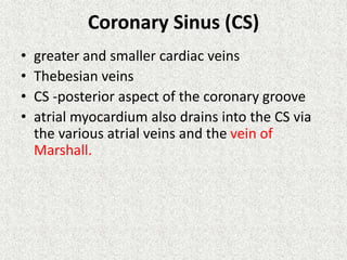
- 1. Coronary Sinus (CS) • greater and smaller cardiac veins • Thebesian veins • CS -posterior aspect of the coronary groove • atrial myocardium also drains into the CS via the various atrial veins and the vein of Marshall.
- 3. • muscular tube • 2 to 3 cm long • 1 cm in caliber • 1 cm above and parallel to the left atrioventricular junction • close contact with the left atrium • enveloped in a sheath of atrial myocardium • small valve (valve of Vieussen) =junction of the great cardiac vein and the CS • CS =posterior and lateral aspects of the heart • oblique entry of these small vessels into the CS =valvular effect
- 5. Histology • endocardium, • striated myocardium, • epicardium and • a specific conduction system • CS =small cardiac chamber that joins the other four chambers at the level of the crux cordis.
- 6. • striated myocardial fibers in the media • cells similar to the P cells of the sinus node • large number of Purkinje-like cells. • Numerous groups of ganglionic neurons = subepicardium
- 7. Function • transfer of blood from the CS to the right atrium during active CS contraction • lowest saturated blood in whole body =30 – 35% • relatively thick • instead of smooth muscle forming a tunica media, consists of striated myocardium continuous with that of the atria
- 8. development
- 19. • Congenital CS morphologic • Diverticulum=inferior aspect at its junction with the middle cardiac vein. ;basis for posterolateral and left posterior accessory pathways • variations in shape, including a wind sock– shaped, filiform, varicoid, or bifid CS;
- 22. Ostial Aneurysm
- 27. Wide CS Zipes, p. 1013
- 28. Small with superior angulation 8 Fr catheter CS Anatomy Variations
- 29. Large with superior angulation 8 Fr catheter CS Anatomy Variations Balloon LAO 20
- 30. CS Anatomy Variations Large with inferior angulation
- 31. CS Anatomy Variations Funnel-shaped with low origin
- 33. CS Anatomy Variations Valve of Vieussens
- 34. CS Anatomy Variations Valve of Vieussens
- 35. Gradual Take-off Branch Anterior Lateral Gradual Lateral Branch AP
- 36. Acute Take-off Branch Acute Lateral Branch AP
- 37. Acute take off (Shepherd's crook) AP
- 38. • diameter of the CS is also variable • Congenital condition • loading conditions • presence and extent of atrial myocardium with the coronary vein • the presence of underlying cardiac disease or prior cardiac surgery.
- 39. CS Ostia Size Non-Heart Failure 3-15 mm. Heart Failure 6-19mm. Hellerstein study of 150 cadaver hearts CS size correlated positively with right atrial size and right atrial pressure, but not with right ventricular pressure or degree of TR atrial fibrillation -larger CS
- 40. How to visualise CS • Non invasive • Echo • CT • MRI • Coronary angiogram – Direct injection(Retrograde venography via conventional balloon occlusion angiography) – During levo phase of arterial angiogram
- 41. Uses • percutaneous techniques for allowing retroinfusion of oxygenated (arterialized) blood in the coronary veins of patients considered to be unsuited for conventional revascularization procedures. • regional application of therapeuticagents such as cardio-protective drugs, cells, or gene vectors. • access route to the myocardium for electrophysiology procedures.
- 42. • retrograde injection of contrast media to the myocardium through the heart’s CS. • administering oxygenated blood to the ischemic myocardium during • USAor • high-risk PTCA • in cardiac surgery, • showing an improvement in ischemia and reducing episodes of chest angina
- 43. Access to the Coronary Sinus • Percutaneous cannulation of the CS • Access =right and left subclavian veins, the right internal jugular vein, and the femoral veins. • Katritsis et al =cannulation of the CS through the femoral vein using a modified 6 Fr Judkins JL5 catheter • complications =arrhythmia, dissection, and perforation with hemopericardium
- 44. Retrograde Venous Coronary Perfusion • benefit of reverse flow in the coronary venous system for oxygenation and nutrition of the myocardial cells • By perfusing the CS=maintain cardiac contractions for 90 minutes in a devascularized feline heart model • increase coronary perfusion while partially occluding the CS in animals. These animals tolerated anterior descendent artery ligation better than control animals that did not have partial CS occlusion.
- 45. • The multiple descriptions that rule out coronary venous system compromise due to atherosclerosis encourage even more research of the coronary venous system as an alternative system for maintaining nutritional flow to the myocardium in patients with severe involvement of arterial circulation. • Prior to the advent of bypass surgery, surgeons experimented with arterializations of the coronary venous system. • Beck et al refined this procedure during the 1940s. In an operation known as the Beck II procedure, they would first obstruct the CS using ligation. After 10 days, during which the CS had fibrosed, an arterial or venous conduit was placed between the aorta and the CS.
- 46. • The initial experimental work on this procedure was carried out on dog animal models. • This revealed two important physiological characteristics: the CS could tolerate arterial pressure for a long period of time, and there was not a broad fistula effect following CS arterialization. • Beck’s focus was termed “global retroperfusion” as the methodfor carrying oxygenated blood to the whole coronary venous system. • However, they found that the permanent obstruction of coronary venous drainage was associated with specific problems, including hemorrhage, fibrosis, and myocardial edema.
- 47. • A second means of venous revascularization, the selective method, in which blood flow is reversed only in precise ischemic areas caused by arterial disease, was proposed by Arealis. • Using this method, only a small portion of venous drainage is reversed, with the epicardial and Thebesian venous systems, and other territories continue to function normally • With the advent of bypass surgery, interest in this technique dwindled; ongoing studies were abandoned and new studies on this topic were relegated.
- 48. • The advent of synchronized diastolic coronary retroperfusion allowed intermittent venous drainage of the myocardium, which is produced during retrograde perfusion. • This technique has been shown to decrease pain episodes in patients with unstable angina and reduce complications in patients undergoing high-risk PTCA
- 49. • In the 1980s, retroperfusion of the CS was used as a way to protect the myocardium during angioplasty in this group of patients. • The technique was able to reduce both anginal symptoms during PTCA as well as the time to appearance of ischemia as seen in ST-segment changes following balloon inflation. • The main reason for its underutilization seem to be that the retroperfusion equipment has technical limitations and is slow to configure; in addition, it is technically impossible in 10%-20% of patients
- 50. Local Drug Delivery • Efficacy of retrograde perfusion of pharmacologically active agents to the myocardium through the coronary sinus • In intact, non-ischemic myocardiums, the medication concentration following retroinfusion is similar to that achieved when the medications are administered intravenously (IV). • However, in the context of myocardial ischemia, retroinfusion achieves higher levels of concentration of the medication in the tissue than when it is administered IV, with the advantage of having lower peak systemic concentrations and fewer systemic side effects • Thrombolytic agents, when administered directly in the coronary veins, act faster, improve functional recovery, and reduce the size of the infarct compared with systemic administration
- 51. Delivery of Genes, Growth Factors and Stem Cells • The genomic and proteomic revolution continues advancing and the underlying molecular biology of several cardiovascular pathologies is increasingly well understood • The success of these treatments depends on many factors, among which is the ability to be delivered adequately to the target tissues • it is assumed that most agents are more effective and better tolerated if they are delivered locally.
- 52. • Delivery of genes to the heart has been attempted using viral and non-viral vectors • Adenovirus is the viral vector that has proven most effective in vivo; however, adverse immune and inflammatory responses may limit its use • Direct injection of vectors based on plasmids to the myocardium has been reported, although the level of gene expression with this method is relatively low • Penta et al showed a plasmid gene delivery vector, hDel-1, known to have angiogenic activity in angiogenesis trials when it is administered to the myocardium through the coronary sinus. It was found in high concentrations in the myocardium, but is absent or at a much lower concentration in peripheral tissues.
- 53. • Growth factors (GF) with angiogenic properties, such as fibroblast basic growth factor (FGF-2) and vascular endothelial growth factor (VEGF), induce growth of collateral coronary vessels. • Various methods for introducing these GFs to the myocardium have been studied, such as direct injection to the myocardium and coronary artery infusion. • Von Degenfeld et al reported the selective and regulated use via venous coronary retroinfusion of FGF in a porcine model of chronic ischemia. • The venous delivery of GF has been favorably compared with intracoronary infusion and has also led to greater levels of meshing of the tissue with FGF-2 and has improved coronary perfusion through collateral vessels
- 54. • Recent studies have emphasized stem cell transplant as an emerging treatment for patients in end-stage heart failure. • The two main methods for delivering stem cells to the myocardium have been researched: these are direct (through intramyocardical injection) and intracoronary infusion. • Both methods have proven to be effective. • However, it has been shown that cells injected directly into the heart to form small islets may be isolated from native myocardium, and intracoronary infusion may not allow the cells to reach the ischemic myocardium • Suzuki et al reported a rat model of retrograde venous perfusion of stem cells in the myocardium. This infusion route has been proven to allow cellular diffusion to all layers of the myocardium with minimal adverse effects seen during or following the retrograde infusion.
- 55. Coronary Venous System in Cardiac Elecrophysiology • The knowledge of coronary venous anatomy is of vital importance for the practice of modern electrophysiology. • The venous system has become a crucial access route for the invasive management of pathologies, such as advanced cardiac failure, atrial fibrillation, and some supraventricular and ventricular tachycardias
- 56. • CS allows percutaneous endocardial access to posterolateral veins that drain the left ventricle, through which guides and catheters can be advanced for placing electrodes used in cardiac resynchronization therapy through programmed biventricular stimulation in patients with advanced heart failure. • This improves their functional class and decreases ventricular dyssynchrony which, in turn, improves the effectiveness of myocardial contractions, increases forward cardiac output and decreases regurgitant mitral volume
- 57. For biventricular pacing, optimal left ventricular pacing lead position is within the posterolateral branch of the coronary sinus, followed by the lateral branch
- 58. • CS approach is also useful in managing atrioventricular (AV) re-entrant arrhythmias, being routinely used as an alternative approach in the ablation of left accessory pathways, as the majority of these anomalous connections tend to have epicardial fibers and insertions which on occasion cannot be eliminated through conventional endocavitary ablation • AV re-entrant tachycardias that use the ligament of Marshall as a re-entry mechanism have been successfully eliminated by catheter ablation through the CS and the vein of Marshall • Some CS fibromuscular tissue bands have electrical conducting properties, and cases of atypical atrial flutter have been described in patients following pulmonary vein Isolation
- 59. • CS endo-epicardial ablation is an important step during radiofrequency ablation for patients with persistent AF today • The ligament of Marshall as an anatomical structure is a remnant of the left superior vena cava and contains nerve fibers with significant electrical conduction properties, connected to the CS muscular tissue in its proximal portion and with the left pulmonary veins in its middle portion. • The vein of Marshall has been a focus point during ablation of paroxysmal AF in young patients, as this structure is an important trigger of arrhythmic events in up to 20% of these cases
- 60. • Knowledge of the exact anatomical location of the CS ostium is paramount during ablation of AV nodal re-entrant tachycardia (AVNRT), as it helps delineate the anatomical boundaries of the triangle of Koch, which is made up in its posteriorportion by the tendon of Todaro, anteriorly by the septal cusp in the tricuspid ring and inferiorly by the CS ostium • The middle portion of this anatomic triangle contains the compact AV node. • In patients with AVNRT, there are at least two (and at times multiple) pathways with different conduction properties that constitute the necessary circuit for this re-entrant arrhythmia (classically labeled as slow-fast for typical AVNRT and fast-slow or slow-slow for atypical AVNRT).
- 61. • The slow pathway, which is usually located at the base of this triangle, is the preferred target of radiofrequency modulation or cryoablation for treating this arrhythmia, as the risk of complete heart block using this approach is <1% • The coronary sinus provides direct access to the cardiac venous system as a potential approach during mapping and ablation of ventricular tachycardias originating from an epicardial substrate. • By placing electrode catheters into the coronary sinus, electrical mapping of low-amplitude diastolic potentials and early local electrograms can guide successful VT ablation in areas of difficult endocardial access such as the mitral annulus (via the greater cardac vein) or the left ventricular summit, at the junction between the greater cardiac and the anterior interventricular veins.
- 62. •The left atrial veins can be used for atrial pacing. When thresholds are poor in the right atrium or precise left atrial-left ventricular synchrony is needed. •Specific cannulation of the CS and then placing the lead into an atrial branch can be done. •Bi-atrial pacing with both a right and left atrial lead has been attempted for pacing related therapy of atrial fibrillation
- 64. Venous System in Valvular Interventions • With the growing interest in structural interventions and the development of percutaneous techniques for the treatment of valvular pathologies through percutaneous routes • The cardiac venous system, and especially the coronary sinus, is attractive because it is a structure located parallel to the posterior portion of the mitral ring and surrounding up to two thirds of the ring, making it possible to implant devices via this route with the capacity to shorten the length of the coronary sinus and in this way carry out a valvular annuloplasty.
- 65. • In a generic way, these devices seek to create a tension or constriction of the sinus, which is transmitted to the mitral valvular apparatus and especially to the valvular ring. • Currently, there are initial experiences in humans with the Cardiac Dimensions Carillon (Cardiac Dimensions), Edwards Monarc System (Edwards Lifesciences), and the Viacor PTMA (percutaneous transvenous mitral annuloplasty)system (Viacor) device
- 66. • The Cardiac Dimensions Carillon system consists of a nitinol guide that has been molded to form a proximal and a distal anchor; these are joined by bridge elements with the ability to shorten the length of the coronary sinus. • The first studies in humans found a decrease in mitral valve insufficiency in at least one degree in all cases, improving left ventricular function and decreasing ventricular diameter and volume. • Likewise, a symptomatic improvement was achieved with a decrease in functional class and improvement after 6 minutes of continuous walking at 6-months follow-up exam.
- 68. • The Edwards Monarc system is made up of proximal and distal anchors formed by self-expanding stents, and a bridge system that joins them. • The distal anchor is released in the anterior interventricular vein and the proximal anchor near the coronary sinus ostium. Once a large part of the mitral ring circumference is captured, the bridge segment is tensed in order to shorten it. • The initial experience with this device showed a one or two degree reduction in insufficiency in most patients. • However, the EVOLUTION II study (Clinical Evaluation of the Edwards Lifesciences Percutaneous Mitral Annuloplasty System for the Treatment of Mitral Regurgitation) was suspended due to patient inclusion difficulties
- 70. • Lastly, the Viacor PTMA system consists of a trilumen catheter that may be implanted through the subclavian, with different nitinol guides that are advanced through the catheter lumen. • Pressure is applied to the central portion of the posterior mitral ring, thus shortening the distance between the septum and the distal wall with a consequent shortening or constriction of the mitral ring.
- 72. • Although the devices are technically feasible to implant in most patients, technical problems have been described secondary to the anatomic variations of the coronary sinus, as well as material fatigue with device fracture, which has been described with the three devices and is related to the complex dynamics of compression and torsion that the device undergoes during the cardiac cycle. • Another significant limitation is the compression of the circumflex artery by the device. Most devices that cause compression of the circumflex artery present dramatic clinical manifestations during the procedure, forcing the removal of the device and cancellation of the procedure. • Evaluation with multidetector scanning is very useful for detecting the anatomic relationship of the coronary sinus to the body of the circumflex artery and the marginal branches in order to decrease this complication
- 73. Thebesian Valve • The ostium of the Coronary Sinus may have a ridge of tissue that is referred to as the Thebesian Valve. • This valve originates from the embryonic Right Venous Valve and may obstruct entry to the CS. • The variations of the thebesian valve may explain the great variations in oxygen content in the blood obtained at different levels in the right atrium and right ventricle.
- 75. • Thebesian valve is a crescent shaped structure often found guarding the mouth of the CS as it opens to the right atrium. • It is highly variable and occasionally may present an obstruction during cannulation of the CS.
- 76. • Makes it difficult to enter the coronary sinus for electrophysiologist • Makes it difficult to administer of retrograde cardioplegia while doing CABG • Studies have demonstrated that valves that obstruct a significant portion of the CS ostium may be successfully transversed by use of modern cannulation techniques (i.e. softer-tipped sheath, preshaped sheath, and anterior approach)
- 77. Divisione di Cardiologia Parma LAO 40° Thebesian Valve Thebesian Valve
- 79. Thebesian Flap
- 80. Thebesian Flap Remnants of flap formed a bar like structure
- 83. Chiari’s Network For a Chiari’s Network, the Network extends out along the Eustachian ridge. 0.03 % or 3 out of 1000 hearts Hellerstein
- 84. • A related but distinct entity is the subthebesian pouch , an outpouching of the right atrial wall inferior to the CS ostium that can serve as a substrate for a reentrant circuit causing atrial flutter . • It is also described as the subeustachian sinus or sinus of Keith, although anatomically it is subthebesian, not subeustachian. • A deep subthebesian pouch has been described as a cause of interventional procedural difficulty and has been reported in 47% of case
- 85. Sub Thebesian Pouch, or a “False CS Ostium” Anderson