More Related Content
Similar to Acs0601 Stroke And Transient Ischemic Attack
Similar to Acs0601 Stroke And Transient Ischemic Attack (20)
More from medbookonline (20)
Acs0601 Stroke And Transient Ischemic Attack
- 1. © 2007 WebMD, Inc. All rights reserved. ACS Surgery: Principles and Practice
6 VASCULAR SYSTEM 1 Stroke and Transient Ischemic Attack — 1
1 STROKE AND TRANSIENT ISCHEMIC
ATTACK
Thomas S. Maldonado, M.D., and Thomas S. Riles, M.D., F.A.C.S.
Assessment and Management of Stroke and TIA
Stroke is defined as any damage to the central nervous system study, as well as to the lower life expectancy of men, especially
caused by interruption of the blood supply. Ischemic strokes result those with vascular risk factors.9 Race and genetic predisposition
from the failure of oxygen and nutrients to reach the affected are also considered independent risk factors, with African
brain. Transient ischemic attacks (TIAs) are defined as transient Americans more than twice as likely to die of stroke as whites.10
neurologic deficits lasting no longer than 24 hours; longer-lasting The underlying mechanism of stroke appears to vary with race as
deficits are considered to be indicative of a cerebral infarction well: intracranial occlusive disease tends to occur more frequently
(“brain attack”). in African and Asian Americans than in whites or in males as a
Infarction from ischemia is typically confined to a vascular ter- whole.11,12 The importance of hereditary risk for stroke has long
ritory, at whose center can be found the injured or obstructed been recognized. In the Framingham Study, both paternal and
artery supplying that parenchyma.The full extent of a stroke may maternal histories of stroke were associated with an increased risk
not become apparent until days or weeks later, when the tenuous of stroke.10,13
peripheral watershed zone, or penumbra, either survives or suc- There are, however, certain risk factors for stroke that are clear-
cumbs to cell death. Smaller infarcts, adequate collateral circula- ly modifiable, including hypertension, smoking, hyperlipidemia,
tion, and prompt intervention and resuscitation are associated asymptomatic high-grade carotid stenosis, and atrial fibrillation
with improved outcome. (AF).14-16 Other purported modifiable risk factors for stroke (e.g.,
Hemorrhagic stroke damages the brain by cutting off connect- obesity, diabetes, oral contraceptive use, and alcohol intake) are
ing pathways and causing localized or generalized pressure injury. more controversial.17-20 Hypertension (systolic as well as diastolic)
Brain edema and hydrocephalus after hemorrhagic stroke may is perhaps the most prominent modifiable risk factor for stroke
also be deleterious. In some cases, hemorrhagic stroke can lead to and is associated with a substantial risk of atherothrombotic, lacu-
ischemia as a consequence of vasospasm, as is seen in the setting nar, or subarachnoid hemorrhage. The Systolic Hypertension in
of subarachnoid hemorrhage (SAH). Likewise, some ischemic the Elderly Program (SHEP) study demonstrated that treating
strokes can undergo hemorrhagic transformation. systolic hypertension in patients 60 years of age or older can
reduce the incidence of stroke by as much as 36%.21 Prompt diag-
nosis and intervention for stroke may be critical, but many author-
Incidence and Risk Factors ities feel that primary prevention is the true cornerstone of thera-
Stroke is the third leading cause of death in the United States: py for this lethal disease.
about 168,000 stroke-related deaths are reported each year.1
Approximately 500,000 first-time strokes are reported annually.
In addition, stroke is the leading cause of serious long-term dis- Clinical Evaluation
ability in the United States and poses a substantial economic bur- When assessing patients
den. For 2003, U.S. expenditure on stroke-related medical costs who present with a neuro-
and disability payments amounted to roughly $51 billion. logic deficit, clinicians must
Although a sharp decline in stroke incidence and mortality was immediately ask themselves
noted throughout most of the 20th century, the decline leveled off the following two questions:
in the early 1990s, and stroke incidence and mortality are now ris- (1) what is the mechanism
ing for the first time since 1915.2,3 of the deficit (i.e., ischemic or hemorrhagic), and (2) where is the
Numerous population studies and stroke registries have been lesion (e.g., cerebral lacunae, the territory around a large vessel, or
designed to examine the incidence, risk factors, and natural histo- a watershed region)? A thorough history and physical examina-
ry of stroke.4-10 Some of the independent risk factors that have tion, in conjunction with brain imaging studies, usually suffice to
been identified—such as age (> 55 years), sex (male), race guide initial management. Nonvascular conditions that can mimic
(African American and Asian), and genetic predisposition—can- stroke (e.g., hypoglycemia, migraine, a postictal state, enceph-
not be modified and therefore carry a fixed level of stroke risk.The alopathy, trauma, and brain tumors or abscesses) must be exclud-
Rochester (Minnesota) population study demonstrated a marked ed. Although these syndromes can all cause focal findings, they
progressive incidence of cerebral infarction with advancing age, as usually lack the abrupt onset of symptoms consistently seen with
well as a nearly 1.5 times greater risk of stroke in males.4 The stroke.
Lausanne Stroke Registry data confirmed the overall higher inci- Ischemic and hemorrhagic stroke must be differentiated early in
dence of stroke in males, though it also demonstrated a female the course of an acute stroke because a number of therapeutic
preponderance in very young (< 30 years) and very old (> 80 interventions that are beneficial for some subtypes of stroke are
years) patients. These latter findings may be attributable to the potentially catastrophic for others. Approximately 71% of all
high frequency of oral contraceptive use in young women in that strokes are ischemic and 26% hemorrhagic; the remaining 3% are
- 2. © 2007 WebMD, Inc. All rights reserved. ACS Surgery: Principles and Practice
6 VASCULAR SYSTEM 1 Stroke and Transient Ischemic Attack — 2
Assessment and Management of
Stroke and TIA
Patient has hemorrhagic stroke
Determine whether stroke is caused by ICH or SAH.
ICH SAH
Reduce BP judiciously. Treat ICP, as for ICH (see left).
Reverse coagulopathy if present. Ligate ruptured aneurysm.
Treat brain edema with hyperosmolar agents, Manage postoperative hydrocephalus,
hyperventilation, or, if necessary, surgical rebleeding, or cerebral vasoconstriction.
evacuation of hematoma. Consider hyperdynamic (triple H)
therapy postoperatively.
- 3. © 2007 WebMD, Inc. All rights reserved. ACS Surgery: Principles and Practice
6 VASCULAR SYSTEM 1 Stroke and Transient Ischemic Attack — 3
Patient presents with neurologic deficit suggestive of stroke
Rule out nonvascular conditions mimicking stroke (e.g., hypoglycemia,
trauma, and encephalopathy).
Perform thorough history and physical examination.
Perform imaging studies of brain (CT and MRI).
Order lab tests (CBC, aPTT, PT, lipid panel, electrolytes, glucose).
Distinguish between hemorrhagic and ischemic stroke.
Patient has ischemic stroke
Establish mechanism of stroke, and identify source
lesion (if one exists).
Systemic hypoperfusion Lacunar infarcts Cardiac embolism Atherothrombosis
Correct pump failure. Establish diagnosis with MRI. Establish diagnosis with ECG, Establish diagnosis with
Treat any underlying concomitant No treatment is available. transthoracic echocardiography, duplex US, MRA, and CT
stenosis. or transesophageal echocardi- angiography; if findings
ography. inconclusive, perform
Initiate early anticoagulation: conventional angiography.
heparin, followed by warfarin (or
other antiplatelet agent if warfarin
is contraindicated).
Consider thrombolysis or surgical
embolectomy if appropriate.
Artery-artery embolization In situ thrombosis
Assess indications for surgery Administer thrombolytic therapy.
(e.g., carotid endarterectomy).
Patient is candidate for carotid endarterectomy Patient is not candidate for carotid
(i.e., has moderate to high-grade stenosis, endarterectomy
TIAs, or stroke in evolution)
Consider embolectomy or thrombolysis.
Perform carotid endarterectomy. (Alternatively, Administer antiplatelet therapy.
consider carotid angioplasty and stenting.)
Administer antiplatelet therapy.
- 4. © 2007 WebMD, Inc. All rights reserved. ACS Surgery: Principles and Practice
6 VASCULAR SYSTEM 1 Stroke and Transient Ischemic Attack — 4
of unknown origin [see Table 1].22 Hemorrhagic strokes result from
subarachnoid or intracerebral bleeding, whereas ischemic strokes
result from systemic hypoperfusion, cardioembolism, or athero-
thrombosis. Although various different laboratory and radiograph-
ic tests are available and should be performed, the diagnosis of
stroke is primarily a clinical one.
HISTORY
A patient who has suffered a stroke typically presents with a his-
tory of sudden onset of a focal brain deficit and a clinical syn-
drome on neurologic examination. A medical history should elic-
it any of a number of risk factors associated with either ischemic
or hemorrhagic stroke. For example, hypertension is often associ-
ated with deep infarcts and hemorrhages, whereas cigarette smok-
ing and hyperlipidemia are more commonly associated with
ischemic infarcts resulting from atherosclerosis.9 (Hypertension
can, however, be associated with infarcts caused by hypertensive
arteriolopathy.) The presence of atrial fibrillation, a recent myo-
cardial infarction (MI) (< 6 weeks old), or a prosthetic heart valve
suggests a possible cardioembolic mechanism.
The onset and course of the neurologic deficit may also be
telling. A steady onset is suggestive of hemorrhage, whereas symp-
toms that progress in a stepwise fashion are more likely to derive
from ischemia. Likewise, accompanying signs are frequently use-
ful in differentiating stroke mechanism subtypes. Headache, vom-
iting, and loss of consciousness constitute the classic picture of
hemorrhagic stroke, whereas a focal deficit preceded by TIAs is
more suggestive of ischemic stroke.23 Although the clinical mani-
festations are generally helpful in differentiating stroke subtypes, it
sometimes happens that large infarcts mimic the classic picture of
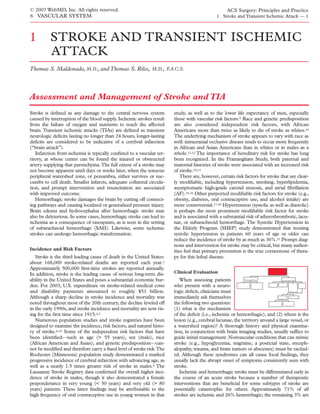
Figure 1 Shown is a CT image of an acute hemorrhagic infarct,
hemorrhage or that lobar or small deep hemorrhages resemble with intraparenchymal bleeding apparent.
infarction resulting from atherosclerosis. Analysis of 1,000 consec-
utive patients from the Lausanne Stroke Registry who experienced
their first stroke confirmed that the classic hemorrhagic picture crepant pulse exams, and carotid bruits should all be noted.
(headaches, progressive neuralgic deficits, and decreased level of Careful examination of the extremities is essential, in that periph-
consciousness) was indeed more common in patients with hem- eral vascular disease is highly correlated with the presence of ath-
orrhage but found that only one third of patients with hemor- erosclerosis of the carotid and vertebral arteries.6,24 An ophthal-
rhagic strokes had this clinical triad.9 mologic examination should detect subhyaloid hemorrhages, as
PHYSICAL AND NEUROLOGIC EXAMINATION
well as cholesterol emboli (Hollenhorst plaques).
Besides localizing the lesion anatomically, the neurologic exam-
A general physical and neurologic examination should detect ination should be able to identify a probable cause.The absence of
vascular and cardiac abnormalities, as well as localize the process major focal neurologic signs is often consistent with SAH; focal
within the CNS. Heart size and sounds, irregular rhythms or dis- deficits localized to the superficial cortex are often attributable to
thromboembolism or ischemia. A number of well-described lacu-
nar syndromes also exist; these typically suggest small-vessel
disease.
Table 1Causes of Stroke as
Recorded in NINCDS Data Bank22
Investigative Studies
Cerebral ischemia 71%
Atherosclerosis 10% IMAGING
Larger-artery stenosis 6% Imaging is a mandatory and integral part of evaluation for acute
Tandem arterial lesions 4% stroke and should be performed expeditiously. Computed tomog-
Lacunae 19%
raphy is the initial test of choice; it is readily available in most hos-
Cardioembolism 14%
Infarct of undetermined cause 27%
pitals and is well tolerated by critically ill patients.25 CT can
Cerebral hemorrhage 26%
promptly identify nonvascular causative conditions (e.g., masses)
Parenchymatous 13% and can readily diagnose intracerebral hemorrhage (ICH) [see
Subarachnoid 13% Figure 1]. Diagnosis of SAH without I.V. contrast can be more dif-
Other 3% ficult, especially if bleeding is minor or occurred more than 1 day
before; the accuracy of CT in detecting SAH decreases after 24
NINCDS—National Institute of Neurological and Communicative Disorders hours.26 SAH should appear as an increased density in the CSF. If
and Stroke SAH is clinically suspected, lumbar puncture may be necessary for
definitive diagnosis.
- 5. © 2007 WebMD, Inc. All rights reserved. ACS Surgery: Principles and Practice
6 VASCULAR SYSTEM 1 Stroke and Transient Ischemic Attack — 5
In cases of acute ischemic stroke, diagnosis of infarction by The high sensitivity of MRI in detecting infarction has been
means of CT can be more difficult. I.V. contrast CT is of limited demonstrated in studies evaluating patients experiencing TIAs.
value in this setting. Signs of infarction can be absent or subtle in When patients without signs or symptoms of infarction underwent
the first few hours after the stroke. A lesion may appear as a slight brain imaging after a TIA, 27% were found to have evidence of so-
hypodensity within the infarcted zone or as a loss of definition called silent infarcts on CT and 73% on MRI.32
between gray and white matter.27 Newer CT scanners may be bet- Diagnosis of ICH can be a more delicate task with MRI than
ter at delineating these nuances.28,29 In the days following an with CT. Careful scrutiny by an experienced observer using dif-
ischemic stroke, an infarct may first appear round or oval and then ferent acquisition techniques is required. The appearance of a
as a hypodense, dark, or wedge-shaped lesion on CT. cerebral hematoma on T1- and T2-weighted sequences varies as the
Magnetic resonance imaging is more expensive and time-con- lesion matures and edema resolves. Acute hematomas are black on
suming than CT and is less well tolerated by critically ill patients. T1-weighted images, and chronic hematomas are white on T1- and
Nonetheless, MRI is more sensitive than CT in detecting early T2-weighted images.
ischemic changes after a stroke. It can be used to distinguish an Normal neuroimaging findings are not uncommon in patients
old stroke from a new one and to assess the size and location of a presenting with neurologic deficits suggestive of acute stroke. A
lesion, especially when the lesion is adjacent to bony structures [see patient with a neurologic deficit and a negative CT scan should
Figure 2].30 The size of the infarct as determined by MRI may be undergo further cerebral imaging with MRI. The absence of
of great importance for prognosis. Although lesion size may not ischemic infarction and hemorrhage on both CT and MRI may
correlate with severity of clinical presentation, larger infarcts in the suggest transient ischemia or persistent ischemia without infarc-
same vascular territory are associated with more severe deficits tion. In such cases, one may have to rely on signs and symptoms
than smaller infarcts in similar anatomic locations are. to localize the ischemic lesion responsible for the stroke.
MRI is especially useful for diagnosing lacunar infarcts. In a Alternatively, electroencephalography (EEG), positron emission
review of 227 patients with lacunar infarcts, 44% were diagnosed tomography (PET), single-photon emission CT (SPECT), or
by CT and 78% by MRI.31 Infarction appears as dark, hypodense xenon-enhanced CT (XeCT) may be employed to help localize
areas on T1-weighted sequences and as bright areas on T2-weight- ischemic foci.33,34
ed sequences. Edema usually develops around the infarct within
LABORATORY TESTS
the first few days and is readily apparent on MRI as a low-density
area surrounding the lesion with mass effect. Sometimes, the Blood should be sent to the laboratory as part of the initial
infarct is small but is still associated with substantial edema. Such assessment of a patient with an acute stroke. Measurement of
edema may be insignificant in an older patient with an atrophied serum electrolyte and glucose levels is essential to rule out nonva-
brain whose cranium is able to accommodate a mass effect but scular conditions mimicking stroke (e.g., hypoglycemia and dehy-
may be life-threatening in a younger patient with little intracranial dration). A complete blood count (CBC), an activated partial
room to spare. thromboplastin time (aPTT), a prothrombin time (PT), and a
a b
Figure 2 Shown are (a) a fluid-attenuated inversion-recovery (FLAIR) MRI image and (b) a T2-
weighted MRI image of an acute ischemic infarct in the left middle cerebral artery distribution.
- 6. © 2007 WebMD, Inc. All rights reserved. ACS Surgery: Principles and Practice
6 VASCULAR SYSTEM 1 Stroke and Transient Ischemic Attack — 6
lipid panel are likewise essential components of the initial assess- brain ischemia. Short-acting antihypertensives should be adminis-
ment. Severe anemia has the potential to exarcerbate or precipitate tered only when the systolic blood pressure is persistently higher
cerebral ischemia in stroke; though this is an uncommon occur- than 180 to 200 mm Hg or when there is evidence of active bleed-
rence, it should be considered and, if present, corrected.35 ing or an enlarging hematoma. Other medical treatment of hemor-
Additional laboratory blood tests (e.g., a cardiac injury panel to rhagic stroke from ICH includes reversal of coagulopathies with
rule out myocardial infarction or an erythrocyte sedimentation transfusions of fresh frozen plasma and platelets when appropriate.
rate to assess the possibility of vasculitis) may be helpful. Mortality from ICH has plummeted from 90% before the
Screening should be performed for hematologic disorders that 1970s to less than 50% in the first decade of the 21st century.2,22,47
result in hypercoagulable states, including homocystinuria,36 This precipitous decline probably reflects improved antihyperten-
antiphospholipid antibody syndrome, protein C and S deficiency, sive therapy, as well as a decreased prevalence of hypertension.48
antithrombin deficiency, and activated protein C resistance from Death from ICH most commonly occurs secondary to herniation
factor V Leiden mutation [see 6:6 Venous Thromboembolism].37,38 from the hematoma itself coupled with brain edema, which can
Hemoglobinopathies (e.g., sickle cell disease and thalassemia) can develop within the first hours after ICH.47,49 Thus, treatment of
also lead to altered blood flow, hypercoagulability, and stroke.39 brain edema is the main focus of treatment. Corticosteroids,
Finally, hyperviscosity syndromes (e.g., polycythemia vera, throm- though indicated for reducing cerebral edema in patients with
bocytosis, and myeloproliferative disorders) should be included in tumors or abscesses, are contraindicated in patients with ICH
the differential diagnosis. If a hyperviscosity syndrome is recog- because they are not beneficial and may in fact be injurious, pre-
nized in the setting of acute stroke, hemodilution therapy may be disposing patients to infection and worsening diabetes.50
warranted. Experimental and clinical trials have shown hemodilu- Hyperosmolar agents (e.g., mannitol or glycerol solutions) may be
tion to increase blood flow in the ischemic brain; however, other more useful for reducing cerebral edema rapidly.51 Alternatively, in
studies have failed to show improvement of neurologic status.40,41 cases of severe brain edema, hyperventilation may reduce ICP by
Hemodilution remains indicated in stroke patients who have a inducing diffuse vasoconstriction in the brain. Any physiologic
high hematocrit or are in a hyperviscosity state; however, close perturbation that might increase cerebral blood volume (e.g.,
monitoring is required in patients with heart disease or cerebral hypercarbia, hypoxia, or vasodilation) or reduce cerebral perfusion
edema. (e.g., hypotension) should be avoided. Finally, surgical evacuation
of a hematoma, though controversial, may be employed as a last
resort for decompression after ICH.52
Recognition and
Management of Specific Subarachnoid
Stroke Subtypes Hemorrhage
Rupture of saccular or
HEMORRHAGIC STROKE
berry aneurysms with sub-
Hemorrhagic stroke can sequent SAH carries signifi-
result from ICH or SAH. cant morbidity and mortali-
The consequences of cerebral bleeding can progress rapidly from ty, depending largely on the
neurologic deficit to coma to death in a significant number of patient’s age, the extent of
patients. the hemorrhage, and the presence and severity of rebleeding, cere-
bral vasospasm, and surgical complications.53 SAH accounts for
Intracerebral Hemorrhage 6% to 10% of all strokes and 22% to 25% of all deaths from cere-
ICH accounts for approximately 10% to 15% of all strokes in brovascular accidents.54 The reported incidence of incidental
the United States and Europe.22,42 Potential causative mechanisms aneurysms discovered in autopsy studies ranges from 0.8% to
include hypertension, trauma, arteriovenous malformations, cere- 18%.55-57 SAH tends to occur more often in patients 50 to 60
bral amyloid angiopathy, brain tumors, blood dyscrasias, and med- years of age than in younger or older patients and in women more
ications (e.g., anticoagulants and thrombolytics). Of these, hyper- often than in men (12.3% versus 7.9%).58 The pathogenesis of
tensive arteriopathy is most commonly responsible for nontrau- saccular aneurysms has not been fully explicated, but the risk fac-
matic ICH.22,43 Hypertensive brain hemorrhages are usually deep tors are well described and include a family history of SAH,59
and are typically located in the lateral ganglionic region, the sub- hypertension, pregnancy,60 and black race.61
cortex, the thalamus, the caudate nucleus, the pons, or the cere- SAH presents with warning signs in as many as 20% of patients
bellum. As early as the 1870s, Charcot and Bouchard correctly within the 3 months preceding a major rupture, presumably
postulated that such events were the result of microaneurysmal because a minor leak develops before the rupture. Such warning
disease of arteries and arterioles penetrating deep into the brains signs include a so-called sentinel headache, oculomotor symp-
of hypertensive patients.44,45 These discrete hemorrhages go on to toms, nausea and vomiting, and loss of consciousness.62 The rup-
compress neighboring capillary networks, causing them to burst ture itself is accompanied by sudden severe headache, nuchal
and bringing about a rapid enlargement of the intracerebral rigidity, back pain, nausea, vomiting, photophobia, lethargy, loss of
hematoma. Indeed, such enlargement is not uncommon: one consciouness, and seizure.60,63
prospective study found that 38% of patients with ICH had a Medical management of SAH includes tranquil bed rest with the
hematoma that was enlarged at 3 hours in comparison with its size head elevated 30°, stool softeners, antiemetics, and analgesics, as
on baseline CT after the initial bleeding event.46 well as deep vein thrombosis (DVT) prophylaxis using pneumatic
Patients presenting with sizable ICH often rely on a marked compression boots [see 6:6 V enous Thromboembolism]. Management
compensatory elevation of blood pressure to maintain a pressure of ICP in SAH patients is similar to that in ICH patients (see
gradient in the setting of acute increases in intracranial pressure above). Furthermore, one must be vigilant for signs of hypothala-
(ICP). It is vital to resist the impulse to lower blood pressure aggres- mic dysfunction manifesting as cardiac dysrhythmias or the syn-
sively in these patients: a rapid drop in blood pressure may induce drome of inappropriate antidiuretic hormone secretion (SIADH).
- 7. © 2007 WebMD, Inc. All rights reserved. ACS Surgery: Principles and Practice
6 VASCULAR SYSTEM 1 Stroke and Transient Ischemic Attack — 7
The timing of microsurgical clip ligation of aneurysms after ure. Certain patients with concomitant underlying stenosis proxi-
SAH is somewhat controversial. The International Cooperative mal to border-zone ischemic territories may benefit from treat-
Study on the Timing of Aneurysm Surgery showed no overall dif- ment of the flow-limiting lesions to eliminate the hemodynamic
ferences in outcome between early surgery (0 to 3 days after SAH) impairment and thus may do better than patients with systemic
and delayed surgery (11 to 14 days after SAH).64 Nonetheless, hypoperfusion.76
patients who were alert preoperatively did better with early surgery
and demonstrated significantly better rates of good recovery. Lacunar Infarcts
Three major neurologic complications affect outcome after Neurologic examination
surgery for SAH: hydrocephalus, rebleeding, and cerebral vaso- of a patient believed to have
constriction. Hydrocephalus may develop acutely as a result of experienced an acute isch-
obstruction of CSF outflow. Ventricular drainage can lead to emic stroke should be atten-
immediate reduction of ICP and improvement in neurologic tive to the possibility of lacu-
symptoms. The risk of rebleeding peaks on postoperative day 1; it nar infarcts, manifested by
is as high as 20% in the first 2 weeks and rises to 50% within 6 pure motor hemiparesis,
months if the aneurysm is not treated.53 Unlike rebleeding, pure sensory syndrome, sensorimotor syndrome, ataxic hemipare-
vasospasm manifests itself gradually over hours to days; it may be sis, and dysarthria–clumsy hand syndrome.77,78 Lacunae are small
associated with up to a threefold increase in mortality during the (1 to 2 cm) subcortical lesions that result from small-vessel disease
2 weeks after SAH.65-67 The diagnosis is initially made via angiog- deep in the brain. They can occur either alone or in groups and
raphy and followed via transcranial Doppler sonography.68,69 may be present in as many as 23% of persons over the age of 65
Hyperdynamic, or triple H, therapy consists of keeping patients years. Lacunar infarcts, unlike other forms of stroke, do not pre-
hyperdynamic (to increase cardiac output), hypervolemic-hyper- sent with headache and are associated with hypertension and dia-
tensive (to augment cerebral perfusion pressure), and hemodilut- betes.79 Although they are asymptomatic (silent) in as many as
ed (to improve cerebral microcirculation by decreasing viscosity). 89% of patients, their benignity is currently in some doubt: there
Because of the risk that the aneurysm will rerupture before oper- is evidence to suggest that they may increase the risk of dementia
ation, triple H therapy is reserved for postoperative patients.70,71 and cognitive decline.80,81 Lacunar syndromes are characteristic
Calcium blockers may also reduce the incidence of symptoms sec- and highly predictive of the presence of lacunae, but they may be
ondary to vasospasm, though they most likely have little effect on less reliable for excluding other mechanisms of stroke. A review of
spasm per se.72 the Northern Manhattan Stroke Study experience demonstrated
that as many as 25% of patients presenting with radiographically
ISCHEMIC STROKE
confirmed lacunar infarcts were ultimately found to have other
If hemorrhagic stroke is mechanisms of ischemic stroke.82 Thus, MRI should be used to
ruled out and ischemic confirm or exclude the presence of lacunar infarcts, as well as to
stroke is diagnosed, the next screen for nonlacunar mechanisms of stroke.
step is to establish the mech- At present, there is no treatment for lacunar infarcts, but the
anism of the stroke and prognosis is quite good. The survival rate is high, the recurrence
identify the source lesion rate is low (mean annual stroke rate, 4% to 7%), and patients gen-
responsible (if one exists). erally achieve relatively good functional recovery, with as many as
Systemic hypoperfusion, cardiac embolism, large-artery athero- 74% experiencing mild or no disability at 1 year.83-86
sclerosis, and small-vessel disease should be systematically consid-
ered as potential causes. Cardiac Embolism
Embolism of cardiac ori-
Systemic Hypoperfusion gin accounts for approxi-
Only a small fraction of all ischemic strokes are attributable to mately 14% of ischemic
systemic hypoperfusion. A quick assessment of vital signs and strokes [see Table 1]. Given
symptoms may provide the first clues that a patient is suffering that many infarcts of unde-
from cerebral ischemia secondary to systemic hypoperfusion. termined cause are probably
Patients are characteristically either unconscious on arrival in the of cardioembolic origin, this
emergency department or awake but exhibiting neurologic symp- figure may in fact be an underestimate.22 Whereas clinical suspicion
toms resembling near-syncope.73 Generally, they are pale, for cardioembolism may be high in the setting of acute stroke, con-
diaphoretic, and hypotensive. Neurologic signs and symptoms are firming the diagnosis can be more difficult. In most instances, car-
varied and are explained by ischemia in the border zone (or water- dioembolic stroke can be diagnosed on the basis of the history, the
shed) between two or three adjacent arterial territories. Difficulty physical examination, and electrocardiography. A number of con-
in reading or identifying visual stimuli (or even frank blindness) ditions are known to predispose to cardioembolism, including AF
may be observed; this may be attributed to ischemia between the or atrial flutter, recent MI (< 6 weeks old), placement of a pros-
middle and posterior cerebral arteries. Bilateral arm weakness or thetic valve, disease of the aortic or mitral valves, and paradoxical
cognitive difficulty may suggest ischemia between the middle and emboli (usually from DVT).87 Key clinical manifestations include
anterior cerebral arteries. Global symptoms usually arise from sudden onset with rapid progression to maximal deficit, infarcts in
bilateral border-zone infarcts, which can develop in association different arterial territories, rapid regression of symptoms, and
with prolonged cardiogenic shock, dysrhythmias, or cardiac decreased consciousness at onset.88-90 Headache and seizure activ-
arrest.74,75 Alternatively, symptoms may be asymmetrical if they ity are not specific for cardioembolic stroke.The rapid regression of
derive from inadequate perfusion distal to a site of severe stenosis symptoms—also known as the spectacular shrinking deficit
or occlusion of major feeding cerebral vessels. (SSD)—is thought to result from rapid recanalization or fragmen-
Treatment should focus primarily on correcting the pump fail- tation and migration of cardioembolus downstream.91 Ironically,
- 8. © 2007 WebMD, Inc. All rights reserved. ACS Surgery: Principles and Practice
6 VASCULAR SYSTEM 1 Stroke and Transient Ischemic Attack — 8
the very mechanism purported to result in rapid regression of Unlike cardioembolism, carotid atheroemboli and thrombosis are
symptoms may trigger the hemorrhagic transformation seen in as more likely to be associated with TIAs. Moreoever,TIAs of carotid
many as 70% of cardioembolic strokes.92,93 The vast majority of arterial origin tend to occur in the same vascular territory, where-
hemorrhagic transformations are caused by cardioembolism. A as TIAs of cardioembolic origin are more haphazard in loca-
2001 study found that delayed recanalization occurring less than 6 tion.88,90 Once the history, the physical examination, and brain
hours after an acute cardioembolic stroke developed in 52.8% of imaging studies are complete, duplex ultrasonography should be
patients and was an independent risk factor for hemorrhagic trans- performed to search for extracranial atherosclerosis of the carotid
formation.93 Other risk factors for such transformation are severe arteries. Duplex examination of the carotid arteries may also iden-
strokes, decreased alertness, and absence of collateral flow.94 tify ulceration and assess plaque echolucency, both of which may
Atrial fibrillation may or may not be detected on ECG, espe- increase the level of risk.109,110 Other important diagnostic modal-
cially if it is paroxysmal; it remains an elusive but increasingly ities commonly used to assess extracranial sources for
important risk factor for cardioembolic strokes in older patients.95 atherothrombotic-embolic mechanisms of stroke are magnetic res-
Holter monitoring and electrophysiologic testing may be especial- onance angiography (MRA) and CT angiography. We routinely
ly useful for diagnosing paroxysmal AF.96 Likewise, transthoracic use these modalities to corroborate the findings of duplex ultra-
echocardiography and transesophageal echocardiography are use- sonography. In patients with carotid arterial stenosis, conventional
ful for detecting mural thrombus caused by AF or another car- angiography is usually limited to cases in which duplex ultra-
diomyopathy, as well as for detecting valvular diseases, vegetations, sonography disagrees with MRA or CT angiography; in patients
tumors, and patent foramina ovalia. with vertebral arterial stenosis, angiography is mandatory before
In the presence of nonrheumatic AF, the overall incidence of any attempt is made at reconstruction.
stroke is nearly five times higher, rising from 1.5% in persons 50 Diagnosis of symptomatic carotid artery lesions in the setting of
to 59 years of age to 23.5% in those 80 to 89 years of age.97 acute ischemic stroke is critical because this subtype of stroke
Moreover, data from the International Stroke Trial indicate that patient stands to benefit significantly from surgical intervention.
mortality is twice as high in patients with AF as in those without The results of three prospective randomized trials from the early
AF (17% versus 7.5% at 2 weeks).98 It is noteworthy that noncar- 1990s comparing medical with surgical treatment of symptomatic
dioembolic strokes are estimated to account for about one third of carotid arterial stenosis demonstrated that carotid endarterectomy
the strokes that occur in AF patients. A 1997 autopsy study of 82 (CEA) had a significant advantage over medical treatment
consecutive patients with symptomatic stroke and nonrheumatic (aspirin).111-113 Perhaps the most convincing of the three studies
AF demonstrated that 29 (35%) of the infarctions in patients with was the North American Symptomatic Carotid Endarterectomy
nonrheumatic AF were in fact of noncardioembolic origin.95 Trial (NASCET), which was terminated prematurely because of
As a rule, acute ischemic stroke of cardioembolic origin is best the clear superiority of the results in the surgical arm.111 In
treated with early anticoagulation to prevent recurrent brain NASCET, the cumulative risk of ipsilateral stroke was 9% with
embolism. Numerous studies have shown that recurrent embolism surgical treatment and 26% with medical treatment at 2-year fol-
to the brain occurs within 2 weeks in 6% to 12% of patients after low-up, for an absolute risk reduction of 17% and a relative risk
an initial ischemic infarct from a cardioembolic source.99-101 A reduction of 65.4%. Patients with carotid arterial stenosis of less
1999 meta-analysis of 16 AF trials indicated that the overall risk of than 50% did not benefit from CEA. Conversely, studies have
stroke could be reduced by an average of 62% with anticoagula- shown that patients who have a completely occluded carotid artery
tion.102 Thus, once a hemorrhagic stroke has been ruled out, in the setting of an acute stroke should not undergo CEA either.
heparinization and treatment with warfarin are indicated. Patients Emergency CEA in an acutely occluded artery can result in con-
with ischemic stroke of cardioembolic origin who are at increased version of an ischemic infarct to a hemorrhagic cerebral infarct,
risk for hemorrhage or who have a contraindication to warfarin with potentially catastrophic results.114
may be treated with aspirin or another antiplatelet agent; however,
such agents are less effective than warfarin in secondary prevention
of cardioembolic ischemic stroke.103,104 Finally, despite the clear Indications for and Timing of Therapy
benefits of early anticoagulation in stroke patients with cardioem-
SURGICAL THERAPY
bolism, such therapy has not been shown to be advantageous in the
general stroke population.
For patients who suffer ischemic strokes of noncardioembolic Carotid Endarterectomy
origin, warfarin appears to offer no additional benefit over aspirin CEA [see 6:9 Treatment Carotid Artery] should be performed
in preventing stroke recurrence.105 Furthermore, most authorities only when a low (< 5%) morbidity and mortality from stroke can
would agree that patients with acute ischemic stroke who present be expected.111,115 When a symptomatic patient with moderate or
within 48 hours of the onset of symptoms should be given aspirin, high-grade stenosis presents with TIAs or a mild stroke, the deci-
160 to 325 mg/day, to reduce mortality and morbidity from sion to perform a CEA is relatively straightforward. Patients who
stroke.106,107 have suffered devastating infarcts, however, usually are not appro-
priate surgical candidates. Generally speaking, the extent to which
Atherothrombosis neurologic function is spared, the presence and severity of medical
Roughly 10% of acute comorbidities, and the patient’s life expectancy should all be
ischemic strokes are believed assessed before the decision is made to embark on CEA.
to occur secondary to large- The timing of CEA after an acute stroke continues to be a sub-
vessel atherosclerosis, throm- ject of controversy. According to some authorities, the risk that an
bosis, or artery-to-artery dis- ischemic infarct will undergo hemorrhagic transformation after
tal embolization.22 Smoking urgent CEA is a contraindication to early surgery.116,117 ICH after
and age are the most impor- endarterectomy is most likely the result of postoperative hyperper-
tant contributors to the development of carotid atheroma.108 fusion. The combination of hypertension (which is common after
- 9. © 2007 WebMD, Inc. All rights reserved. ACS Surgery: Principles and Practice
6 VASCULAR SYSTEM 1 Stroke and Transient Ischemic Attack — 9
stroke) and diminished vasomotor regulation in the penumbra 13,000 CEAs found that cardiopulmonary comorbid conditions
may predispose small vessels in this region to hemorrhage. Other did not increase the risk of perioperative stroke, death, or cardiac
authorities, however, argue that delaying surgery exposes the events.126 Finally, though many investigators have considered
patient to a substantial risk of recurrent stroke or carotid occlusion patients over the age of 80 (who were ineligible for NASCET and
and that early intervention is therefore warranted.118 Although it the Asymptomatic Carotid Atherosclerosis Study [ACAS]) to be at
would probably be generally agreed that waiting an obligatory 30 increased risk with open surgery, the interim results from the lead-
days before surgery is prudent, there are numerous small series in in phase of the Carotid Revascularization Endarterectomy versus
the literature supporting the idea that early CEA after an acute Stent Trial (CREST), which is currently in progress, indicated that
ischemic stroke is indeed safe and may be indicated.119,120 the risk of periprocedural stroke and death for patients undergoing
Furthermore, documentation of the location and size of the lesions CAS, surprisingly, was substantially higher (12.1%) in the 99
may help identify infarcts that are at increased risk for hemorrhag- patients who were over the age of 80.127 The reason for this increas-
ic transformation after CEA.120 Ultimately, the optimal timing of ing risk in older patients is not clear; the CREST results were not
surgery after an acute ischemic stroke must be determined on the affected by adjustment for potential confounding factors such
basis of prospective, randomized clinical trials.
as symptomatic status, the use of antiembolic devices, gender,
the degree of carotid stenosis, or the presence of distal arterial
Stroke in evolution A patient with neurologic deficits that
tortuosity.
worsen progressively in a stuttering fashion is considered to have a
The goal of reliably predicting which patients may be at high
stroke in evolution.This state is associated with the highest level of
risk, whether it is treated medically or surgically.121 A patient with risk with either stenting or open surgery has impelled some inves-
a stroke in evolution should promptly undergo CT to rule out tigators to develop risk stratification scales for CAS. A 2006
hemorrhagic stroke, followed immediately by systemic anticoagu- prospective study of 606 consecutive patients who underwent CAS
lation with heparin and urgent operation. In a 1981 study of identified the following independent risk factors: diabetes mellitus
patients with stroke in evolution or crescendo TIAs that compared with inadequate glycemic control (hemoglobin A1c level > 7%),
medical treatment (N=31) with urgent CEA (N=24), nonopera- advanced age ( 80 years), ulceration of the carotid artery stenosis,
tive management of stroke in evolution yielded a significantly high- and a significant contralateral stenosis ( 50%). Patients with two or
er mortality than surgical management (15% versus 6%).122 more of these risk factors had an 11% risk of a periprocedural
Furthermore, 70% of the patients who underwent CEA achieved complication, whereas patients with none or only one had a 2%
complete or near-complete recovery, compared with 19% of those risk.128
managed medically.
Assessment of results The results of various clinical trials
Carotid Angioplasty and Stenting comparing CAS with CEA, some already completed and some still
In keeping with the continuing growth of minimally invasive under way, will help determine whether there is clinical equipoise
surgery, carotid angioplasty and stenting (CAS) has come to play between the two procedures.127,129-133 The SAPPHIRE (Stenting
an emerging role in the management of both symptomatic and and Angioplasty with Protection in Patients at HIgh Risk for
asymptomatic carotid stenosis [see 6:10 Carotid Angioplasty and Endarterectomy) trial randomly compared CAS with CEA in 334
Stenting].The advent of neuroprotection techniques using balloon patients with coexisting conditions that potentially increased the
occlusion and aspiration of debris, filter wires placed distal to the risk associated with endarterectomy.130 Analysis of 30-day and 1-
culprit lesion, or reversal-of-flow technology has allowed interven- year outcomes (including death, stroke, and MI) found that CAS
tionalists to perform CAS more safely, with complication rates was not inferior to CEA. However, the overall risk levels were dis-
approximating those of CEA.123,124 Nevertheless, the precise role turbingly high in both arms of this trial: 12.2% for CAS patients
of carotid artery stenting in treatment or prevention of stroke has and 20.1% for CEA patients. The CaRESS (Carotid Revas-
not yet been defined.The two primary questions that remain unre- cularization using Endarterectomy or Stenting Systems) trial was
solved are (1) which patients benefit most from carotid stenting as a multicenter prospective study that compared the two techniques
opposed to open surgery, and (2) are the immediate and long-term in 397 patients on a nonrandom basis.132 The results indicated that
results of CAS as good as those of CEA? Determination of the part both the 30-day risk and the 1-year risk of death, stroke, or MI
CAS will eventually play in the management of cerebrovascular
were essentially the same in CAS patients as in CEA patients.The
disease awaits the results of ongoing clinical trials comparing CAS
Carotid And Vertebral Artery Transluminal Angioplasty Study
with CEA.
(CAVATAS) randomly assigned 504 patients to the two treat-
ments.131 The incidence of major stroke or death in the 30 days fol-
Selection of patients Initially, CAS was considered a proce-
dure that would be most beneficial for high-risk patients—for lowing the procedure did not differ significantly between the two
example, patients who were poor surgical candidates because of groups: 6.4% for CAS patients and 5.9% for CEA patients. At 1
substantial medical comorbidities and patients with so-called hos- year, severe carotid restenosis was noted more frequently in the
tile anatomy (such as an extremely high or low bifurcation or a CAS group (14% versus 4%). Finally, the Stent-supported
neck that had previously been irradiated or operated on). Indeed, Percutaneous Angioplasty of the Carotid artery versus End-
one clear benefit of CAS over CEA is that the risk of nerve injury arterectomy (SPACE) trial randomly assigned 1,200 patients with
for patients undergoing the former procedure is 0%. A precise def- symptomatic carotid artery stenosis to undergo either CAS or
inition of a high-risk patient, however, has proved elusive. One CEA. At 30 days, the incidence of stroke or death was 6.9% in the
study reviewed CEAs that were performed in 228 patients who, CAS group and 6.3% in the CEA group.133 The investigators con-
because of their increased level of risk, would not have met the cluded (1) that the results failed to prove the noninferiority of CAS
NASCET inclusion criteria.125 This study was unable to demon- in comparison with CEA and (2) that the results did not justify the
strate that these supposedly high-risk patients actually had inferior widespread use of CAS in the short term for the treatment of
outcomes with CEA. A subsequent study that included more than carotid artery stenosis.
- 10. © 2007 WebMD, Inc. All rights reserved. ACS Surgery: Principles and Practice
6 VASCULAR SYSTEM 1 Stroke and Transient Ischemic Attack — 10
MEDICAL THERAPY
come measures at 90 days between placebo and rt-PA in an inten-
tion-to-treat analysis.136 Similarly, the Alteplase ThromboLysis for
Thrombolysis Acute Noninterventional Therapy in Ischemic Stroke
Any patient who presents with an acute ischemic stroke, regard- (ATLANTIS) trial reported no benefit in patients treated with rt-
less of subtype, is potentially a candidate for thrombolytic therapy. PA within 3 to 5 hours of the onset of symptoms.137,138 However,
However, ICH documented on the initial head CT is a clear patients treated within the golden 3-hour window were more like-
absolute contraindication to I.V. thrombolysis. Other considera- ly to have a favorable outcome at 90 days than patients treated with
tions affecting the decision whether to administer thrombolytic placebo (60.9% versus 26.3%).
agents include a history of GI or urologic hemorrhage, recent Unfortunately, most patients are ineligible for I.V. rt-PA because
major surgery, and rapidly improving neurologic signs, any of of delays in obtaining treatment. Indeed, studies show that only 1%
which may constitute a clinical contraindication to medical treat- to 2% of ischemic stroke patients receive I.V. rt-PA.139,140
ment. Vigilant monitoring of the aPTT, the PT (or the interna- Compared with I.V. thrombolysis, intra-arterial thrombolytic
tional normalized ratio [INR]), the platelet count, and the fib- therapy ought, in theory, to be able to deliver a higher local con-
rinogen level is essential throughout the course of thrombolytic centration of the agent where it is needed while minimizing the sys-
treatment. temic concentration. Proponents of intra-arterial thrombolysis
It has been suggested that maximal benefit is derived from I.V. hope that it may lengthen the 3-hour treatment window. The
thrombolytic therapy for acute ischemic stroke when it is delivered PROACT II trial provided the best evidence to date that intra-
within a “golden 3-hour window” starting from the onset of symp- arterial thrombolysis can improve patient outcomes.141 This ran-
toms. The National Institute for Neurological Disorders and domized, open-label, multicenter study with blinded follow-up
Stroke (NINDS) trial (parts 1 and 2) randomly assigned 624 randomly assigned 180 patients with stroke of less than 6 hours’
patients to receive either recombinant tissue plasminogen activator duration to receive either heparin with recombinant prourokinase
(rt-PA), 0.9 mg/kg I.V. to a maximum of 90 mg/kg, or placebo.134 (r-proUK), 9 mg intra-arterially, or heparin alone. Intra-arterial
Patients with all types of ischemic stroke were eligible, provided thrombolysis resulted in significantly better recanalization rates
that they could be treated within 3 hours of the onset of symptoms. than heparin alone did (66% versus 18%). In addition, more of the
Outcome at 3 months was better with rt-PA than with placebo on r-proUK group had no neurologic deficit or only a slight deficit at
each of the four outcome measures studied. The odds ratio for a 90 days (40% versus 25%). However, intra-arterial thrombolysis
favorable outcome in the rt-PA group was 1.7. The overall rate of did result in significantly increased rates of ICH (35% versus
symptomatic hemorrhage was 6.4% in the rt-PA group and 0.6% 13%). Symptomatic ICH with neurologic deterioration within 24
in the placebo group. The beneficial effects of rt-PA were similar hours occurred in 10% of r-proUK patients and 2% of control
for all stroke subtypes and persisted for up to 12 months after the patients. Patients who experienced ICH after r-proUK therapy
stroke.135 Patients treated with rt-PA were at least 30% more like- had a high mortality (83%).142 It is noteworthy that only 2%
ly to have minimal or no disability at 12 months than patients treat- (180/12,333) of the screened patients in the PROACT II trial were
ed with placebo were. Mortality at 1 year was comparable in the randomized according to inclusion criteria, which suggests that
two groups. intra-arterial thrombolysis may be of limited applicability. Finally,
Other randomized, double-blind, placebo-controlled trials of rt- intra-arterial thrombolyis requires an experienced staff capable of
PA for treatment of acute ischemic stroke have examined the effect performing cerebral angiography and navigating a microcatheter
of thrombolytic therapy given within the first 6 hours after the to the clot. At present, I.V. thrombolysis is certainly more practical
onset of symptoms. The European Cooperative Acute Stroke than intra-arterial thrombolysis; more important, it can be done
Study (ECASS) found no significant differences in functional out- earlier in the course of the stroke.
References
1. Association AS Stroke Facts 2003: All Harvard Cooperative Stroke Registry: a prospec- vascular risk factors. J Neurol 249:507, 2002
Americans. CDC/NCHS 2001–2003 tive registry. Neurology 28:754, 1978 16. Goldstein LB, Adams R, Becker K, et al: Primary
2. Broderick JP, Phillips SJ, Whisnant JP, et al: 9. Bogousslavsky J, Van Melle G, Regli F: The prevention of ischemic stroke: a statement for
Incidence rates of stroke in the eighties: the end Lausanne Stroke Registry: analysis of 1,000 con- healthcare professionals from the Stroke Council
of the decline in stroke? Stroke 20:577, 1989 secutive patients with first stroke. Stroke of the American Heart Association. Stroke
3. Gillum RF, Sempos CT: The end of the long- 19:1083, 1988 32:280, 2001
term decline in stroke mortality in the United 10. Sacco RL, Benjamin EJ, Broderick JP, et al: Ameri- 17. Jorgensen H, Nakayama H, Raaschou HO, et al:
States? Stroke 28:1527, 1997 can Heart Association Prevention Conference: IV. Stroke in patients with diabetes.The Copenhagen
4. Matsumoto N, Whisnant JP, Kurland LT, et al: Prevention and rehabilitation of stroke. Risk fac- Stroke Study. Stroke 25:1977, 1994
Natural history of stroke in Rochester, tors. Stroke 28:1507, 1997
18. Tegos TJ, Kalodiki E, Daskalopoulou SS, et al:
Minnesota, 1955 through 1969: an extension of 11. Wong KS, Huang YN, Gao S, et al: Intracranial Stroke: epidemiology, clinical picture, and risk
a previous study, 1945 through 1954. Stroke stenosis in Chinese patients with acute stroke. factors (part I of III). Angiology 51:793, 2000
4:20, 1973 Neurology 50:812, 1998
19. Stadel BV: Oral contraceptives and cardiovascu-
5. Sacco RL, Wolf PA, Kannel WB, et al: Survival 12. Caplan LR, Gorelick PB, Hier DB: Race, sex lar disease (first of two parts). N Engl J Med
and recurrence following stroke: the Framingham and occlusive cerebrovascular disease: a review. 305:612, 1981
study. Stroke 13:290, 1982 Stroke 17:648, 1986
20. Gill JS, Zezulka AV, Shipley MJ, et al: Stroke and
6. Wolf PA, D’Agostino RB, Belanger AJ, et al: 13. Kiely DK, Wolf PA, Cupples LA, et al: Familial alcohol consumption. N Engl J Med 315:1041,
Probability of stroke: a risk profile from the aggregation of stroke: the Framingham Study. 1986
Framingham Study. Stroke 22:312, 1991 Stroke 24:1366, 1993
21. Prevention of stroke by antihypertensive drug
7. Sobel E, Alter M, Davanipour Z, et al: Stroke in 14. Amarenco P: Blood pressure and lipid lowering treatment in older persons with isolated systolic
the Lehigh Valley: combined risk factors for in the prevention of stroke: a note to neurolo- hypertension: final results of the Systolic
recurrent ischemic stroke. Neurology 39:669, gists. Cerebrovasc Dis 16(suppl 3):33, 2003 Hypertension in the Elderly Program (SHEP).
1989 15. Leys D, Deplanque D, Mounier-Vehier C, et al: SHEP Cooperative Research Group. JAMA
8. Mohr JP, Caplan LR, Melski JW, et al: The Stroke prevention: management of modifiable 265:3255, 1991
- 11. © 2007 WebMD, Inc. All rights reserved. ACS Surgery: Principles and Practice
6 VASCULAR SYSTEM 1 Stroke and Transient Ischemic Attack — 11
22. Foulkes MA, Wolf PA, Price TR, et al: The Stroke 44. Cole FM,Yates P: Intracerebral microaneurysms 66. Corsten L, Raja A, Guppy K, et al: Contemporary
Data Bank: design, methods, and baseline charac- and small cerebrovascular lesions. Brain 90:759, management of subarachnoid hemorrhage and
teristics. Stroke 19:547, 1988 1967 vasospasm: the UIC experience. Surg Neurol
23. Gorelick PB, Hier DB, Caplan LR, et al: Head- 45. Caplan L: Intracerebral hemorrhage revisited. 56:140, 2001
ache in acute cerebrovascular disease. Neurology Neurology 38:624, 1988 67. Torner JC, Kassell NF, Wallace RB, et al:
36:1445, 1986 46. Brott T, Broderick J, Kothari R, et al: Early hem- Preoperative prognostic factors for rebleeding and
24. Kannel WB, McGee DL: Diabetes and cardio- orrhage growth in patients with intracerebral survival in aneurysm patients receiving antifibri-
vascular disease. The Framingham study. JAMA hemorrhage. Stroke 28:1, 1997 nolytic therapy: report of the Cooperative
241:2035, 1979 Aneurysm Study. Neurosurgery 9:506, 1981
47. Schuetz H, Dommer T, Boedeker RH, et al:
25. Welch KM, Levine SR, Ewing JR:Viewing stroke Changing pattern of brain hemorrhage during 68. Sloan MA, Burch CM, Wozniak MA, et al:
pathophysiology: an analysis of contemporary 12 years of computed axial tomography. Stroke Transcranial Doppler detection of vertebrobasi-
methods. Stroke 17:1071, 1986 23:653, 1992 lar vasospasm following subarachnoid hemor-
rhage. Stroke 25:2187, 1994
26. Adams HP Jr, Kassell NF, Torner JC, et al: CT 48. Ueda K, Hasuo Y, Kiyohara Y, et al: Intracerebral
and clinical correlations in recent aneurysmal hemorrhage in a Japanese community, 69. Newell DW, Winn HR: Transcranial Doppler in
subarachnoid hemorrhage: a preliminary report Hisayama: incidence, changing pattern during cerebral vasospasm. Neurosurg Clin North Am
of the Cooperative Aneurysm Study. Neurology long-term follow-up, and related factors. Stroke 1:319, 1990
33:981, 1983 19:48, 1988 70. Tommasino C, Picozzi P: Physiopathological cri-
27. von Kummer R, Nolte PN, Schnittger H, et al: 49. Silver FL, Norris JW, Lewis AJ, et al: Early mor- teria of vasospasm treatment. J Neurosurg Sci
Detectability of cerebral hemisphere ischaemic tality following stroke: a prospective review. 42(1 suppl 1):23, 1998
infarcts by CT within 6 h of stroke. Neuroradio- Stroke 15:492, 1984 71. Treggiari MM, Walder B, Suter PM, et al:
logy 38:31, 1996 50. Poungvarin N, Bhoopat W, Viriyavejakul A, et al: Systematic review of the prevention of delayed
28. Hunter GJ, Hamberg LM, Ponzo JA, et al: Effects of dexamethasone in primary supratento- ischemic neurological deficits with hypertension,
Assessment of cerebral perfusion and arterial rial intracerebral hemorrhage. N Engl J Med hypervolemia, and hemodilution therapy follow-
anatomy in hyperacute stroke with three-dimen- 316:1229, 1987 ing subarachnoid hemorrhage. J Neurosurg
sional functional CT: early clinical results. AJNR 98:978, 2003
51. Steiner T, Ringleb P, Hacke W: Treatment
Am J Neuroradiol 19:29, 1998 options for large hemispheric stroke. Neurology 72. Feigin VL, Rinkel GJ, Algra A, et al: Calcium
29. von Kummer R, Allen KL, Holle R, et al: Acute 57(5 suppl 2):S61, 2001 antagonists in patients with aneurysmal sub-
stroke: usefulness of early CT findings before 52. Ziai WC, Port JD, Cowan JA, et al: Decompressive arachnoid hemorrhage: a systematic review.
thrombolytic therapy. Radiology 205:327, 1997 craniectomy for intractable cerebral edema: expe- Neurology 50:876, 1998
30. Maeda M, Abe H, Yamada H, et al: Hyperacute rience of a single center. J Neurosurg Anesthesiol 73. Caplan LR: Diagnosis and treatment of ischemic
infarction: a comparison of CT and MRI, includ- 15:25, 2003 stroke. JAMA 266:2413, 1991
ing diffusion-weighted imaging. Neuroradiology 53. Kassell NF, Torner JC, Haley EC Jr, et al: The 74. Torvik A: The pathogenesis of watershed infarcts
41:175, 1999 International Cooperative Study on the Timing in the brain. Stroke 15:221, 1984
31. Arboix A, Marti-Vilalta JL, Garcia JH: Clinical of Aneurysm Surgery: part 1. Overall manage-
75. Angeloni U, Bozzao L, Fantozzi L, et al: Internal
study of 227 patients with lacunar infarcts. ment results. J Neurosurg 73:18, 1990
borderzone infarction following acute middle
Stroke 21:842, 1990 54. Sacco RL, Wolf PA, Bharucha NE, et al: Sub- cerebral artery occlusion. Neurology 40:1196,
32. Nicolaides AN PK, Grigg M, et al: Amaurosis arachnoid and intracerebral hemorrhage: natural 1990
Fugax. Springer, New York, 1988 history, prognosis, and precursive factors in the
76. Bogousslavsky J, Regli F: Borderzone infarctions
33. Kilpatrick MM,Yonas H, Goldstein S, et al: CT- Framingham Study. Neurology 34:847, 1984
distal to internal carotid artery occlusion: prog-
based assessment of acute stroke: CT, CT 55. McCormick WF, Acosta-Rua GJ: The size of nostic implications. Ann Neurol 20:346, 1986
angiography, and xenon-enhanced CT cerebral intracranial saccular aneurysms: an autopsy
77. Mori E, Tabuchi M, Yamadori A: Lacunar syn-
blood flow. Stroke 32:2543, 2001 study. J Neurosurg 33:422, 1970
drome due to intracerebral hemorrhage. Stroke
34. Green JB, Bialy Y, Sora E, et al: High-resolution 56. Inagawa T, Hirano A: Autopsy study of unrup- 16:454, 1985
EEG in poststroke hemiparesis can identify ipsi- tured incidental intracranial aneurysms. Surg
lateral generators during motor tasks. Stroke Neurol 34:361, 1990 78. Fisher CM: A lacunar stroke: the dysarthria-
30:2659, 1999 clumsy hand syndrome. Neurology 17:614, 1967
57. Dell S: Asymptomatic cerebral aneurysm: assess-
35. Kim JS, Kang SY: Bleeding and subsequent ane- ment of its risk of rupture. Neurosurgery 10:162, 79. Mast H, Thompson JL, Lee SH, et al:
mia: a precipitant for cerebral infarction. Eur 1982 Hypertension and diabetes mellitus as determi-
Neurol 43:201, 2000 nants of multiple lacunar infarcts. Stroke 26:30,
58. Kojima M, Nagasawa S, LeeYE, et al: Asymptoma- 1995
36. Eikelboom JW, Hankey GJ, Anand SS, et al: tic familial cerebral aneurysms. Neurosurgery
Association between high homocyst(e)ine and 43:776, 1998 80. Longstreth WT Jr, Bernick C, Manolio TA, et al:
ischemic stroke due to large- and small-artery Lacunar infarcts defined by magnetic resonance
59. Nakagawa T, Hashi K, Kurokawa Y, et al: Family imaging of 3660 elderly people: the Cardiovas-
disease but not other etiologic subtypes of history of subarachnoid hemorrhage and the
ischemic stroke. Stroke 31:1069, 2000 cular Health Study. Arch Neurol 55:1217, 1998
incidence of asymptomatic, unruptured cerebral
37. Kenet G, Sadetzki S, Murad H, et al: Factor V aneurysms. J Neurosurg 91:391, 1999 81. Vermeer SE, Prins ND, den Heijer T, et al: Silent
Leiden and antiphospholipid antibodies are sig- brain infarcts and the risk of dementia and cog-
60. Dias MS, Sekhar LN: Intracranial hemorrhage nitive decline. N Engl J Med 348:1215, 2003
nificant risk factors for ischemic stroke in chil- from aneurysms and arteriovenous malforma-
dren. Stroke 31:1283, 2000 tions during pregnancy and the puerperium. 82. Gan R, Sacco RL, Kargman DE, et al: Testing
38. Madonna P, de Stefano V, Coppola A, et al: Neuro surgery 27:855, 1990 the validity of the lacunar hypothesis: the
Hyperhomocysteinemia and other inherited pro- Northern Manhattan Stroke Study experience.
61. Broderick JP, Brott T, Tomsick T, et al: The risk Neurology 48:1204, 1997
thrombotic conditions in young adults with a of subarachnoid and intracerebral hemorrhages
history of ischemic stroke. Stroke 33:51, 2002 in blacks as compared with whites. N Engl J Med 83. Clavier I, Hommel M, Besson G, et al: Long-
39. Brass LM, Prohovnik I, Pavlakis SG, et al: 326:733, 1992 term prognosis of symptomatic lacunar infarcts:
Middle cerebral artery blood velocity and cere- a hospital-based study. Stroke 25:2005, 1994
62. Bassi P, Bandera R, Loiero M, et al: Warning
bral blood flow in sickle cell disease. Stroke signs in subarachnoid hemorrhage: a cooperative 84. Salgado AV, Ferro JM, Gouveia-Oliveira A:
22:27, 1991 study. Acta Neurol Scand 84:277, 1991 Long-term prognosis of first-ever lacunar
40. Asplund K: Haemodilution for acute ischaemic strokes: a hospital-based study. Stroke 27:661,
63. Hart RG, Byer JA, Slaughter JR, et al: Occurrence
stroke. Cochrane Database Syst Rev (4): 1996
and implications of seizures in subarachnoid hem-
CD000103, 2002 orrhage due to ruptured intracranial aneurysms. 85. Gandolfo C, Moretti C, Dall’Agata D, et al:
41. Strand T: Evaluation of long-term outcome and Neurosurgery 8:417, 1981 Long-term prognosis of patients with lacunar
safety after hemodilution therapy in acute syndromes. Acta Neurol Scand 74:224, 1986
64. Haley EC Jr, Kassell NF, Torner JC: The Interna-
ischemic stroke. Stroke 23:657, 1992 tional Cooperative Study on the Timing of 86. Hier DB, Foulkes MA, Swiontoniowski M, et al:
42. Sivenius J, Heinonen OP, Pyorala K, et al: The Aneurysm Surgery: the North American experi- Stroke recurrence within 2 years after ischemic
incidence of stroke in the Kuopio area of East ence. Stroke 23:205, 1992 infarction. Stroke 22:155, 1991
Finland. Stroke 16:188, 1985 65. Pasqualin A: Epidemiology and pathophysiology 87. Special report from the National Institute of
43. Wityk RJ, Caplan LR: Hypertensive intracerebral of cerebral vasospasm following subarachnoid Neurological Disorders and Stroke Classification
hemorrhage: epidemiology and clinical pathology. hemorrhage. J Neurosurg Sci 42(1 suppl 1):15, of cerebrovascular diseases III. Stroke 21:637,
Neurosurg Clin North Am 3:521, 1992 1998 1990
- 12. © 2007 WebMD, Inc. All rights reserved. ACS Surgery: Principles and Practice
6 VASCULAR SYSTEM 1 Stroke and Transient Ischemic Attack — 12
88. Arboix A, Oliveres M, Massons J, et al: Early dif- both, or neither among 19435 patients with Defining the high-risk patient for carotid
ferentiation of cardioembolic from atherothrom- acute ischaemic stroke. International Stroke Trial endarterectomy: an analysis of the prospective
botic cerebral infarction: a multivariate analysis. Collaborative Group Lancet 349:1569, 1997 National Surgical Quality Improvement
Eur J Neurol 6:677, 1999 108. Lees RS: The natural history of carotid artery Program database. J Vasc Surg 43:285, 2006
89. Timsit SG, Sacco RL, Mohr JP, et al: Brain disease. Stroke 15:603, 1984 127. Hobson RW 2nd, Howard VJ, Roubin GS, et al:
infarction severity differs according to cardiac or Carotid artery stenting is associated with
109. Kardoulas DG, Katsamouris AN, Gallis PT, et
arterial embolic source. Neurology 43:728, 1993 increased complications in octogenarians: 30-
al: Ultrasonographic and histologic characteris-
day stroke and death rates in the CREST lead-in
90. Kittner SJ, Sharkness CM, Sloan MA, et al: tics of symptom-free and symptomatic carotid
phase. J Vasc Surg 40:1106, 2004
Infarcts with a cardiac source of embolism in the plaque. Cardiovasc Surg 4:580, 1996
NINDS Stroke Data Bank: neurologic examina- 128. Hofmann R, Niessner A, Kypta A, et al: Risk
110. el-Barghouty N, Nicolaides A, Bahal V, et al: The
tion. Neurology 42:299, 1992 score for peri-interventional complications of
identification of the high risk carotid plaque. Eur
carotid artery stenting. Stroke 37:2557, 2006
91. Minematsu K,Yamaguchi T, Omae T: ‘Spectacular J Vasc Endovasc Surg 11:470, 1996
shrinking deficit’: rapid recovery from a major 129. Gray WA, Hopkins LN, Yadav S, et al: Carotid
111. Beneficial effect of carotid endarterectomy in
hemispheric syndrome by migration of an embo- stenting in high-surgical-risk patients: the
symptomatic patients with high-grade carotid
lus. Neurology 42:157, 1992 ARCHeR results. J Vasc Surg 44:258, 2006
stenosis. North American Symptomatic Carotid
92. Hornig CR, Bauer T, Simon C, et al: Hemorrhagic Endarterectomy Trial Collaborators N Engl J 130. Yadav JS,Wholey MH, Kuntz RE, et al: Protected
transformation in cardioembolic cerebral infarc- Med 325:445, 1991 carotid-artery stenting versus endarterectomy in
tion. Stroke 24:465, 1993 high-risk patients. N Engl J Med 351:1493, 2004
112. MRC European Carotid Surgery Trial: interim
93. Molina CA, Montaner J, Abilleira S, et al: results for symptomatic patients with severe 131. Endovascular versus surgical treatment in
Timing of spontaneous recanalization and risk of (70–99%) or with mild (0–29%) carotid stenosis. patients with carotid stenosis in the Carotid and
hemorrhagic transformation in acute cardioem- European Carotid Surgery Trialists’ Collaborative Vertebral Artery Transluminal Angioplasty Study
bolic stroke. Stroke 32:1079, 2001 Group Lancet 337:1235, 1991 (CAVATAS): a randomised trial. Lancet
357:1729, 2001
94. Alexandrov AV, Black SE, Ehrlich LE, et al: 113. Mayberg MR, Wilson SE, Yatsu F, et al: Carotid
Predictors of hemorrhagic transformation occur- endarterectomy and prevention of cerebral 132. Carotid revascularization using endarterectomy
ring spontaneously and on anticoagulants in ischemia in symptomatic carotid stenosis. or stenting systems (CARESS): phase I clinical
patients with acute ischemic stroke. Stroke Veterans Affairs Cooperative Studies Program trial. J Endovasc Ther 10:1021, 2003
28:1198, 1997 309 Trialist Group. JAMA 266:3289, 1991 133. The SPACE Collaborative Group: 30 day results
95. Yamanouchi H, Nagura H, Mizutani T, et al: 114. Blaisdell WF, Clauss RH, Galbraith JG, et al: from the SPACE trial of stent-protected angio-
Embolic brain infarction in nonrheumatic atrial Joint study of extracranial arterial occlusion. IV. plasty versus carotid endarterectomy in sympto-
fibrillation: a clinicopathologic study in the A review of surgical considerations. JAMA matic patients: a randomized non-inferiority
elderly. Neurology 48:1593, 1997 209:1889, 1969 trial. Lancet, epub ahead of print, August 10,
2006
96. Peters NS, Schilling RJ, Kanagaratnam P, et al: 115. Easton JD, Sherman DG: Stroke and mortality
Atrial fibrillation: strategies to control, combat, rate in carotid endarterectomy: 228 consecutive 134. Tissue plasminogen activator for acute ischemic
and cure. Lancet 359:593, 2002 operations. Stroke 8:565, 1977 stroke. The National Institute of Neurological
Disorders and Stroke rt-PA Stroke Study Group.
97. Wolf PA, Abbott RD, Kannel WB: Atrial fibrilla- 116. Bruetman M, Fields W, Crawford E, et al: N Engl J Med 333:1581, 1995
tion as an independent risk factor for stroke: the Cerebral hemorrhage in carotid artery surgery.
Arch Neurol 9:458, 1963 135. Kwiatkowski TG, Libman RB, Frankel M, et al:
Framingham Study. Stroke 22:983, 1991
Effects of tissue plasminogen activator for acute
98. Saxena R, Lewis S, Berge E, et al: Risk of early 117. Wylie E, Hein M, Adams J: Intracranial hemor- ischemic stroke at one year. National Institute of
death and recurrent stroke and effect of heparin rhage following surgical revascularization for Neurological Disorders and Stroke Recombinant
in 3169 patients with acute ischemic stroke and treatment of acute strokes. J Neurosurg 21:212, Tissue Plasminogen Activator Stroke Study
atrial fibrillation in the International Stroke Trial. 1964 Group. N Engl J Med 340:1781, 1999
Stroke 32:2333, 2001 118. Dosick SM, Whalen RC, Gale SS, et al: Carotid 136. Hacke W, Kaste M, Fieschi C, et al: Intravenous
99. Immediate anticoagulation of embolic stroke: a endarterectomy in the stroke patient: computer- thrombolysis with recombinant tissue plasmino-
randomized trial. Cerebral Embolism Study ized axial tomography to determine timing. J gen activator for acute hemispheric stroke. The
Group Stroke 14:668, 1983 Vasc Surg 2:214, 1985 European Cooperative Acute Stroke Study
100. Cardiogenic brain embolism. Cerebral Embolism 119. Whittemore AD, Ruby ST, Couch NP, et al: (ECASS). JAMA 274:1017, 1995
Task Force Arch Neurol 43:71, 1986 Early carotid endarterectomy in patients with 137. Clark WM, Albers GW, Madden KP, et al: The
101. Cardiogenic brain embolism. The second report small, fixed neurologic deficits. J Vasc Surg rtPA (Alteplase) 0- to 6-hour acute stroke trial,
of the Cerebral Embolism Task Force Arch 1:795, 1984 part A (276g): results of a double-blind, placebo-
Neurol 46:727, 1989 120. Toni D, Fiorelli M, Bastianello S, et al: Hemor- controlled, multicenter study. Thrombolytic
rhagic transformation of brain infarct: predictabil- Therapy in Acute Ischemic Stroke Study
102. Hart RG, Benavente O, McBride R, et al: Investigators. Stroke 31:811, 2000
Antithrombotic therapy to prevent stroke in ity in the first 5 hours from stroke onset and influ-
patients with atrial fibrillation: a meta-analysis. ence on clinical outcome. Neurology 46:341, 138. Clark WM, Wissman S, Albers GW, et al:
Ann Intern Med 131:492, 1999 1996 Recombinant tissue-type plasminogen activator
121. Moore WS, Mohr JP, Najafi H, et al: Carotid (Alteplase) for ischemic stroke 3 to 5 hours after
103. Warfarin versus aspirin for prevention of throm- symptom onset. The ATLANTIS Study: a ran-
boembolism in atrial fibrillation: Stroke endarterectomy: practice guidelines. Report of
the Ad Hoc Committee to the Joint Council of domized controlled trial. Alteplase Thrombolysis
Prevention in Atrial Fibrillation II Study. Lancet for Acute Noninterventional Therapy in
343:687, 1994 the Society for Vascular Surgery and the North
American Chapter of the International Society Ischemic Stroke. JAMA 282:2019, 1999
104. Petersen P, Boysen G, Godtfredsen J, et al: for Cardiovascular Surgery. J Vasc Surg 15:469, 139. Hacke W, Brott T, Caplan L, et al: Thrombolysis
Placebo-controlled, randomised trial of warfarin 1992 in acute ischemic stroke: controlled trials and
and aspirin for prevention of thromboembolic clinical experience. Neurology 53(7 suppl 4):S3,
complications in chronic atrial fibrillation. The 122. Mentzer RM Jr, Finkelmeier BA, Crosby IK, et
1999
Copenhagen AFASAK study. Lancet 1:175, al: Emergency carotid endarterectomy for fluctu-
ating neurologic deficits. Surgery 89:60, 1981 140. Katzan IL, Furlan AJ, Lloyd LE, et al: Use of tis-
1989
sue-type plasminogen activator for acute
105. Mohr JP, Thompson JL, Lazar RM, et al: A com- 123. Ohki T, Veith FJ, Grenell S, et al: Initial experi-
ischemic stroke: the Cleveland area experience.
parison of warfarin and aspirin for the prevention ence with cerebral protection devices to prevent
JAMA 283:1151, 2000
of recurrent ischemic stroke. N Engl J Med embolization during carotid artery stenting. J
Vasc Surg 36:1175, 2002 141. Furlan A, Higashida R, Wechsler L, et al: Intra-
345:1444, 2001
arterial prourokinase for acute ischemic stroke.
106. CAST: randomised placebo-controlled trial of 124. Parodi JC, Ferreira LM, Sicard G, et al: Cerebral
The PROACT II study: a randomized controlled
early aspirin use in 20,000 patients with acute protection during carotid stenting using flow
trial. Prolyse in Acute Cerebral Thrombo-
ischaemic stroke. CAST (Chinese Acute Stroke reversal. J Vasc Surg 41:416, 2005
embolism. JAMA 282:2003, 1999
Trial) Collaborative Group Lancet 349:1641, 125. Gasparis AP, Ricotta L, Cuadra SA, et al: High-
142. Kase CS, Furlan AJ, Wechsler LR, et al: Cerebral
1997 risk carotid endarterectomy: fact or fiction. J Vasc hemorrhage after intra-arterial thrombolysis for
107. The International Stroke Trial (IST): a ran- Surg 37:40, 2003 ischemic stroke: the PROACT II trial. Neurology
domised trial of aspirin, subcutaneous heparin, 126. Stoner MC, Abbott WM, Wong DR, et al: 57:1603, 2001