1. 1
HORMONES AND THEIR ACTIONS
The body has two message bearing systems, the electric rapidly reacting nervous
system and a slower chemical system using the hormones. The nervous system and
the endocrine system interface at the pituitary or at the adrenal medulla.
A hormone is a chemical messenger substance which when carried to a target tissue,
influences its functional activity. The hormonal (endocrine) ductless glands secrete
internally, usually into the blood (eccrine glands such as sweat and mammary glands
secrete externally, usually via ducts). Some glands have both endocrine and eccrine
functions. The pancreas for example secretes enzymes into the gut and insulin and
glucagon into the blood. Hormones are usually targeted towards specific cells or
tissues of the body (thyroid hormones and adrenaline which may affect many systems
are exceptions).
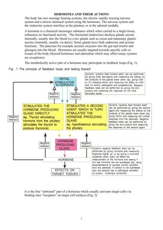
The metabolically active part of a hormone may participate in feedback loops (Fig. 1).
It is the free “unbound” part of a hormone which usually activates target cells via
binding sites “receptors” on target cell surfaces (Fig. 2).
2. 2
One of the best understood intracellular targets for hormonal action is an enzyme,
adenylate cyclase, whose catalyst action can either be stimulated or inhibited by the
appropriate hormone to vary the intracellular levels of cyclic AMP which is “the
second messenger,” which can then modify (usually increasing) some of the general
or specific actions of the cell involved (Fig. 3). Using this and other mechanisms
thyroid stimulating hormone increases the production of thyroid hormone,
adrenocorticotrophic hormone stimulates part of the adrenal cortex to produce
cortisol, and follicle stimulating hormone stimulates the female ovarian follicles.
Adrenaline however affects several target cells, stimulating the actions of adenylate
cyclase in some sites but inhibiting it in others by binding onto different receptor sites
- alpha sites (to stimulate smooth muscle to contract) or beta sites (to stimulate
smooth muscle contraction and to increase the rate and force of heart muscle
contraction).
3. 3
Some hormones are metabolised within minutes, and thus have a short half-life, but
others have intrinsically longer half-lives. Some hormones are partially protected
from breakdown by the liver and/or excretion by the kidneys by binding to proteins
(corticosteroid binding globulin and sex-hormone binding globulin for example).
Obviously diagnosis of hormone dysfunction initially depends upon clinical suspicion
and for many hormone disorders a blood specimen taken at an appropriate time may
be highly suggestive, but levels of certain hormone levels may vary through the
course of a day (corticosteroids) or menstrual cycle (oestrogens). Often dynamic tests
(giving stimulating or inhibiting stimuli) are used, particularly if feedback
mechanisms involving the hypothalamus, pituitary and peripheral organs are involved
(Figs. 1 and 2).
Hormones produced by tissues derived from embryonic ectoderm or endoderm
(including the pituitary, parathyroids and pancreas) are proteins, peptides, or amino
acids and cannot be given by mouth as they would be digested. Hormones secreted
from mesodermally derived tissues (gonads, adrenal cortex and placenta) are steroids
and can usually be given by mouth.
4. 4
The Pituitary
Figure 4 presents a simplified account of the sites of production of the major
hormones from the hypothalamus, pituitary, peripheral glands and their actions on
peripheral tissues. Hormone-related diseases may be caused several mechanisms
(Fig. 5)
Pituitary tumours may secrete trophic (=stimulating) hormones which then cause
abnormal function of their target organs. Additionally, both secretory and non-
secretory pituitary tumours hormones, may present with:
• Signs from pressure on the optic nerves (classically a bitemporal hemianopia
• Pituitary hypofunction caused by compression with underexcretion of other
trophic hormones
• Headache
5. 5
The anterior pituitary
Growth hormone
Underproduction of growth hormone by the anterior pituitary during childhood leads
to retarded growth and dwarfism, whereas overproduction in childhood leads to
giantism with overgrowth of the long bones of the limbs. Overproduction of growth
hormone in adults leads to overgrowth of the bones of the hands, feet, and head to
produce acromegaly (Fig. 6).
6. 6
Prolactin
Prolactin stimulates milk secretion and reduces ovarian secretion of oestrogens or
testicular secretion of testosterone. It can block luteinising hormone effects to
produce hypogonadism. In females hyperprolactinaemia usually presents with
amenorrhoea and in males with lack of libido.
The posterior pituitary
The posterior pituitary is derived from Rathke’s pouch, originally an outpouching of
the fish mouth which presumably responded to changes in water, and indeed the
human posterior pituitary retaines water balance as one of its functions by production
of antidiuretic hormone (vasopressin). Antidiuretic hormone causes the distal
convoluted kidney tubules and collecting ducts to retain water. Failure of antidiuretic
hormone production (or insensitivity of the kidneys to it) causes diabetes insipidus
with production of large volumes of dilute urine. Oxytocin, the other hormone
produced by the posterior pituitary, has an important role in the physiology of
childbirth and in the milk letdown reflex of lactation.
PERIPHERAL GLAND DYSFUNCTION
Thyroid
Iodine is taken up by the thyroid ( = shield shaped) gland and is eventually processed
along with tyrosine (an amino acid) to produce thyroxine and triiodothronine which
are general metabolic stimulants. Thyroid stimulating hormone is produced by the
anterior pituitary (Fig. 7) and is the main stimulator of this process. Both thyroxine
and triiodothyronine are bound to proteins in the blood and it is the unbound (free)
levels that are physiologically important. Thyroxine is converted to the more active
triiodothyronine in peripheral tissues. In areas where iodine deficiency is common
enlarged thyroid (a goitre) may result (Fig. 8).
7. 7
Hyperthyroidism (Fig. 9)
Hyperthyroidism results when the thyroid produces excessive thyroxine and
triiodothyronine. If the hyperthyroidism is caused by primary thyroid hyperactivity
the thyroxine and/or triiodotyronine levels are increased which supresses thyroid
stimulating hormone (Fig. 7). In contrast if the hyperactivity is secondary to pituitary
overproduction the thyroid stimulating hormone level will be raised (this is very rare).
Hyperthyroidism is usually caused by immune mediated mechanisms and less
commonly caused by focal oversecretion by thyroid nodules (toxic adenomas).
Hyperthyroidism caused by diffuse thyroid overactivity (Grave’s disease) may be
associated with protrusion of one or both eyes (exophthalmos), retraction of the upper
eyelid, and delay in descent of the upper eyelid as the eye is rotated downwards -
lidlag (Fig.10). These signs are caused by swelling of the intraorbital muscles and
protrusion of the eyeball.
8. 8
Figure 10.
Treatment of hyperthyroidism is with anti-thyroid drugs, radioactive iodine or
surgery. Beta-blocking drugs are useful for initial treatment as they block the
“pseudo-adrenergic” overdrive caused by high levels of thyroid hormones.
Hypothyroidism (Fig. 9)
In primary hypothyroidism the thyroxine and triiodothyronine levels are low. The
thyroid stimulating hormone levels are high as the pituitary tries drive the failing
thyroid harder. Indeed a raised thyroid stimulating hormone may be the first
indication of impending hypothyroidism. Hypothyroidism may be present at, or
develop shortly after, birth and cause cretinism (Fig. 11).
10. 10
The endocrine pancreas (Fig. 12)
The pancreas produces insulin which increases peripheral tissue utilisation of glucose.
Too little insulin, or too great a demand for insulin leads to diabetes mellitus with a
high plasma glucose which functions as an osmotic diuretic which, if left untreated,
may produce dehydration and coma. Glucagon, which causes glucose release from
glycogen, is also produced by the pancreas.
Parathyroids (Fig. 13)
Parathormone and vitamin D affect calcium and phosphorus levels. If free ionised
calcium falls or phosphate increases the parathyroids excrete extra parathormone
which tries to restore the free ionised calcium to normal by:
• Stimulating release of calcium from bone
• Increasing the loss of phosphate in the urine and increasing calcium reabsorption
by the kidney
• Favouring of active forms of Vitamin D, which promote gastrointestinal absorption
of calcium
11. 11
Hyperparathyroidism results in increased calcium concentrations with borderline/low
phosphorus. Mood changes, muscle weakness, muscle pain, increased risk of renal
stones (caused by augmented urinary calcium excretion) and osteoporosis may result.
12. 12
THE ADRENALS (Fig.14)
Adrenal cortex
The adrenal cortex secretes three main classes of hormones:
• Glucocorticoids (including cortisol) which affect carbohydrate, protein and fat
metabolism
• Mineralocorticoids (including aldosterone) which affect electrolyte and water
regulation
• Androgens which are anabolic (building up) and virilising in action
Adrenal cortical hyperfunction (Fig. 15 and 16)
Adrenal cortical hyperfunction may be caused by adrenal cortical overdevelopment
(hyperplasia), benign glandular tumours (adenomas), or adenocarcinomas. Cushing’s
syndrome is caused by overproduction of cortisol by the adrenal cortex, either primary
or secondary to pituitary overproduction of adrenocorticotrophic hormone.
14. 14
Treatment can be with surgery or radiotherapy or with drugs which block adrenal
production of cortisol.
Aldosterone overproduction causes excessive sodium retention and excessive
potassium excretion by the distal convoluted tubules of the kidney. In primary
hyperaldosteronism rennin concentrations are low and aldosterone/renin ratios are
high. Excessive production of androgens causes virilism.
Cortisol deficiency (Fig. 17)
The principle features of cortisol deficiency are sodium depletion, water depletion, a
high serum potassium, and a raised urea (caused by dehydration). With severe
deficiencies there is a fast heart rate, a low blood pressure and dehydration with signs
of hypovolaemic shock.
If hypocortisolaemia is caused by adrenal cortex failure (Addison’s disease) rather
than by pituitary failure there may be increased pigmentation of pressure points, skin
creases, and lining of the cheeks. This is caused by increased adrenocorticotrophic
hormone production by the pituitary (as it attempts to drive the failing adrenal cortex)
and adrenocortocotrophic hormone is co-secreted with melanocyte stimulating
hormone which stimulates melanocytes ( pigment producing cells of the skin) to
produce a brownish discolouration. These effects do not occur if the cause is primary
pituitary failure.
If a low cortisol is caused by pituitary failure (with lack of adrenocorticotrophic
hormone) the signs are usually less dramatic - mostly because the continued
production of aldosterone by the adrenal cortex continues.
The adrenal medulla (medulla = pith)
The adrenal medulla is innervated by the sympathetic nervous system and produces
several catecholamines, notably adrenaline (epinephrine) and nonadrenaline.
Catecholamines are responsible for “adrenergic” responses to physical and mental
stress and produce a fast heart rate, nervousness, and mobilisation of energy.
15. 15
SEX HORMONES
Male sexual development (Fig. 18)
Functional testes are necessary for male pattern growth, sexual differentiation and
function, penile growth, secondary sexual characteristics and behaviour. The
secondary male characteristics are mediated by testosterone (or dihydrotestosterone).
In the male the production of follicle stimulating hormone and luteinising hormone
are constant whereas production in the female is cyclical.
After puberty in the male (usually between 10-15 years) the testes produce androgens
(testosterone and dihydrotestosterone) in response to pituitary-derived luteininsing
hormone. Spermatozoa are produced in response to the combination of follicle
stimulating hormone and testosterone. The androgens (=male makers) along with
growth hormone cause the pubertal growth spurt. The sex hormone binding globulins
decrease at puberty which releases more free hormones.
16. 16
Female sexual development
Interestingly it is the lack of testes that is a requirement for development of female
characteristics (Fig. 19).
At puberty, which usually occurs two years earlier in the female, follicle stimulating
hormone causes maturation of ovarian follicles (follicle = small sac or gland) and
luteinising hormone causes ovarian follicles to rupture and the corpus luteum (the
mass formed in the uterine wall after the follicle has discharged the ovum) to develop.
Figure 20 details the various feedback systems and actions of these hormones in
relationship to the menstrual cycle.
17. 17
Following menstruation follicle stimulating hormone rises, follicle development is
thereby stimulated, with follicles secreting oestrogen. One follicle becomes
dominant, matures and secretes oestradiol which suppresses competing follicles and
inhibits pituitary secretion of follicle stimulating hormone. Just before menstruation
the oestradiol level falls and luteinising hormone increases which induces the ripe
follicle to ovulate - “burst” - and the remaining tissue to form the corpus luteum
which secretes progesterone and oestradiol to maintain a receptive endometrium in
case fertilization and implantation occurs. In the absence of fertilisation the corpus
luteum withers, progesterone levels fall, and the inner surface layers of the
endometrium are shed (menstruation).
If fertilisation occurs the fertilised ovum penetrates the uterine inner wall
(endometrium) and it and the placenta secretes chorionic gonadotrophins (oestrogens,
18. 18
progesterone, and gonadotrophic hormones) which constitute the ovum’s version of
luteinising hormone. These stop the corpus luteum degenerating and thus the early
pregnancy is maintained. Progesterone also stimulates the oestrogen primed breast in
the luteal phase and prepares the breast for lactation. Prolactin has a more important
role in lactation, later in pregnancy, and in parturition.
Hypogonadism
There are several syndromes which are associated with hypogonadism. Rather than
present the numerous syndromes the following is a brief review of the principle
mechanisms and manifestations.
Male hypogonadism
Male hypogonadism results from inadequate testosterone production from the testes,
pituitary dysfunction or hypothalamic dysfunction. Male hypogonadism, particularly
if occurring before puberty, may result in lack of height, lack of bodily hair, an
unbroken voice, small testes, poor muscle development, and a small penis and
scrotum (Fig 18). In such patients bone epiphyses fail to fuse and heights are often
greater than average. In postpubertal males hypogonadism is usually of slow onset
with low mood, decreased libido, muscle weakness, osteoporosis and, when severe,
less need to shave and regression of secondary sexual characteristics.
Female hypogonadism
If there is primary ovarian hypofunction the pituitary-derived follicle stimulating
hormone and/or the luteinising hormone are often raised in an attempt to stimulate the
failing ovaries. If the ovarian dysfunction is secondary to other causes (such as
weight loss in anorexia nervosa) the follicle stimulating hormone and luteinising
hormone are low with lowoestradiol and progesterone levels leading to absence of
periods (amenorrhoea).
During the normal cessation of menstruation, the menopause, oestradiol levels
become low and follicle stimulating hormone levels rise - as if to stimulate the failing
ovaries. The lack of oestrogen is associated with vaginal dryness, infertility,
amenorrhoea, lack of libido, breast atrophy and bone loss (the traditional hot flushes
are caused by sympathetic nervous system activation).
Problems of sexual differentiation
These require specialist assessments and investigations outwith the scope of this
account.
------
There are numerous other tissues which secrete hormones including the pineal (which
secreates melatonin which is responsible for light-adapted body systems), the thymus,
the atria of the heart, the stomach, the small intestine, and areas adjacent to the kidney
glomerulus (which produce renin).