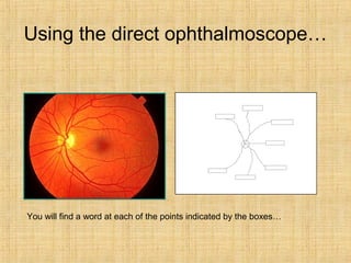
1. Using the direct ophthalmoscope…
You will find a word at each of the points indicated by the boxes…
2. A patient standing six metres from a standard Snellen Chart can
only see the top line. How is this recorded in snellen notation?
3. E
5 minutes of arc: 6/6 acuity
10 minutes: 6/12
20 minutes: 6/24
30 minutes: 6/36
6 metres
E 50 minutes of arc: 6/60 acuity
The human eye is just able to discern separate objects if the angle between
them is 30 seconds of arc.
10. What abnormality is shown here?
Suggest two important differential diagnoses
Suggest two useful investigations
11. This patient suddenly lost all useful vision in her
right eye a few hours before the photo was taken.
What is the diagnosis?
What tests would you perform
What is the commonest accompanying systemic
disease?
What pupil abnormality would be present?
14. Extra-ocular muscles
Medial Rectus Third nerve
Sixth nerve
Third nerve
Third nerve
Fourth nerve
Third nerve
Lateral rectus
Superior Rectus
Inferior rectus
Superior Oblique
Inferior Oblique
Muscle: Nerve supply:
35. Answers
• Slide 11 - Painless sudden loss of vision =
central retinal artery occlusion/thrombosis.
Tests – BP, cholesterol, glucose. Patient would
be a vascularpath. Pupil abnormality is relative
afferent pupillary defect, as retina not able to
detect light
• Slide 13 – Complete ptosis, “down & out” eye,
dilated pupil = 3rd
nerve palsy. Possible cause =
posterior communicating artery aneurysm
pressing on nerve, or vascualr occlusion along
the length of the 3rd
nerve.
36. Answers
• Slide 15
– 2 – right eye loss of vision
– 3 – bitemporal hemianopia “tunnel vision”
– 5 – right homonymous hemianopia
– 8 – right homonymous hemianopia
– 9 – right homonymous hemianopia with central
sparing
• Slide 17 – Horners, see partial ptosis,
constricted pupil & decreased sweating. Causes
anything along the length of the sympathetic
chain, e.g. tumour, vascular origin
38. Answers
• Slide 24 – central retinal vein occlusion,
see multiple haemorrhages
• Slide 25 – internal carotid artery atheroma
and consequent embolus seen on retina
• Slide 26 – diagnosis = diabetic retinopathy
• Slide 28 – Stevens Johnson with
decreased tear production & scarring
39. Answers
• Slide 29 – areas of depigmentation of
retina
• Slide 30 – RA and see deterioration of the
sclera called scleromalacia perforans
• Slide 31 – sturge weber syndrome
• Slide 32 – inverting eye lid with cotton bud
Editor's Notes
Answer: Cataract – phaco-emulsification
Answer: Entropion, where eye lash folds in towards eye. Main symptom is a gritty scratchy “there’s something in my eye” kind of pain
Painless sudden loss of vision = central retinal artery occlusion/thrombosis
Tests – BP, cholesterol, glucose
Patient would be a vascularpath
Pupil abnormality is relative afferent pupillary defect, as retina not able to detect light
2 – right eye loss of vision
3 – bitemporal hemianopia “tunnel vision”
5 – right homonymous hemianopia
8 – right homonymous hemianopia
9 – right homonymous hemianopia with central sparing