SlideShare a Scribd company logo
1 of 21
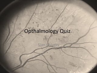
Opthalmology Quiz
Sian Chivers
Question 1
•What’s This?
•Name 3 Causes?
•What Symptoms may a
person complain of?
•How is it Treated
Answer 1
• Cataract
• Causes
– Senile (Commonest)
– Diabetes, Recurrent Uveitis, Previous ocular surgery
– Drugs – steroids, phenothiazines
– Trauma and foreign bodies
– Ionising radiation – Xray, UV
– Congenital
– Inherited – Myotonic Dystrophy, Marfan’s, Lowe’s, Rubella, high
myopia
• Symptoms
– Reduced acuity, Glare in sunshine, Distortion of lines, Diplopia,
Altered colours
• Mydriatic drops, sunshades and sunglasses. Surgery –
Phacoemulsification and Lens implant (US breaks up lens)
Question 2
•What is this?
•What investigation is being
carried out?
•What is the causative organism?
•What are the clinical features?
•What is the treatment?
Answer 2
• Dendritic ulcer
• Eye examination under fluorescein stain
• Herpes Simplex virus
• Irritable red eye, watering, photophobic
• Refer to an opthalmologist, Aciclovir 3% eye
ointment 5x daily
Question 3
•What is this?
•Name 3 pathological features
shown in the picture and
describe how they occur?
•Name 2 complications of this
eye condition?
•How can this eye condition be
treated and what would you
see when you looked at the
retina post procedure?
Answer 4
• Background Diabetic retinopathy
• Microaneurysms – outpouching of retinal caps
Cotton wool spot – microinfarct of retinal nerve fibre
layer
Retinal oedema and exudate – leakage of serum into the
neural retina and lipid accumulation
Neovascularisation (proliferative) new vessels grow
outside the retina along posterior surface of vitreous
• Complications – blindness, vitreous haemorrhage and
retinal detachment. Cataract as part of DM
Answer 4 Cont
•Treatment
•Good Glycaemic control
prevents complications
•Laser photocoagulation
indicated for proliferative DR
via argon laser
•Vitrectomy – vitreous is
removed via a trans-pars
incision – to clear vitreous
haemorrhage, to relieve
retinal traction and to treat
diffuse macular oedema
Question 5
•What abnormality is seen here?
•Name 3 causes?
•What may be seen when
examining the pupils?
Answer 5
• Optic Atrophy
• Causes
– Hereditary – Autosomal dominant/recessive, Leber’s
hereditary
– Retinal Dystrophy – Cone Dystrophy, retinitis pigmentosa
– Vascular – central retinal artery occlusion
– Nutritional – VitB12 deficiency, tobacco, alcohol, drugs –
ethambutol chloramphenicol
– Inflammatory – Sarcoid, PAN
– Demyelination (common)
– Compressive (glioma or meningioma)
• Relative afferent pupillary defect
Question 6
This patient has AIDS
What is shown in fig 1
What organism cause this
What other organisms commonly
infect the retina of a patient with
AIDS
What is shown in fig 2
Would you expect lesions elsewhere
What other infections affect the lids
and conjunctiva of a patient with
AIDS
Answer 6
• CMV retinitis
• Millions – CMV, EBV, Toxoplasmosis, Syphilis,
Cryptococcus, M.TB, M.aviumintracellulare, Candida,
Aspergillus, histoplasmosis
• Kaposi Sarcoma
• Yes
• Trichomegaly (long eyelashes), molluscum
contagiosum, conjunctivitis, conjunctival granulomas ,
SCC assoc w HPV, herpes zoster opthalmitis
Question 7
•What intervention has been carried
out here
•What underlying condition does
this patient have
•What symptoms may the patient
have experienced in the acute phase
•What signs may have been
apparent on examination
•What is the treatment for the acute
phase of the illness?
Answer 7
• A peripheral iridotomy
• Glaucoma. Iridotomy is the intervention of choice
to prevent angle closure glaucoma or after an
attack has been broken by medical treatment
• Symps – sudden onset, severely painful red eye,
blurred vision, halos around lights, headache,
nausea and vomiting
• Signs – reduced visual acuity, brick red eye, hazy
cornea, vertically mid-dilated fixed pupil, high IOP
• Treatment – Pilocarpine 2-4% 2h, acetazolamide
500mg iv stat then 250mg/8h, mannitol 20% by
ivi. Topical steroids and antihypertensive drops
may be used
Question 8
•What is shown here?
•What symptoms may the patient
complain of?
•What associated condition may the
patient have
•What signs are found on
examination?
•What is the management?
•Complication?
Answer 8
• Anterior Uveitis
• Painful red eye, photophobia, blurred vision or
floaters
• Autoimmune disease – ank spond, inflam bowel
disease, sarcoidosis
• Signs – ciliary injection, reduced visual acutiy,
sluggish or irregular pupil, hazy iris details, high
IOP
• refer to opthalmologist, reducing regimen of
topical steroid (dexamethosone 0.1%) and
cycloplegic and dilating drops (cyclopentolate
1%)
• Cataract in recurrent or chronic cases
Name to following
Conjunctivitis Subconjunctival
haemorrhage
Pterygium
Question 10
•Diagnosis
•Signs
•Main cause
•Management
•Diagnosis
•Inherited underlying condition that
may predispose
•Other features of that condition
Answer 10
• Central retinal vein occlusion
• Signs – afferent pupillary defect common, retinal haemorrhages,
optic disc swelling, venous dilatation and tortuosity, cotton wool
spots
• Up to 33% are ischaemic assoc with rubeosis iridis or
neovascularisation of disc
• Detect any underlying systemic disease and treat, check IOP, screen
for new vessels, treaat cv risk factors, laser new vessels.
• Lens dislocation
• Marfan’s syndrome
• Aortic dissection or dilatation, dural ectasia, arachnodactly,
armspan greater than height, pectus deformity, pes planus,
scoliosis high arched palate, mitral valve prolapse, joint
hypermobility.
Well done, that’s enough
opthalmology for now!!

More Related Content

What's hot

What's hot (20)

Refrective surgery ppt
Refrective surgery pptRefrective surgery ppt
Refrective surgery ppt
 
Corneal dystrophies
Corneal dystrophiesCorneal dystrophies
Corneal dystrophies
 
Anatomy of uvea
Anatomy of uveaAnatomy of uvea
Anatomy of uvea
 
Macular hole
Macular holeMacular hole
Macular hole
 
Macular hole
Macular holeMacular hole
Macular hole
 
Lamellar keratoplasty
Lamellar keratoplastyLamellar keratoplasty
Lamellar keratoplasty
 
Clinical - Traumatic cataract
Clinical  - Traumatic cataractClinical  - Traumatic cataract
Clinical - Traumatic cataract
 
Eales disease
Eales diseaseEales disease
Eales disease
 
Vitreous hemorrhage
Vitreous hemorrhageVitreous hemorrhage
Vitreous hemorrhage
 
AMBLYOPIA AND ITS MANAGEMENT
AMBLYOPIA AND ITS MANAGEMENTAMBLYOPIA AND ITS MANAGEMENT
AMBLYOPIA AND ITS MANAGEMENT
 
My Clouding Cornea
My Clouding CorneaMy Clouding Cornea
My Clouding Cornea
 
Secondary open angle glaucoma
Secondary open angle glaucomaSecondary open angle glaucoma
Secondary open angle glaucoma
 
Evaluation of squint
Evaluation of squint Evaluation of squint
Evaluation of squint
 
Keratoconus
KeratoconusKeratoconus
Keratoconus
 
Branch retinal vein occlusion (BRVO)
Branch retinal vein occlusion (BRVO)Branch retinal vein occlusion (BRVO)
Branch retinal vein occlusion (BRVO)
 
Vitreous hemorrhage
Vitreous hemorrhageVitreous hemorrhage
Vitreous hemorrhage
 
treatment of non healing corneal ulcer
treatment of non healing corneal ulcertreatment of non healing corneal ulcer
treatment of non healing corneal ulcer
 
Physiology of lens and cataractogenesis sivateja
Physiology of lens and cataractogenesis sivatejaPhysiology of lens and cataractogenesis sivateja
Physiology of lens and cataractogenesis sivateja
 
Cataract evaluation ppt
Cataract evaluation pptCataract evaluation ppt
Cataract evaluation ppt
 
Lid diseases ii
Lid diseases iiLid diseases ii
Lid diseases ii
 

Viewers also liked

Viewers also liked (20)

Ophthalmology ppt
Ophthalmology pptOphthalmology ppt
Ophthalmology ppt
 
Ophthalmology Revision
Ophthalmology RevisionOphthalmology Revision
Ophthalmology Revision
 
Austin Ophthalmology
Austin OphthalmologyAustin Ophthalmology
Austin Ophthalmology
 
Opthalmology in the ED - Dr Andrew White (June 2013)
Opthalmology in the ED - Dr Andrew White (June 2013)Opthalmology in the ED - Dr Andrew White (June 2013)
Opthalmology in the ED - Dr Andrew White (June 2013)
 
Fundoscopy OSCE
Fundoscopy OSCEFundoscopy OSCE
Fundoscopy OSCE
 
Eye osce
Eye osceEye osce
Eye osce
 
Ophthamology Revision
Ophthamology RevisionOphthamology Revision
Ophthamology Revision
 
Most common opthalmology slides with anwers in uhs ospe
Most common opthalmology slides with anwers in uhs ospeMost common opthalmology slides with anwers in uhs ospe
Most common opthalmology slides with anwers in uhs ospe
 
Clinical ptosis
Clinical   ptosisClinical   ptosis
Clinical ptosis
 
Concurrency in database system
Concurrency in database systemConcurrency in database system
Concurrency in database system
 
The red and painful eye
The red and painful eyeThe red and painful eye
The red and painful eye
 
Episcleritis
Episcleritis Episcleritis
Episcleritis
 
Glaucoma RCSI
Glaucoma RCSIGlaucoma RCSI
Glaucoma RCSI
 
Het oog
Het oogHet oog
Het oog
 
Cataract and refractive surgery
Cataract and refractive surgeryCataract and refractive surgery
Cataract and refractive surgery
 
Ophthalmology
OphthalmologyOphthalmology
Ophthalmology
 
A medical student guide to Red Eye
A medical student guide to Red EyeA medical student guide to Red Eye
A medical student guide to Red Eye
 
Retinal detachment
Retinal detachmentRetinal detachment
Retinal detachment
 
Corneal pachymetry by ben okeeffe
Corneal pachymetry by ben okeeffeCorneal pachymetry by ben okeeffe
Corneal pachymetry by ben okeeffe
 
paediatric ophthalmology and strabismus
paediatric ophthalmology and strabismuspaediatric ophthalmology and strabismus
paediatric ophthalmology and strabismus
 

Similar to Opthalmology Quiz

Neuroophth Emergencies MDS - 2019
Neuroophth Emergencies MDS - 2019Neuroophth Emergencies MDS - 2019
Neuroophth Emergencies MDS - 2019neurophq8
 
Gradual loss of vision (opthalmology)
Gradual loss of vision (opthalmology)Gradual loss of vision (opthalmology)
Gradual loss of vision (opthalmology)Ma Wady
 
Abc's of vision impairments and communication styles
 Abc's of vision impairments and communication styles Abc's of vision impairments and communication styles
Abc's of vision impairments and communication stylesshelleterp
 
Abc's of vision impairments and communication styles
 Abc's of vision impairments and communication styles Abc's of vision impairments and communication styles
Abc's of vision impairments and communication stylesshelleterp
 
Approach to a patient with bilateral vision loss
Approach to a patient with bilateral vision lossApproach to a patient with bilateral vision loss
Approach to a patient with bilateral vision lossNeurologyKota
 
ABC's of vision impairments and communication styles
 ABC's of vision impairments and communication styles ABC's of vision impairments and communication styles
ABC's of vision impairments and communication stylesshelleterp
 
Abc's of vision impairments and communication styles
 Abc's of vision impairments and communication styles Abc's of vision impairments and communication styles
Abc's of vision impairments and communication stylesshelleterp
 
830_Optic_nerve_final copy 2.pptx
830_Optic_nerve_final copy 2.pptx830_Optic_nerve_final copy 2.pptx
830_Optic_nerve_final copy 2.pptxHarshika Malik
 
20-eye-diseases.ppt diseae\ses of the eye
20-eye-diseases.ppt diseae\ses of the eye20-eye-diseases.ppt diseae\ses of the eye
20-eye-diseases.ppt diseae\ses of the eyeMamataStephen
 
comunity ophthalmology - 2.pptx
comunity ophthalmology - 2.pptxcomunity ophthalmology - 2.pptx
comunity ophthalmology - 2.pptxMukhtarJamac3
 
Clinical approach to acute vision loss
Clinical approach to acute vision loss  Clinical approach to acute vision loss
Clinical approach to acute vision loss neurophq8
 
Protocol for differential diagnosis of common ocular diseases
Protocol for differential diagnosis of common ocular diseasesProtocol for differential diagnosis of common ocular diseases
Protocol for differential diagnosis of common ocular diseasesPuneet
 
830_Optic_nerve_final.ppt
830_Optic_nerve_final.ppt830_Optic_nerve_final.ppt
830_Optic_nerve_final.pptDarshuBoricha
 
vi_psa_pres_common_eye_conditions_aug10.ppt
vi_psa_pres_common_eye_conditions_aug10.pptvi_psa_pres_common_eye_conditions_aug10.ppt
vi_psa_pres_common_eye_conditions_aug10.pptshahilagaz
 
vi_psa_pres_common_eye_conditions_aug10.ppt
vi_psa_pres_common_eye_conditions_aug10.pptvi_psa_pres_common_eye_conditions_aug10.ppt
vi_psa_pres_common_eye_conditions_aug10.pptkevinguadilla
 

Similar to Opthalmology Quiz (20)

Neuroophth Emergencies MDS - 2019
Neuroophth Emergencies MDS - 2019Neuroophth Emergencies MDS - 2019
Neuroophth Emergencies MDS - 2019
 
Gradual loss of vision (opthalmology)
Gradual loss of vision (opthalmology)Gradual loss of vision (opthalmology)
Gradual loss of vision (opthalmology)
 
Abc's of vision impairments and communication styles
 Abc's of vision impairments and communication styles Abc's of vision impairments and communication styles
Abc's of vision impairments and communication styles
 
Abc's of vision impairments and communication styles
 Abc's of vision impairments and communication styles Abc's of vision impairments and communication styles
Abc's of vision impairments and communication styles
 
Approach to a patient with bilateral vision loss
Approach to a patient with bilateral vision lossApproach to a patient with bilateral vision loss
Approach to a patient with bilateral vision loss
 
ABC's of vision impairments and communication styles
 ABC's of vision impairments and communication styles ABC's of vision impairments and communication styles
ABC's of vision impairments and communication styles
 
Abc's of vision impairments and communication styles
 Abc's of vision impairments and communication styles Abc's of vision impairments and communication styles
Abc's of vision impairments and communication styles
 
830_Optic_nerve_final copy 2.pptx
830_Optic_nerve_final copy 2.pptx830_Optic_nerve_final copy 2.pptx
830_Optic_nerve_final copy 2.pptx
 
20-eye-diseases.ppt diseae\ses of the eye
20-eye-diseases.ppt diseae\ses of the eye20-eye-diseases.ppt diseae\ses of the eye
20-eye-diseases.ppt diseae\ses of the eye
 
comunity ophthalmology - 2.pptx
comunity ophthalmology - 2.pptxcomunity ophthalmology - 2.pptx
comunity ophthalmology - 2.pptx
 
Clinical approach to acute vision loss
Clinical approach to acute vision loss  Clinical approach to acute vision loss
Clinical approach to acute vision loss
 
Protocol for differential diagnosis of common ocular diseases
Protocol for differential diagnosis of common ocular diseasesProtocol for differential diagnosis of common ocular diseases
Protocol for differential diagnosis of common ocular diseases
 
Cataract-Ppt.pptx
Cataract-Ppt.pptxCataract-Ppt.pptx
Cataract-Ppt.pptx
 
Ophthalmology
OphthalmologyOphthalmology
Ophthalmology
 
Ocular signs in medicine/ neurology
Ocular signs in medicine/ neurologyOcular signs in medicine/ neurology
Ocular signs in medicine/ neurology
 
830_Optic_nerve_final.ppt
830_Optic_nerve_final.ppt830_Optic_nerve_final.ppt
830_Optic_nerve_final.ppt
 
Cataract
CataractCataract
Cataract
 
Congenital retinal anomalies
Congenital retinal anomaliesCongenital retinal anomalies
Congenital retinal anomalies
 
vi_psa_pres_common_eye_conditions_aug10.ppt
vi_psa_pres_common_eye_conditions_aug10.pptvi_psa_pres_common_eye_conditions_aug10.ppt
vi_psa_pres_common_eye_conditions_aug10.ppt
 
vi_psa_pres_common_eye_conditions_aug10.ppt
vi_psa_pres_common_eye_conditions_aug10.pptvi_psa_pres_common_eye_conditions_aug10.ppt
vi_psa_pres_common_eye_conditions_aug10.ppt
 

More from meducationdotnet

More from meducationdotnet (20)

No Title
No TitleNo Title
No Title
 
Spondylarthropathy
SpondylarthropathySpondylarthropathy
Spondylarthropathy
 
Diagnosing Lung cancer
Diagnosing Lung cancerDiagnosing Lung cancer
Diagnosing Lung cancer
 
Eczema Herpeticum
Eczema HerpeticumEczema Herpeticum
Eczema Herpeticum
 
The Vagus Nerve
The Vagus NerveThe Vagus Nerve
The Vagus Nerve
 
Water and sanitation and their impact on health
Water and sanitation and their impact on healthWater and sanitation and their impact on health
Water and sanitation and their impact on health
 
The ethics of electives
The ethics of electivesThe ethics of electives
The ethics of electives
 
Intro to Global Health
Intro to Global HealthIntro to Global Health
Intro to Global Health
 
WTO and Health
WTO and HealthWTO and Health
WTO and Health
 
Globalisation and Health
Globalisation and HealthGlobalisation and Health
Globalisation and Health
 
Health Care Worker Migration
Health Care Worker MigrationHealth Care Worker Migration
Health Care Worker Migration
 
International Institutions
International InstitutionsInternational Institutions
International Institutions
 
Haemochromotosis brief overview
Haemochromotosis brief overviewHaemochromotosis brief overview
Haemochromotosis brief overview
 
Ascities overview
Ascities overviewAscities overview
Ascities overview
 
Overview of the Liver
Overview of the LiverOverview of the Liver
Overview of the Liver
 
Overview of Antidepressants
Overview of AntidepressantsOverview of Antidepressants
Overview of Antidepressants
 
Gout Presentation
Gout PresentationGout Presentation
Gout Presentation
 
Review of orthopaedic services: Prepared for the Auditor General for Scotland...
Review of orthopaedic services: Prepared for the Auditor General for Scotland...Review of orthopaedic services: Prepared for the Auditor General for Scotland...
Review of orthopaedic services: Prepared for the Auditor General for Scotland...
 
Sugammadex - a revolution in anaesthesia?
Sugammadex - a revolution in anaesthesia?Sugammadex - a revolution in anaesthesia?
Sugammadex - a revolution in anaesthesia?
 
Dermatology Atlas
Dermatology AtlasDermatology Atlas
Dermatology Atlas
 

Opthalmology Quiz

  • 2. Question 1 •What’s This? •Name 3 Causes? •What Symptoms may a person complain of? •How is it Treated
  • 3. Answer 1 • Cataract • Causes – Senile (Commonest) – Diabetes, Recurrent Uveitis, Previous ocular surgery – Drugs – steroids, phenothiazines – Trauma and foreign bodies – Ionising radiation – Xray, UV – Congenital – Inherited – Myotonic Dystrophy, Marfan’s, Lowe’s, Rubella, high myopia • Symptoms – Reduced acuity, Glare in sunshine, Distortion of lines, Diplopia, Altered colours • Mydriatic drops, sunshades and sunglasses. Surgery – Phacoemulsification and Lens implant (US breaks up lens)
  • 4. Question 2 •What is this? •What investigation is being carried out? •What is the causative organism? •What are the clinical features? •What is the treatment?
  • 5. Answer 2 • Dendritic ulcer • Eye examination under fluorescein stain • Herpes Simplex virus • Irritable red eye, watering, photophobic • Refer to an opthalmologist, Aciclovir 3% eye ointment 5x daily
  • 6. Question 3 •What is this? •Name 3 pathological features shown in the picture and describe how they occur? •Name 2 complications of this eye condition? •How can this eye condition be treated and what would you see when you looked at the retina post procedure?
  • 7. Answer 4 • Background Diabetic retinopathy • Microaneurysms – outpouching of retinal caps Cotton wool spot – microinfarct of retinal nerve fibre layer Retinal oedema and exudate – leakage of serum into the neural retina and lipid accumulation Neovascularisation (proliferative) new vessels grow outside the retina along posterior surface of vitreous • Complications – blindness, vitreous haemorrhage and retinal detachment. Cataract as part of DM
  • 8. Answer 4 Cont •Treatment •Good Glycaemic control prevents complications •Laser photocoagulation indicated for proliferative DR via argon laser •Vitrectomy – vitreous is removed via a trans-pars incision – to clear vitreous haemorrhage, to relieve retinal traction and to treat diffuse macular oedema
  • 9. Question 5 •What abnormality is seen here? •Name 3 causes? •What may be seen when examining the pupils?
  • 10. Answer 5 • Optic Atrophy • Causes – Hereditary – Autosomal dominant/recessive, Leber’s hereditary – Retinal Dystrophy – Cone Dystrophy, retinitis pigmentosa – Vascular – central retinal artery occlusion – Nutritional – VitB12 deficiency, tobacco, alcohol, drugs – ethambutol chloramphenicol – Inflammatory – Sarcoid, PAN – Demyelination (common) – Compressive (glioma or meningioma) • Relative afferent pupillary defect
  • 11. Question 6 This patient has AIDS What is shown in fig 1 What organism cause this What other organisms commonly infect the retina of a patient with AIDS What is shown in fig 2 Would you expect lesions elsewhere What other infections affect the lids and conjunctiva of a patient with AIDS
  • 12. Answer 6 • CMV retinitis • Millions – CMV, EBV, Toxoplasmosis, Syphilis, Cryptococcus, M.TB, M.aviumintracellulare, Candida, Aspergillus, histoplasmosis • Kaposi Sarcoma • Yes • Trichomegaly (long eyelashes), molluscum contagiosum, conjunctivitis, conjunctival granulomas , SCC assoc w HPV, herpes zoster opthalmitis
  • 13. Question 7 •What intervention has been carried out here •What underlying condition does this patient have •What symptoms may the patient have experienced in the acute phase •What signs may have been apparent on examination •What is the treatment for the acute phase of the illness?
  • 14. Answer 7 • A peripheral iridotomy • Glaucoma. Iridotomy is the intervention of choice to prevent angle closure glaucoma or after an attack has been broken by medical treatment • Symps – sudden onset, severely painful red eye, blurred vision, halos around lights, headache, nausea and vomiting • Signs – reduced visual acuity, brick red eye, hazy cornea, vertically mid-dilated fixed pupil, high IOP • Treatment – Pilocarpine 2-4% 2h, acetazolamide 500mg iv stat then 250mg/8h, mannitol 20% by ivi. Topical steroids and antihypertensive drops may be used
  • 15. Question 8 •What is shown here? •What symptoms may the patient complain of? •What associated condition may the patient have •What signs are found on examination? •What is the management? •Complication?
  • 16. Answer 8 • Anterior Uveitis • Painful red eye, photophobia, blurred vision or floaters • Autoimmune disease – ank spond, inflam bowel disease, sarcoidosis • Signs – ciliary injection, reduced visual acutiy, sluggish or irregular pupil, hazy iris details, high IOP • refer to opthalmologist, reducing regimen of topical steroid (dexamethosone 0.1%) and cycloplegic and dilating drops (cyclopentolate 1%) • Cataract in recurrent or chronic cases
  • 19. Question 10 •Diagnosis •Signs •Main cause •Management •Diagnosis •Inherited underlying condition that may predispose •Other features of that condition
  • 20. Answer 10 • Central retinal vein occlusion • Signs – afferent pupillary defect common, retinal haemorrhages, optic disc swelling, venous dilatation and tortuosity, cotton wool spots • Up to 33% are ischaemic assoc with rubeosis iridis or neovascularisation of disc • Detect any underlying systemic disease and treat, check IOP, screen for new vessels, treaat cv risk factors, laser new vessels. • Lens dislocation • Marfan’s syndrome • Aortic dissection or dilatation, dural ectasia, arachnodactly, armspan greater than height, pectus deformity, pes planus, scoliosis high arched palate, mitral valve prolapse, joint hypermobility.
  • 21. Well done, that’s enough opthalmology for now!!