1. SPECIAL SENSES
Animals, unlike plants, tend to seek out rapid interaction with the environment. For example plants
respond to light but animals look, and some plants respond to vibration but animals listen. Often
detector organs of animals, particularly the eyes and ears, are able to “tune in” to events of interest.
Indeed some animals send out specifically targeted impulses to which their specifically tuned detectors
can respond - porpoises and bats probably “see” by echolocation.
The special senses are, sight, hearing, taste, and smell. All are situated in the head.
In evolutionary terms the organs of special sense, smell, hearing and vision, were all useful for detecting
food at a distance and presumably evolved with the integrative centers at the head end of the nervous
system so that purposeful action could follow detection.
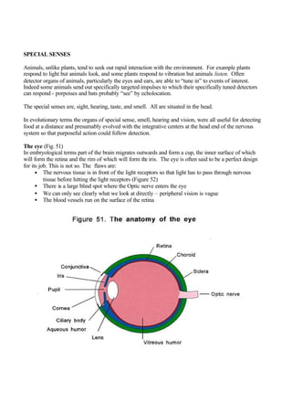
The eye (Fig. 51)
In embryological terms part of the brain migrates outwards and form a cup, the inner surface of which
will form the retina and the rim of which will form the iris. The eye is often said to be a perfect design
for its job. This is not so. The flaws are:
• The nervous tissue is in front of the light receptors so that light has to pass through nervous
tissue before hitting the light receptors (Figure 52)
• There is a large blind spot where the Optic nerve enters the eye
• We can only see clearly what we look at directly – peripheral vision is vague
• The blood vessels run on the surface of the retina
2.
3. The lens is derived from ectoderm and the sclera, choroid, cornea and ciliary apparatus derive from
mesenchyme. In humans eyes are paired, allowing stereoscopic vision.
The retina is derived from the lining of the third ventricle and it appears that the light detectors in the
retina, the rods and cones, are modifications of cilia.
There are two types of photoreceptor cells, rods and cones. There are about 120 million rods which:
• Are all alike
• Contain rhodopsin
• Are more able to respond to low level light and movements in the visual field than cones
• Do not respond to colour
There are about six million cones which:
• Are used for fine discrimination
• Respond to light of different wavelengths (colour)
• Each contain one of three visual pigments, each to responding to short, medium or long
wavelengths
Humans have a macula, an area of maximum acuity, which contains cones and very few rods. The
central macular depression is the fovea (=a pit) centralis on which images of interest are focused (Fig.
53).
4. The peripheries of the retina contain rods and few cones whereas the fovea contains only cones. The
visual object of maximum interest is focussed onto the fovea for maximum definition. It takes 0.02 of a
second for retinal impulses to reach the visual cortex: this time is prolonged if there is dysfunction of
myelinated nerves (as may occur in multiple sclerosis).
Curiously the light has to travel through nerve tissues of the retina before it reaches the receptors. The
probable explanation of this apparent inefficiency is shown in Figure 52.
The sclera is a firm yet flexible housing for the eye. The sclera merges with the cornea, which partially
bends light before the lens performs the major role in the focusing of images onto the retina. Humans
focus by changing the shape of the lens by using the radial muscle to stretch the lens (Fig. 22) which
has to return to its previous shape by virtue of its intrinsic elasticity (which declines with age).
The iris surrounds the pupil and, by contraction of its circular or radial muscle attempts to ensure that a
constant amount of light reaches the retina. Accordingly the pupils are small in bright light and large in
dim light.
The aqueous humour (the fluid in the anterior chamber) is formed by the ciliary body. The aqueous
humour is drained (into the canal of Schlemm) in the filtration angle. If this angle is too acute the canal
cannot drain the humour and closed angle glaucoma (glaucoma = grayish blue, describing the
appearance of the cornea in this disorder) results from raised intraocular pressure.
The conjunctivae (=to connect) is a thin transparent membrane which can be considered to be part of the
skin which lines the inner lids and the exposed areas of the eye to protect them against the elements.
5. Figure 54 illustrates the visual losses produced by damage at various sites in the optic pathways.
6. The ear(Fig. 55).
Amphibians first dragged themselves out of the sea onto the land, probably from estuary shores, 200-
300 million years ago. Initially their heads were in contact with the ground and they used bone
conduction for hearing. Later the head and body were lifted off the ground which encouraged the
development of other hearing mechanisms with part of the lateral line of our fish ancestors sinking into
the head to become a new organ, the ear, (presumably this new organ developed in the head presumably
because it was away from the jogging influence of the limbs). Sound appreciation is thus a modification
of aquatic “tactile vibration sense.”
Vibration sense is a “crude hearing” relayed by all general somatic sensory nerves. If someone has
complete nerve deafness then vibration sense can still be appreciated by trigeminal innervation
(Beethoven rested a ruler, one end of which was held in his teeth, on his piano to “hear” his playing
when he became deaf)
The ear has to collect sound waves varying in frequency between 20-20,000 cycles per second, funnel
them toward the sensory detectors, “concentrate” the sound by mechanical means and then convert this
concentrated movement via vibrations in the various ear lymphs into nerve impulses for onward
transmission to the brain. The sensitivity of the system is so great that the two ears together can localise
sound using the delay in the time taken for sound to travel the distance of the head diameter and the
slight differences in volume. The scale used for measuring volume (decibels) is logarithmic (each
change in 10 units represents a tenfold change in volume). The ear’s range of useful volume appreciation
is 1-140 decibels – a range of about 1013
.
7. The inner ear (Fig. 56) is made up of the utricle (=little leather bag), saccule (=small sac), the
semicircular canals, and the vestibule (which is responsible for hearing).
The outer ear focuses air borne vibrations onto the tympanic (= drum) membrane. These vibrations are
then greatly amplified by three middle ear ossicles (= small bones), the malleus (= hammer), incus (=
anvil) and stapes (= stirrup). The stapes depresses the oval window and creates a pressure wave in the
tube containing perilymph (the scala vestibuli) which then doubles back on itself (as the scala tympani)
with the scala media (which bears the spiral organ of Corti) in between. The tubes are coiled to form the
cochlea (= snail shell). Pressure waves in the scala vestibuli are transmitted across the thin membrane to
the endolymph of the scala media and from there to the basilar membrane of the scala tympani which
bears the hair cells which detect vibrations in the fluid.
To maintain orientation and balance, the effects of acceleration, deceleration, rotation, and gravity have
to be assessed. Vision became the predominant modality for orientation in our ancestors, and to do this
stable retinal images had to occur, and to do this the head had to be stabilized. To achieve this the hair
cells in the semicircular canals of the ear are utilized in two ways to detect changes in acceleration or
rotation.
8. Firstly, the semicircular canals comprise three loops, each lying in different planes and each filled with
endolymph. At one end of each canal there is a swelling, the ampulla (= flask) within which there are
sensory cells, the crista (= crest), bearing the hair cells which detect movement in the endolymph.
Interestingly the orientation of the semicircular canals is such that acceleration in a horizontal plane
causes a flow of endolymph such that the brain thinks that the body is moving upwards. This is why
pilots flying in mist have to rely on their instruments if they are not to crash into the ground: if they
cannot use sight for orientation their ears tell them they are going slightly upwards and they therefore
react by flying slightly downwards, ultimately into the ground. When flying in fog pilots have to rely on
their instruments and not their sensation of what is horizontal.
Secondly, to assess orientation when there is no head movement there are small speckles of calcium
carbonate (otoliths) that are found in the otolithic membrane of the utricle and saccule. Gravity acts on
otoliths and hair cells which assess the direction of the pull of gravity. Movement of the perilymph will
also affects the otoliths but to a lesser extent.
9. Taste and smell
Appreciation of food depends upon the detection of dissolved substances by chemical detectors on the
tongue (Fig. 57). Smell depends on detectors, situated at the top of the nose, of airborne substances
which have to be dissolved before they can be smelt. Smell is thus a tasting of airborne substances. In
our marine ancestors taste probably developed first - in the sea everything was dissolved and not
airborne - and smell developed later when the land was invaded. Information about taste is conveyed to
the medulla, then to the thalamus and then to the cerebral hemispheres. Each taste bud is most
responsive to one of the four basic tastes - sweet, sour, salt and bitter.