
Recommended
More Related Content
What's hot
What's hot (20)
Similar to Gingival enlargement
Similar to Gingival enlargement (20)
More from Mehul Shinde
More from Mehul Shinde (20)
Recently uploaded
Recently uploaded (20)
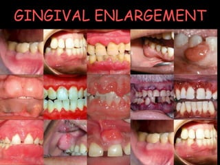
Gingival enlargement
- 2. CONTENTS • Introduction • Classification • Indices • Inflammatory Enlargement • Enlargements associated with systemic diseases • Neoplastic enlargement • Drug-induced enlargement • Idiopathic gingival enlargement • False enlargement • Conclusion • References
- 3. INTRODUCTION Hypertrophic gingivitis Gingival hyperplasia Gingival enlargement Gingival overgrowth
- 4. CLASSIFICATION A] Based on etiologic factors and pathologic changes I. Inflammatory Enlargement - Acute - Chronic II. Drug-induced enlargement III. Enlargements associated with systemic diseases IV. Neoplastic enlargement (gingival tumors) Benign tumors Malignant tumors V. False enlargement ii) Systemic diseases causing gingival enlargement 1. Leukemia 2.Granulomatous diseases (Wegener's granulomatosis, sarcoidosis) i) Conditioned enlargement 1. Pregnancy 2. Puberty 3. Vitamin C deficiency 4. Plasma cell gingivitis 5. Nonspecific conditioned enlargement
- 5. B] Based on location and distribution I. Localized II. Generalized III. Marginal IV. Papillary V. Diffuse VI. Discrete
- 6. INDICES Bokenkamp & Bohnhorst 1994 Grade 0 No signs of gingival enlargement Grade 1 Enlargement confined to IDP Grade 2 Enlargement involves IDP & marginal gingiva Grade 3 Enlargement covers three quarters / more of crown
- 7. Degree of gingival hyperplasia according to modified index by Angelopoulos & Goaz 1972 Grade Hyperplasia Size Tooth coverage 0 No Normal No 1 Minimal <2 mm Cervical 3rd or less 2 Moderate 2-4 mm Middle 3rd 3 Severe >4 mm More than 2/3rd
- 8. Gingival overgrowth index- Mc Gaw et al 1987 Grade 0 No overgrowth, feather edge gingival margin Grade 1 Blunting of gingival margin Grade 2 Moderate gingival overgrowth (one third crown length) Grade 3 Marked gingival overgrowth (more than one thirds of crown)
- 9. Clinical index for drug induced gingival overgrowth Ingles et al 1999 G r a d e 0 G r a d e 1 G r a d e 2 G r a d e 3 G r a d e 4 1.No overgrowth 2. slight stippling, no/slight granular appearance 3. knife edge margin 4. no increase in the density or size 1. Early overgrowth slight increase in density 2.marked stippling and granular appearance 3.tip of the papillae is round 4.probing depth ≤ 3mm 1.Moderate overgrowth increase in size of the papillae and rolled gingival margins 2. The contour concave or straight 3. buccolingual dimension of upto 2mm 4. Probing ≤ 6mm 5. Papillae retractable 1. Marked overgrowth encroachment on the clinical crown 2. The contour convex 3. Buccolingual dimension ≥ 3 mm 4. The probing depth ≥ 6mm 5. Papillae retractable 1. Severe overgrowth, profound thickening of gingiva 2. Large % of the clinical crown is covered 3. Papillae - retractable 4. Probing depth ≥ 6mm 5.buccolingual dimension 3mm
- 11. a. Chronic inflammatory enlargement Etiology Plaque accumulation & retention Poor oral hygiene Anatomic abnormalities Improper restoration Orthodontic appliances Malocclusion
- 12. Clinical Features 1. Slight ballooning of IDP & marginal gingiva Life preserver shaped bulge Smooth , edematous , bleed easily Localised / generalised Progress- slowly and painlessly Pseudopockets
- 13. Discrete sessile or pedunculated tumor like mass Interproximal / marginal or attached gingiva Slow growing and painless Clinical Features 1. 2.
- 14. Histopathology 1.Soft and friable 2.Firm and resilient
- 15. B. ACUTE INFLAMMATORY ENLARGEMENT 1. GINGIVALABSCESS Etiology Foreign substances Clinical features Marginal gingiva or IDP Red swelling smooth shiny surface Fluctuant and pointed with a surface orifice expresses purulent exudate
- 16. 2. PERIODONTAL ABSCESS Lateral abscess / parietal abscess Depending on location - Gingival - Periodontal (Acute / Chronic) - Pericoronal Meng et al ’99 Depending on number - Single - Multiple
- 17. Etiology PERIODONTITIS RELATED Extension of infection from PD pocket Lateral extension of inflammation Pocket with a tortuous course Incomplete removal of calculus
- 18. Etiology NON PERIODONTITIS RELATED Impaction of foreign bodies Endodontic perforation Lateral cyst infection Factors affecting morphology of root
- 19. Signs and symptoms Acute abscess - Mild to severe discomfort - Localized red, ovoid swelling - Periodontal pocket - Mobility - Tooth elevation in the socket - Tenderness to percussion or biting - Suppuration - Elevated temperature - Regional lymphadenopathy Chronic abscess - No pain or dull pain - Localized inflammatory lesion - Slight tooth elevation - Intermittent exudation - Fistulous tract often associated with deep pocket - Usually without systemic involvement
- 20. ENLARGEMENTS ASSOCIATED WITH SYSTEMIC DISEASES Two mechanisms 1. Magnification of an existing inflammation initiated by dental plaque - conditioned enlargement 2. Manifestation of the systemic disease independently of the inflammatory status of the gingiva - systemic disease causing enlargement
- 21. 1. Conditioned enlargement • Systemic condition exaggerates or distorts usual gingival response to plaque • Bacterial plaque Types 1. Hormonal – pregnancy , puberty 2. Nutritional – vitamin C deficiency 3. Allergic Non specific conditioned
- 22. 1. Marginal and generalised enlargement 2. Single or multiple tumor like masses Hormonal changes - Progesterone and estrogen - Vascular permeability – edema , inflammatory response Subgingival microbiota – P. intermedia a. Enlargement in pregnancy
- 23. 1. Marginal enlargement - Generalised , more prominent interdentally - Bright red or magenta colour - Friable , smooth & shiny surface - Bleeding – spontaneously or on slight provocation “ Pregnancy rhinitis”
- 24. 2. Tumor like gingival enlargement “Pregnancy tumor” - Discrete mushroomlike , flattened spherical mass - Dusky red or magenta , smooth glistening surface - Doesnot invade underlying bone - Semifirm – soft , friable - sessile or pedunculated - Painless unless its size and shape foster accumulation of debris
- 25. Angiogranuloma Thickened epilthelium Newly formed, engorged capillaries Fibrous stroma Inflammatory infiltrate Histopathology
- 26. Treatment • Removal of plaque and calculus • Tumor like gingival enlargement - surgical excision and SRP • Recurrence • Spontaneous reduction – termination of pregnancy
- 27. • Male and female adoloscents • Areas of plaque accumulation • Facial surface • Marginal and interdental Histopathology - Similar to Chronic inflammation Difference b. Enlargement in puberty
- 28. c. Enlargement in vitamin C deficiency Clinical features - Bluish red , soft , friable, boggy - smooth & shiny surface - Haemorrhage – spontaneous / slight provocation - Surface necrosis with pseudomembrane formation • Classic description of scurvy • Acute deficiency – hemorrhage , collagen degeneration , edema • modify response to plaque
- 29. d. Plasma cell gingivitis • Atypical gingivitis / plasma cell gingivostomatitis • Plasma cell granuloma – localised form • Allergic in origin Clinical features
- 30. • Pyogenic granuloma Clinical features • Discrete spherical , tumorlike mass , pedunculated , smooth surface • Bright red or purple , friable or firm • Painless • Hemorrhage e. Nonspecific conditioned enlargement
- 31. 2.Systemic Disease That Cause Gingival Enlargement 1. Leukemia malignant neoplasia of WBC precursors - diffuse replacement of bone marrow – proliferating leukemic cells - abnormal number and forms of immature WBCs - widespread infiltrates Acute myeloid leukemia
- 32. Clinical features • Diffuse / marginal • Localised / generalised • Overextension of marginal gingiva • Discrete tumorlike interproximal mass • Bluish red , shiny surface • Firm • Hemorrhage
- 33. Leukemic infiltration Leukemic cell infiltration of gingival corium Gingival thickness Gingival pockets Plaque accumulation Secondary inflammatory lesion
- 34. Histopathology - Connective tissue – dense mass of immature and proliferating leukocytes , engorged capillaries , edema - Epithelium – degree of leukocytic infiltration and edema
- 35. 2. Granulomatous disease a.Wegeners granulomatosis - Acute granulomatous necrotising lesions of respiratory tract , nasal and oral defects - Acute necrotising vasculitis Clinical features -oral mucosal ulcerations -delayed healing -Papillary enlargement – reddish purple , - bleeds easily -Strawberry gingiva
- 36. Etiology - Unknown - Immunologically mediated tissue injury Histopathology
- 37. b. Sarcoidosis - Unknown etiology - Involve any organ Clinical features
- 38. NEOPLASTIC ENLARGEMENT 1. Benign tumors of gingiva Epulis a. Fibroma i) Giant cell fibroma ii) Peripheral ossifying fibroma
- 39. b. Papilloma - Proliferations of surface epithelium associated with HPV - HPV 6 & 11
- 40. c. Peripheral Giant Cell Granuloma Peripheral giant cell tumors d.Central Giant Cell Granuloma - Arise within the jaw – central cavitation
- 41. e. Leukoplakia • WHO: White patch or plaque that does not rub off & cannot be diagnosed as any other disease • Associated use of tobacco Other probable factors: Candida, HPV-16, HPV-18 & Trauma
- 42. Gingival Cyst: Develop from odontogenic epithelium or traumatically implanted sulcular epithelium
- 43. 2. Malignant tumors of gingiva Squamous cell carcinoma: • 90% of all Oral cancer • 6th –most common cancer in males • 12th - females • Most common malignant tumor of gingiva
- 44. Malignant melanoma: • Rare tumor hard palate, maxillary gingiva -older persons • Darkly pigmented, rapid growth, early metastasis
- 45. Thankyou
- 47. Contents • Introduction • Classification • Indices • Inflammatory Enlargement • Enlargements associated with systemic diseases • Neoplastic enlargement • Drug-induced enlargement • Idiopathic gingival enlargement • False enlargement • Conclusion • References
- 48. Drug induced gingival enlargement • Side effect – non dental treatment • First case – Kimball 1939 Drugs associated with gingival overgrowth Anticonvulsants Phenytoin Sodium valproate Phenobarbitone Vigabatrin Immunosuppressants Cyclosporin Calcium channel blockers Dihydropyridines Nifedipine Felopdipine Amlodipine Phenylalkylamine Verapamil Benzothiazepine Diltiazem
- 49. Prevalence of DIGO • 50 % - phenytoin ( Angelopoulous & Goaz 1972) • 30% - Cyclosporine • 10% - Nifedipine (Seymour 1987 , Barclay 1992) • In India, 57% of epileptic children - aged 8-13 years - phenytoin therapy Prasad et al 2002
- 50. Risk factors for DIGO Risk factors Age Sex Drug variables Concomitant medication Genetic factors Periodontal variables
- 51. •Early studies on phenytoin – teenagers , hospitalised or institutionalised •Two community based studies – 1. mean age 40.6 years – Thomason 1992 2.Younger age – Casetta 1997 •Cyclosporin - children (Daley 1994) •Calcium channel blockers – not applicable •Middle age and older Circulating androgen + gingival fibroblasts Testosterone – 5α dihydrotestosterone PHT – enhances metabolism Circulating androgen – adoloscents and teenagers Age
- 52. •Phenytoin – no difference Hassell 1981 •CsA- Male •CCBs – male 3 times more Sex Concomitant medication •Nifedipine + cyclosporin – increases prevalence but not the severity •Polypharmacy – PHT – metabolised by P450 •other anticonvulants – induce P450 isoenzyme
- 53. Drug variables 1. Drug dosage – poor predictor •Dose / pts body weight •PHT & CCB – therapeutic drug level 7-10 days •Cyclosporin – trough concentration •Area under plasma/ serum concentration time curve (AUC) 2. Type of preparation CsA – solution - 37 % - early onset – higher in saliva capsules – 43% (Wondimu 1996) 3. Salivary concentration PHT &CsA – salivary concentration positive correlation with OG 4. GCF – nifedipine
- 54. Genetic factors Cytochrome P450 gene polymorphism HLA- DR1 – protection against OG HLA-DR2 – OG susceptible Pernu 1994 Periodontal variables Plaque scores & gingival inflammation – exacerbate OG
- 55. General features of DIGO Painless beadlike enlargement of IDP Marginal gingiva Massive tissue fold Plaque control difficult Secondary inflammatory process Combined enlargement
- 56. • Generalised • Not in edentulous areas • Chronic , slow • Recurs • Discontinuation of drug – spontaneous reduction
- 57. Histopathology
- 58. Anticonvulsants • Epilepsy – • First antiepileptic drug – phenytoin – DOC • Merritt & Putman 1938 • 1st DIGO case • Active metabolite – 5 parahydroxyphenyl – 5 phenylhydantoin • Other hydantoins – ethotoin , mephenytoin • Other anticonsulvant – succinimides , valproic acid
- 59. Anticonvulsant properties 1. Reduces excessive discharge 2. Reduces spread of excitation Stabilising neuronal membrane Na – prevents influx K – blocks outward flow Ca – decreases calcium influx
- 60. • Clinical features 1. Esthetic disfigurement, 2. Malpositioning of teeth, 3. Interfere masticatory function, 4. Speech, 5. Oral hygiene
- 61. Theories of pathogenesis 1. Gingival fibroblasts 1. High activity 2. Low activity Hasell 1983
- 62. 2. Lack of collagen breakdown • FBS – inactive collagenase • mRNA collagenase levels are diminished • Gene expression of MMP-1, 2, and 3 was reduced by phenytoin administration, • the TIMP-1 mRNA was markedly augmented 2005, Kato et al • macrophages pretreated with phenytoin - lower production of MMPs • intracellular pathway - related to a lower expression of α2β1-integrin
- 63. 3. Non collagenous matrix • Non collagenous matrix – 20% of dry weight • Increased hexoamine , uronic acid • Increased sulphated GAG • Higher volume density of non collagenous protein compared to collagenous Dahllof et al 1984
- 64. 4. Role of growth factors • TGF-β - stimulating collagen biosynthesis • latency-associated protein (LAP) - TGF-β inactive • CTGF levels are increased • Epithelial mesenchymal transition • PDGF – PHT facilitated expression of PDGF B – 6 times
- 66. 5.Immunosuppression sIgA - decreased Repair process Gingival overgrowth
- 67. 6. PHT and Adrenal gland Suppression of ACTH production Suppression of adrenocortical function Reduction of glucocorticoid synthesis compensatory increase in the Somatotrophic hormone Fibroblast proliferation
- 68. 7. PHT and folic acid depletion Decreases absorption of folic acid Blocks transport - intestinal epithelium Enzyme folate reductase Folic acid – DNA synthesis Impaired maturation – sulcular epithelium CT susceptible to inflammation
- 69. Cyclosporin induced gingival overgrowth • Cyclosporin (CsA) - 1972 James Borel • Organ transplantation , autoimmune disease • Monotherapy - CsA • 2 drugs – CsA + cortisone / dihydropyridine • 3 drugs – CsA + cortisone + azathiprine • Cyclosporin-induced gingival overgrowth – 1983 Rateitschak- Plu¨ss et al
- 70. Cyclosporin and T cells • a) Inhibits T cell helper function to accessory cells - interleukin 1 • b) Prevents the formation of receptors to interleukin I on the membrane of the T-cell. • c) Renders T-cells unresponsive to - interleukin 2 .
- 72. Pathogenesis of Cs GO Cyclosporin Cytokines Extracellular matrix metabolism Cell proliferation Apoptosis Synthesis Degradation -I/C pathway -E/C pathway
- 73. • More in labial aspect • Soft, red or bluish-red, extremely fragile and bleed easily, more hyperemic than PIGO
- 74. Histopathology
- 75. Calcium channel blockers • CCB’s introduced in 1980’s • Used extensively in the management of CV disorders(HTN, angina, coronary artery spasm, cardiac arrythmia) • NIFEDIPINE angina, mild to moderate HTN • Relaxes smooth muscles and dilates the coronary arteries • NIGO 1984 by Lederman et al
- 76. Pathogenesis: Nifedipine • Affects calcium metabolism similar to phenytoin • Role of TGF beta • Heparan GAG Verapamil Subpopulation of fibroblast
- 77. • IDPmarginal attached • Lobulated and nodular • Anteriorly, facial surface • Inflammation Combination enlargement
- 79. Idiopathic gingival enlargement • Gingivomatosis , Elephantiasis, Idiopathic fibromatosis, Hereditary gingival hyperplasia , Congenital familial fibromatosis , Hereditary gingival fibromatosis • rare oral disease • autosomal dominant • hypertrichosis, mental retardation and epilepsy
- 80. • Nodular form • Symmetric form- most common type • During eruption of permanent teeth
- 81. • most common effects • diastemas, • Malpositioning of teeth • prolonged retention of primary teeth • cover the dental crowns • the alveolar bone is not affected (Bittencourt et al. 2000).
- 83. TGF 1 Increased proliferation HGF cells Low levels of MMP 1, MMP 2 Myofibroblasts High level of extracellular matrix proteins (collagen) TGF 1 Gingival overgrowth
- 84. False enlargement a. Underlying osseous lesions • Commonly Tori, Exostosis • Also seen in Paget’s disease, Fibrous dysplasia, Cherubism, Central giant cell granuloma, Ameloblastoma, Osteoma and Osteosarcoma
- 85. b. Underlying dental lesions • Various stages of eruption of primary dentition labial gingiva • Developmental enlargement
- 86. Conclusion Gingival enlargement are multifactorial and complex in nature , which may be in respone to various interaction between host and environment. GO considerably reduce the quality of life and may result in serios emotional and social problems due to esthetics and functionality hence the prevention and treatment based on the understanding the cause and underlying pathologic changes ,
- 87. References • Newman MG , Takei HH , Klokkevold PR , Carranza FA . Carranza’s Clinical Periodontology, 10th edition • Marshall R , Bartold M A clinical review of drug induced gingival overgrowth Australian dental journal 1999 ;44:4 219- 232 • Seymour RA, Ellis JS, Thomason JM: Risk factors for drug- induced gingival overgrowth.J Clin Periodontol 2000; 27: 217– 223. • Seymour RA , Thomasan JM Pathogenesis of Drug Induced Gingival Overgrowth- J Clin Periodontol 1996;23:165-175 • Strawberry –like gingival tumor as first sign of Wegener’s Granulomatosis. J Periodontol 2008; 79: 1297-1303 • Seymour RA and Heasman PA: Drugs and the periodoniium. J Clin Periodontol 1988: 15: 1-16
- 88. • Jˆoice Dias Corrˆea et al Phenytoin-Induced Gingival Overgrowth: A Review of the Molecular, Immune, and Inflammatory Features ISRN Dentistry 2011,1-8 • Williamw . Hallmo&n J Effrey A. Rossmann The role of drugs in the pathogenesis of gingival overgrowth A collective review of current concepts Periodontology 2000, Vol. 21, 1999, 176-196 • Paulom. Camargo, Philip R.Melnick, Flavia Q. M. Pirih, Rodrigo Lagos & Henry H. Takei Treatment of drug-induced gingival enlargement: aesthetic and functional considerations Periodontology 2000, Vol. 27, 2001, 131–138 • Dustin Tedesco and Lukas Haragsim Cyclosporine: A Review Journal of Transplantation Volume 2012 • Bitu CC, Sobral LM, Kellermann MG, Martelli-Junior H, Zecchin KG, Graner E, Coletta RD. Heterogeneous presence of myofibroblasts in hereditary gingival fibromatosis. J Clin Periodontol 2006; 33: 393–400
- 89. Thankyou
Editor's Notes
- In the past Hyperplasia - the abnormal multiplication or increase in the number of cells Hypertrophy- increase in the size of the individual cell These terrms are not prescise esription of gingival enlargement because these are strictly histological diagnosis , and require a microscopic analysis of tissue sample Since these identification cannot be prformed wit clinical examination alone , the accepted terminology is .. Thus g. enlargemet- An overgrowth or increase in size of the gingiva.
- Limited to a single tooth or a group of teeth Involving throughout the mouth Confined Confined idp MG +IDP + Attached An isolated sessile or pedunculated tumorlke enlargement
- Chronc – more common Secondary complication to other types of enlargemet – combined enlargement
- Poor oral hygiene – Anatomic abnormalities Improper restoration Orthodontic appliances Malocclusion
- Originates as around the teeth - Can incresase – covers part of crowns Making oral hygiene difficult
- Inflammatory cells Vascular engorgement New capillary formation Degenerative changes Abundance of fibroblasts and collagen
- Localised , painful , rapidly expanding lesion , sudden onset – impaction of - tooth brush bristle, piece of apple core , lobster shell fragment
- As a localised purulent infection affecting the tissues surrounding a periodontal pocket that can lead to destruction of supportng structures
- And localisation of suppurative inflammatory process along the lateral aspect of the root from inner surface of periodontal pocket into connective tissue of pocket wall Reslut in abscress formanation in the culde sac , the deep end of which is shut off from the surface Gingival wall shrins occluding the pocket orifive
- Popcorn kernal , dental floss Of tooth wall Furction involvement , invaginated root
- -
- – necessary for initiation , not the sole determinant
- Estrogen – regulates cell proliferation , differentiation , Progesteronr – permeability , alters pattern of collagen destruction
- Aggravation of previous inflammation Incidence 10% - 70 % Preg rhinitis – anterior site inflammation maay be exacerbated by increased mouth breathing from preg rhinitis
- Pregnancy tumor – not a neoplasm , it is an inflammatory response to bacterial plaque and is modified pts condition Appears after 3rd month of pregnancy gingival margin or IDP –. Flattened – tend to expand laterall & pressyre from tongue n cheek Often exhibits numerous deep red pinpoint markings
- With some degree of intracellular n exracellular edema 3. With degree of edema
- – incomplete elimination of local irritants in pregnancy emphasis should be 1 . Preventing gingival disease before it occurs 2. Treating before it worsens
- H/p Prominent edema , Degenerative changes 1.Degree of enlargement , 2. massive recurrence in presence of scanty plaque 3.After puberty – spontaneous reduction
- – in areas of plaque accumulation – in the presence of scanty deposits H/p Prominent edema , Degenerative changes Degree of enlargement , massive recurrence After puberty – spontaneous reduction
- Enlargement of gingiva – Itself doiesnt cause gingival inflammation but …. – inhibit normal defensive delimiting reaction , exaggeration of inflammation Hp Epithelium – thinning Blood exudes through break in epithelium Lamina propria – thin walled leaky blood vessels , chronic inflammatory cell infiltrate , poorly formed collagen fibres Scattered areas of hemorrhage with engorged capillaries
- Allergy – spices like cardamom , red peper Flavouring agents - cinnamom in chewing gums and dentifices c/f Edematous and inflammed gingiva on the labial aspect Red , friable, bleeds esily sometimes granular Facial aspect of attached gingiva – differs from plaque induced gingivitis H/P Epithelium – mild hyperplsia with focal areas of liquefaction forming microvesicles , spongiosis, inflammatory infiltrate Connective tissue – dense infiltrate of plasma cells
- Is a pedunculated hemorrhagic tumorlike enlargement that is considered as an exaggerated conditioned response to minor trauma Involute spontaneously to become fibroepithelial papilloma -hp Epi – thin and atropic Vast number of vascular spaces and extreme proliferation of FBs CT - fasciculi of colagen fibres -Chronic inflammatory cell infiltrate
- True leukemic gingival enlargement – Diffuse enlargement
- Typical granular appearance – pathognomic sign
- Chronic inflammation Scattered giant cells Foci of acute inflammation microabscess
- Gingiva- red , smooth , painless enlargement Sarcoid granulomas – noncaseating whorls of epitheloid cells multinucleated foreign body type giant cells
- Epulis - is a generic term used to clinically designate all discrete tumors and tumorlike masses of the gingiva Serves to locate the tumor but doesn’t describe it Arise from gingival connective tissue or PDL Slow growing spherical tumors Firm and nodular Pedunculated Histopathology Bundles of collagen fibres Fibrocytes and variable vascularity
- Gingival papilloma – solitary , wart like or cauliflower like Small & discrete or broad , hard elevations Histopathology Fingerlike projections of stratifed squamous epithelium Central core of fibrovascular connective tissue
- -arise interdentally / marginal gingiva Sessile / pedunculated Smooth , regularly / irregularly outlined masses Multilobed protruberances with surface indentation Painless Firm and spongy , pink – deep purplish Hp Multinucleated giant cells woith haemosiderin particles Scattered areas of chronic inflammatiopn
- Gingiva grayish white, flattened, scaly lesion to a thick, irregularly shaped, keratinous plaque 80% benign… 20% malignant or premalignant… 3% of them are invasive carcinomas H/P: Hyperkeratosis and acanthosis premalignant and malignant cases – atypical epithelial changes Dysplastic changes involve all layers- carcinoma in situ Basement membrane is breached – invasive carcinoma Inflammatory involvement of CT – common finding
- Localised enlargement marginal, attached gingiva Mandibular canine, premolar area painless D/D : lateral periodontal cyst developmental in origin arises within alveolar bone adjacent to root H/P: lined by thin, flattened epithelium Unkeratinized stratified squamous, Keratinized stratified squamous, parakeratinized epithelium with palisading basal cells
- Exophytic,-irregular growth or Ulcerative- flat erosive lesion Often symptom free Locally invasive Metastasis usually confined to the region above clavicle More extensive involvement may include, lungs, liver, or bone
- Flat or nodular Arises from melanoblasts in gingiva, palate or cheek Infiltration into underlying bone , metastasis to cervical , axillary nodes
- For which gingival tissue is notthe intended target organ Associated with chronic usage of antiepileptic drug phenytoin
- years who were undergoing - gingival overgrowth within 6 months of treatment.
- Criticised for sampling technique….. Did not representtrue refection of the problem Csa- age is reported as arisk factor – 52% Since the use of this drug is usually confine midd Active metabolite act on subpopulation of ging FB – increase in collagen synthesis or decrease in collagenasw
- Serum threshold above which overgrowth occurs is lower in males Phenobarbitone and carbamazepine
- Some baseline or threshold concentrationis required to initiate gingival changes It would be more appropriate to relate dos eto Bioavailability , volume of distribution , drug concentration in relation to time Is a measure of total con of drug over a specific period of time Trough Level is the amount of a DRUG in the BLOOD circulation at the drug’s lowest therapeutic concentration. Generally the trough level occurs immediately before the person is due to take the next DOSE of the drug secreted in saliva
- Pts who expressed HLA DR1
- Growth starts as…. And extend to facial and lingual gingival margind As the condition progreses the marginal & IDP unite to develop…. Covering a considerable portion of crown and may interfere witj occlusion 1 pic – uncomplicated by inflammation – mulberry shaped , firm , pale pink , with minutely lobulaated surface and no tendency to bleed 2 pic – red or bluish re d discolouration incresed bleeding tendency
- But is more severe in max and mand ant region After surgical removal
- Acanthosis of epithelium Elongated retepegs extend deep inte ct dCT – densely arranged collagen fibres , increased amorphous grounds ubstance An increase in the number of cells in the prickle cell layer of stratified squamous epithelium, with thickening of the entire epithelial cell layer and a broadening and fusing of rete pegs
- Group of central nervous system disorders which have in common the occurrence of sudden and transitory episodess of abnormal phenomena of motor , sensory , automomic or psychic origin Grand mal , temporal lobe
- Voltage-gated sodium channels are responsible for depolarisation of the nerve cell membrane and conduction of action potentials across the surface of neuronal cells Voltage-gated calcium channels contribute to the overall electrical excitability of neurones, are closely involved in neuronal burst firing, and are responsible for the control of neurotransmitter release at pre-synaptic nerve terminals Voltage-gated potassium channels Voltage-gated potassium channels are primarily responsible for repolarisation of the cell membrane in the aftermath of action potential firing and also regulate the balance between input and output in individual neurones
- …. Average 50%
- Initiates at interdental papillae Granular or pebbly surface, extending facially or lingually Affected enlarged papillae pseudoclefts overgrowth diminishes as it approaches the MGJ, coronally - partially or totally obscure the crowns Esthetic disfigurement, malpositioning of teeth, interfere masticatory function, speech, oral hygiene
- different subpopulations of fibroblasts, 1. some of which are capable of high protein and collagen synthesis 2.only capable of low protein synthesis (low activity fibroblasts). Hassell has suggested that high activity fibroblasts in the presence of certain predisposing factors (i.e.. inflammation) become sensitive to phenytoin and there is a subsequent increase in collagen production. Phenytoin or its metabolites has no effect on other (low activity) gingival fibroblasts. phenytoin or its metabolites may be cytotoxic to low activity gingiva! fibroblasts thus facilitating an increase in the population of high activity fibroblasts. It has been demonstrated that certain gingival fibroblasis have the ability to metabolise phenytoin. This metabolic activity may determine the susceptibility of a patient to phenytoin- induced gingival overgrowth
- and then exposed to LPS h phenytoin significantly decreased collagen endocytosis,ad Alpha-2-Beta-1-integrin functions as a specific receptor for collagen type I in fibroblasts and acts in the initial step of collagen phagocytosis, providing an adhesive interaction between fibroblasts and collagen
- Only 7% in normal tissue
- is a cytokine secreted by several cell types, including macrophages, and with an important role in regulating the collagen metabolism in the connective tissues by… TGF-β is stored within the cell as a homodimer, noncovalently bound to a protein called ,,,,which maintains The dissociation of TGF-β and LAP is catalyzed by several agents, such as cathepsins and MMPs CTGF was also shown to stimulate fibroblast proliferation and ECM deposition epithelial mesenchymal transition (EMT) [57]. EMT is a process in which epithelial cells trans-differentiate into fibroblast- like cells. TGF-β1 is a potent inducer of EMT in a variety of tissues and CTGF expression is increased in cells undergoing EMT
- Several mechanisms are involved in the development of gingivalovergrowth. 1. Phenytoin induces a decrease in the Ca2+ cell influx leading to a reduction in the uptake of folic acid, thus limiting the production of active collagenase. 2The drug decreases collagen endocytosis through induction of a lower expression of α2β1-integrin by fibroblasts. 3Myofibroblasts seem to be stimulated by phenytoin. 4 Phenytoin-activated fibroblasts produce large amounts of IL-6, IL-1, and IL-8. Suchmediators are capable of activating the proliferation of T cells and the recruitment of neutrophils to the involved tissues, establishing a direct interaction between the immune system and the connective tissue. This interaction seems to be highly associated with fibrotic diseases. 5Growth factors such as CTGF, PDGF, FGF and TGF-β are found in higher levels in fibrotic tissues and play a role in PGO. 6.Phenytoin may affect the production of IL-13 by an activation of Th2 cells, IL- 13 induces the formation of latent TGF-β and also the production of both cathepsins and MMPs that cleave LAP and activate TGF-β 7 as well as it may induce the release of TGF-β, CTGF and other growth factors bymacrophages, which leads, synergistically, to fibroblast proliferation, collagen biosynthesis, activation of TIMPs, inhibition of MMPs and ECM synthesis, characteristic processes observed in fibrotic lesions.
- has complex effects on the immune system and it was already observed an Experimental studies in animals also have demonstrated a role for Th2-immune responses and cytokines IL-4, IL-13, IL-5, and IL-21 in fibrotic processes [47].
- S Iga is first line of defence against bacterial plaque,,, Renders tissue more susceprtible to inflamm Bodys attempt to deal c infll via repair
- Effect on T lymphocytes Inhibit macrophage activation and IL1 production Prevents production of IL 1 receptor Inhibits IL2 synthesis
- a, cells (i.e., macrophages) for the synthesis of……I (previously known as lymphocyte activation factor). b. Activation of interleukin I receptors is an essential stage in the production of interleukin 11 (T-cell growth factor). Production of the latter is therefore suppressed. (c ) As a result of these three mechanisms, there is a suppression of Tcell activity. Cyclosporin does not directly inhibit the killer (N.K.) cells, thus maintaining the role of these cells in tumour surveillance (Landergren et al. 1981). However, the drug indirectly affects the activity of N.K. cells by interfering with T-cel! production of gamma interferon. a positive modulator of N.K. cells (Gidlund et al. 1978).
- The cytoplasmic target for cyclosporine is calcineurin. After binding to cyclophillin (Cyp), cyclosporine interacts with calcineurin, inhibiting its catalytic domain. Thus dephosphorylation of transcription factors is prevented, as exemplified by the nuclear factor of activated T lymphocyte (NF-AT).. Because phosphorylated transcription factors cannot cross the nuclear membrane, the production of key factors for lymphocyte activation and proliferation (ie, interleukin- 2, tumor necrosis factor-, interferon, c-myc, and others) is nhibited [1]. NF-ATc—nuclear factor of activated T-lymphocyte cytoplasmic form; P—phosphorus; Ca—calcium.
- up regulation of both collagen 1 protein and gene expression along with increased deposition of decorin- a proteoglycan known for its inhibitory effects of collagen 1 internalization, thus impeding collagen phagocytosis . increased expression of chondroitin-4-sulphate 2. CsA down-regulated both MMP-1 gene and protein production in gingival fibroblasts at a c, decreasing of both cathepsin L expression oncentration of 500-2000ng/ml , CsA inhibited MMP-2’s gelatinolytic activity Decreased levels of α2β1 integrin expression have been reported in gingival fibroblasts derived from CsA-hyperplastic gingiva
- Irregular, multilayered, parakeratinized epithelium varying in thickness Epithelial ridges penetrate deep into CT Highly vascular, focal accumulations of infiltrating inflammatory cells Plasma cells present predominantly Increased ground substance
- The underlying mechanism for the pathogenesis of this gingival over-growth remains to be fully understood. These drugs which affect intracellular calcium metabolism or transport may in some patients stimulate gingival fibroblasts to cause increased deposition of extracellular matrix components, such as glycosaminoglycans (2). The other proposed non-inflammatory mechanisms include defective collagenase activity, blockage of aldosterone synthesis in adrenal cortex which is also calcium dependent and causes a consequent feedback increase in ACTH level (9), and up-regulation of the keratinocyte growth factor (10). Alterations in the cytokine balances may contribute more significantly to the development and maintenance of gingival overgrowth. Proliferation and differentiation of connective tissue cells and production of extracellular matrix are controlled by cytokines that initiate signaling cascades mediated by specific receptors. Recent studies have demonstrated abnormally high levels of specific cytokines such as IL-6, IL-1beta, platelet derived growth factor (PDGF-B), Fibroblast growth factor (FGF-2), Transforming growth factor (TGF-beta) and connective tissue growth factor (CTGF) in gingival overgrowth tissues (11).
- Epithelium: parakeratosis, elongantion of rete ridges, thickening of spinous layer. Ten fold increase in epithelial width . Inflammatory changes: edema, infiltrates of lymphocytes & plasma cell, fibroblastic proliferation
- Drug substitution –PHT – vigabatrin , gabapentin , lomatrigene Cs – tacrolimus, rapamycin Nifedipine – isradipine , ACE inhibitors
- (HGF) is a …. Characterized by a slow and progressive enlargement of both the maxilla and mandible gingiva (Bozzo et al. 1994) The enlarged gingiva is of with variable penetrance and expressivity (Martelli- Junior et al. 2005). The most prominent pathologic manifestation of this disease is an excessive accumulation of extracellular matrix, predominantly type I collagen mode of inheritance
- localized, is characterized by the presence of multiple enlargements in the gingiva. The symmetric form, the …of the disorder, results in uniform enlargement of the gingiva. Both forms vary in shape and volume and may cover the dental crownsnormal colour, firm consistency, with abundant stippling Buccal n lingual tissues involved non-haemorragic, asymptomatic an isolated finding or associated with other features such as hypertrichosis, mental retardation, and epilepsy (Ramon et al. 1967; Horning et al. 1985).
- More severe lesions may , resulting in both aesthetic and functional problems.
- the fibrous connective tissue presents bundles of coarse collagenous fibres and a high degree of differentiation with young fibroblasts and scarce blood vessels. Moreover, the epithelium is dense, with elongated papillae and hyperkeratosis
- Myofibroblasts are cells related to fibroblasts and exhibit a hybrid phenotype between fibroblasts and smooth muscle cells (Gabbiani 1992). These cells are characterized by expression of the specific smooth muscle isoform of a-actin (a-SMA) and, when activated, synthesizehigh levels of extracellular matrix proteins, particularly collagen myofibroblasts are the main cellular type involved in extracellular matrix deposition during tissue repair. transforming growth factor-b1 (TGF-b1) stimulates myofibroblast transdifferentiation it has been proposed that gingival overgrowth develops through activation or selection of the resident tissue fibroblasts, phenotypically characterized by increased proliferation, low levels of extracellular matrix-degrading metalloproteinases (MMP-1 and MMP-2), and abnormally high collagen production (Coletta et al. 1998, 1999a, b). Furthermore, the autocrine stimulation by excessive amounts of TGF-b1 produced by HGF cells seems to contribute to these phenotypes
- Enlargement of bone subjacent to gingival area mos often Gingival tissue appear normal
- Labial gingiva may show a bulnbous marginal distortion caused by superimposition og bulk of gingiva on the normal prominence of crown Persists until the JE has migrated from enamel to CEJ These are physiologic
- Gingival enlargement are multifactorial and complex in nature , which may be in respone to various interaction between host and environment. GO considerably reduce the quality of life and may result in serios emotional and social problems due to esthetics and functionality hence the prevention and treatment based on the understanding the cause and underlying pathologic changes ,