

Recommended
More Related Content
What's hot
What's hot (20)
Similar to Oral epithelial tumor
Similar to Oral epithelial tumor (20)
More from memoalawad
More from memoalawad (20)
Recently uploaded
Recently uploaded (20)
Oral epithelial tumor
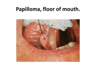
- 1. Papilloma, floor of mouth.
- 3. Papilloma
- 8. Oral wart.
- 9. wart
- 10. Oral wart.
- 11. Oral wart. Immunohistochemical stain for common human papillomavirus in an oral wart. Positive, brown-staining nuclei are seen in upper level keratinocytes.
- 13. Focal epithelial hyperplasia of the lip.
- 14. Focal epithelial hyperplasia of the buccal mucosa.
- 15. Biopsy specimen showing carcinoma in situ.
- 16. in situ carcinoma extending into salivary duct.
- 17. Squamous cell carcinoma. Small, crusted ulcer of the lower lip vermilion.
- 18. Squamous cell carcinoma. Ulcerated mass of the lower lip vermilion.
- 19. Squamous cell carcinoma of the gingiva.
- 20. Squamous cell carcinoma, floor of mouth.
- 21. Advanced squamous cell carcinoma of the posterior-lateral tongue.
- 22. Squamous cell carcinoma, gingiva.
- 23. Squamous cell carcinoma of the lip.
- 24. Exophytic squamous cell carcinoma of the lip.
- 25. Squamous cell carcinoma of the lateral tongue in a 34-year-old man.
- 26. Squamous cell carcinoma of the lateral tongue.
- 27. Squamous cell carcinoma of the ventral surface of the tongue.
- 28. Early squamous cell carcinoma of the floor of mouth.
- 29. Early squamous cell carcinoma of the floor of mouth.
- 30. Squamous cell carcinoma of the floor of mouth.
- 31. Squamous cell carcinoma of the floor of mouth.
- 32. Second primary squamous cell carcinoma of the palate in a 34-year-old man.
- 33. Metastasis of squamous cell carcinoma of the tongue to a submandibular lymph node.
- 34. Squamous ce ll carcinoma. Metastatic deposits within cervical lymph nodes present as firm, painless enlargements as seen in this patient with metastasis to a superior jugular node froma posterior lateral tongue carcinoma.
- 35. Well-differentiated squamous cell carcinoma. l ow-power photomicrograph showing islands of malignant squamous epithelium invading into the lamina properia.
- 37. Well-differentiated squamous cell carcinoma. High-power view showing dysplastic epithelial cells with keratin pearl formation.
- 40. Moderately differentiated squamous cell carcinoma. Although no keratinization is seen in this medium-power view, these malignant cells are still easily recognizable as being of squamous epithelial origin.
- 43. Poorly differentiated squamous cell carcinoma. The numerous pleomorphic cells within the lamina properia represent anaplastic carcinoma.
- 48. Verru cous carcinoma. Extensive papillary, white lesion of the maxillary vestibule.
- 49. Verrucous carcinoma. Large, exophytic, papillary mass of the maxillary alveolar ridge.
- 50. Verrucous carcinoma. Low-power photomicrograph showing marked epithelial hyperplasia with a rough, papillary surface and keratin plugging.
- 51. Verrucouscarcinoma. High-power view showing bulbous rete ridges without significant dysplasia.
- 53. Erythroleukoplakia. Mixed red-an d-white lesion of the lateral borde r of the tongue. Biopsy revealed carcinoma in situ.
- 54. l eu koplakia. Extensive ventra l and lateral tongue lesion containing multiple areas representi ng various possible pha ses or clinical appearances
- 55. Moderate epithelial dysplasia. Dysplast ic changes extend to the midpo int of the ep ithelium and are character ized by nuclear hyperchromatism. pleomorphism, and ce llular crowding.
- 58. Ductal dysplasia. Salivary gland duct exhibiting squamous metaplasia and dysplasia t hat originated from an overlying surface epit helial dysplasia
- 60. Basal cell carcinoma. Noduloulcerat ive lesion of the upper lip demonstrating telangiectasia and small ulceration.
- 62. Basal cell carcinoma. Pigmented basal cell carcinoma of the cheek.
- 64. Basal cell carcinoma. Note solid tumor (left) and nested tumor (right).
- 65. Basal cell carcinom a. low- power photomicrograph showing ulceration of the epidermal surface associated with an invading tumor of hyperchromatic epithelial cells. Inset demonstratesislands of basophilic epithelium with peripheral palisading.