Ventricular septal defect (vsd)
•
94 likes•37,419 views

this is my case presentation when i was assigned in NICU.

Recommended
More Related Content
What's hot
What's hot (20)
Viewers also liked
Viewers also liked (20)
Similar to Ventricular septal defect (vsd)
Similar to Ventricular septal defect (vsd) (20)
Recently uploaded
Recently uploaded (20)
Ventricular septal defect (vsd)
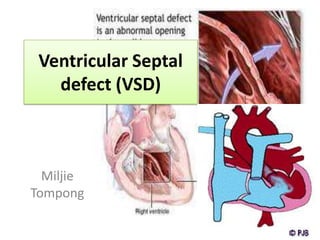
- 1. Ventricular Septal defect (VSD) Miljie Tompong
- 2. Ventricular septal defect (VSD)? • a heart malformation present at birth. "congenital" condition • a type of congenital heart disease (CHD).
- 3. Anatomy and physiology • The heart with a VSD has a hole in the wall (the septum) between its two lower chambers (the ventricles).
- 5. How do VSDs cause problems?
- 6. How is a VSD diagnosed? • chest X-ray
- 7. • Echocardiogram (used to define the anatomy and evaluate the characteristics (amount and pressures) of the shunted blood)
- 8. Electrocardiogram • is a noninvasive test that is used to reflect underlying heart conditions by measuring the electrical activity of the heart.
- 9. What are the symptoms of a VSD? • A murmur is a sound generated by abnormally turbulent flow of blood through the heart. • symptomless at birth usually develop until later in the first week of life. • no signs of cyanosis.
- 10. • Small defects (less than 0.5 square cm) are common • small VSDs close spontaneously (on their own). • closure is due to the small VSD being located between heart fibers that increase in size in time
- 11. • large VSD (usually one greater than 1 cm2 • Infant will fail to thrive and become sweaty and tachypnoiec (breathe faster) with feeds • increase in the blood pressure of the lungs called "pulmonary hypertension.
- 12. Medical Management • Digoxin (LANOXIN) • works directly on the heart to increase the velocity of contraction and the force of the contraction. • FUROSEMIDE (LASIX) • Diuretics remove edema and act to lower blood pressure. This helps reduce the extra load on the pulmonary system.
- 13. Treatment • PA Banding a palliative treatment particularly reserved for those patients who are unable to withstand an open heart procedure for total correction.
- 14. Surgical Treatment • median sternotomy • Surgical access to the VSD depends upon it's location and the presence of other cardiac defects. • The right ventricle is often opened to gain access to perimembranous or inlet defects. • The pulmonary artery may be used to close outlet septum VSDs
- 15. • Large defects may be repaired using a pericardium patch sutured into place to occlude the space.
- 16. Preoperative and postoperative • prophylactic antibiotics are often required to prevent infectious endocarditis. • The child should be assessed postoperatively for dysrhythmia, since edema in the septum may interfere with conduction.
- 17. Nursing Diagnosis “Decrease in cardiac output associated with heart malformations” Objective: improve the heart Rainfall Outcome criteria: signs of improvement in cardiac output
- 18. Nursing Intervention : • Observation of the quality and strength of heart rate, peripheral pulse, skin color and warmth. • Set the degree of cyanosis (mucous membranes, clubbing) • Monitor signs of CHF (anxiety, tachycardia, tachipnea, cramped, tired while drinking milk, periorbital edema, and hepatomegaly Oliguria. • Collaboration for the administration of drugs (diuretics, to reduce after load) as indicated.