SlideShare a Scribd company logo
1 of 70
1
PEDIATRIC DENTISTRY REVIEW
I. CHRONOLOGY OF THE HUMAN DENTITION
A. PRIMARY DENTITION: INITIATION AND CALCIFICATION
TOOTH LETTER INITIATION CALCIFICATION ERUPTION
CENTRAL A 7 WEEKS (IU) 14 WEEKS (IU) ~ 6 - 9 MONTHS
LATERAL B 7 WEEKS (IU) 16 WEEKS (IU) ~ 7 - 10 MONTHS
CANINE C 7 WEEKS (IU) 17 WEEKS (IU) ~ 18 MONTHS
1ST MOLAR D 8 WEEKS (IU) 15 WEEKS (IU) ~ 14 MONTHS
2ND MOLAR E 10 WEEKS (IU) 19 WEEKS (IU) ~ 24 MONTHS
- PRIMARY ROOT COMPLETION 18 MONTHS POST ERUPTION. (12-18 MOS.)
- TEETH ERUPT WHEN THE ROOT IS 2/3 COMPLETE.
- MOST FAVORABLE ERUPTION SEQUENCE FOR PRIMARY TEETH BOTH MAX & MAN:
ABDCE
NATAL TEETH - BORN WITH TEETH.
NEONATAL - 30 DAYS AFTER BIRTH
- DON'T REMOVE.(UNLESS VERY MOBILE AND WORRIED ABOUT ASPIRATION
- 85% OF TIME TRUE PRIMARY TEETH.
PRIMARY TEETH:
- GERMS FORM AT 6-8 WKS IU
- ENAMEL FORMS 4-6 MONTHS
- ROOTS COMPLETE APPROX 1 YR AFTER ERUPTION
B. PERMANENT DENTITION: INITIATION ( MAX AND MAND )
MAXILLARY MANDIBULAR
CENTRAL ~ 5 MONTHS (IU) ~ 5 MONTHS (IU)
LATERAL ~ 5 MONTHS (IU) ~ 5 MONTHS (IU)
CUSPID 5 - 6 MONTHS (IU) 5 - 6 MONTHS (IU)
1ST PREMOLAR BIRTH BIRTH
2ND PREMOLAR 7 - 8 MONTHS (PP) 7 - 8 MONTHS (PP)
1ST MOLAR 3.5 - 4 MONTHS (IU) 3.5 - 4 MONTHS (IU)
2ND MOLAR 8.5 - 9 MONTHS (PP) 2 YEARS (PP)
3RD MOLAR 3.5 - 4 YEARS (PP) 5 YEARS (PP)
2
C. PERMANENT DENTITION CALCIFICATION:
MAXILLARY MANDIBULAR
CENTRAL 3 - 4 MONTHS 3 - 4 MONTHS
LATERAL 10 - 12 MONTHS 3 - 4 MONTHS
CUSPID 4 - 5 MONTHS 10 -12 MONTHS
1ST PREMOLAR 1.5 - 2 YEARS 1.5 - 2 YEARS
2ND PREMOLAR 2 - 2.5 YEARS 2 - 2.5 YEARS
1ST MOLAR BIRTH BIRTH
2ND MOLAR 2.5 - 3 YEARS 2.5 - 3 YEARS
3RD MOLAR 7 - 9 YEARS 8 - 10 YEARS
D. PERMANENT DENTITION ERUPTION SEQUENCE:
- MAX: 61245378
- MAN: 61234578
EMERGENCE WHEN 2/3 ROOT PRESENT
ROOT COMPLETION 3 YEARS POST ERUPTION (2-3 YRS.)
ERUPTION SEQUENCE:
- MANDIBULAR INCISORS
- 1ST PERMANENT MOLARS, UPPER INCISORS
- CANINE (MAND)
- 1ST PREMOLAR
- MAX CANINE
- 2ND PREMOLAR
ALSO:
MAXILLARY: 1ST MOLAR, CI, LI, 1ST PM, 2ND PM, CUSPID, 2ND MOLAR, 3RD MOLAR
MANDIBULAR: 1ST MOLAR, CI, LI, CUSPID, 1ST PM, 2ND PM, 2ND MOLAR, 3RD MOLAR
PREMATURE ERUPTION:
- NOT ENOUGH ROOT FOR STABILITY IN PRIMARY TOOTH
- GROSS CARIES PRIMARY MOLARS, LOST EARLY.
- ABSCESS CAUSES BONE RESORPTION BENEATH PRIMARY TOOTH
RESULTS IN EARLY LOST OF PRIMARY TOOTH AND PREMATURE ERUPTION OF PERMANENT
REVIEW OF EMBRYOLOGY
1. INITIATION (BUD STAGE)
A. PRIMARY DENTITION: 6-8 WKS IN UTERO
3
B. PERMANENT DENTITION: FROM 5 MOS IN UTERO (ALL ANTERIORS, 1ST BICUSPIDS, 1ST
MOLAR) TO 5 YRS OF AGE (THIRD MOLARS)
2. PROLIFERATION (CAP STAGE)
3. MORPHODIFFERENTIATION / HISTODIFFERENTIATION (BELL STAGE)
4. APPOSITION
5. CALCIFICATION
VARIABLES THOUGHT TO INFLUENCE PERMANENT TOOTH ERUPTION
1. GENETIC
A. FAMILIAL
B. RACE (BLACKS SLIGHTLY EARLIER THAN WHITES)
C. SEX (FEMALES SLIGHTLY EARLIER THAN MALES)
2. ENVIRONMENTAL
A. LOW BIRTH WEIGHT BABIES HAVE DELAYED ERUPTION, PARTICULARLY IN THE PRIMARY
DENTITION.
B. NUTRITION HAS LITTLE OR NO EFFECT ON ERUPTION
3. SYSTEMIC FACTORS/HERITABLE CONDITIONS THAT AFFECT EXFOLIATION/ERUPTION (ALL TEETH
AFFECTED)
A. ENDOCRINE - DELAYED EXFOLIATION/ERUPTION
- HYPOPITUITARISM
- HYPOTHYROIDISM
- LOW LEVELS OF HUMAN GROWTH HORMONE
B. ENDOCRINE - PREMATURE EXFOLIATION
- HYPERPITUITARISM
- HYPERTHYROIDISM
C. OTHER - PREMATURE EXFOLIATION
- CHERUBISM
- JUVENILE PERIODONTITIS
- PAPILLON LEFEVRE SYNDROME
- HISTIOCYTOSIS X
- LEUKEMIA
- CYCLIC NEUTROPENIA
II. ORTHODONTICS AND OCCLUSION REVIEW
OCCLUSION CLASSIFICATION
THE LOWER FACE IS THE LAST PART OF THE HEAD TO COMPLETE ITS GROWTH.
4
- PRIMARY 2ND MOLAR TERMINAL PLANE RELATIONSHIP ( 75 % OF FIRST MOLAR END TO END
RELATIONSHIPS WILL DEVELOP INTO CLASS 1)
PRIMARY TERMINAL
PLANE
49% MESIAL STEP
37% FTP
14 % DISTAL STEP
INITIAL PERMANENT
MOLAR RELATIONSHIP
1% CLASS III
27% CLASS I
49% END ON
23% CLASS II
FINAL OCCLUSION
3% CLASS III
59% CLASS I
39% CLASS II
LEEWAY SPACE: DIFFERENCE BETWEEN THE SUMS OF THE MESIAL - DISTAL WIDTHS OF C, D,
AND E AND THE SUCCEEDING PERMANENT TEETH 3, 4 AND 5.
- 0.9 MM /QUADRANT IN MAX (PER SIDE)
- 1.7 MM /QUADRANT IN MAND (PER SIDE)
PRIMATE SPACES:
- MESIAL TO PRIMARY MX CANINE AND DISTAL TO PRIMARY MAND CANINES
- EARLY SHIFT ( 1ST PERM MOLARS CLOSE POSTERIOR SPACES )
- LATE SHIFT INTO CLASS I HAPPENS WITH LEEWAY SPACE.
INCISOR LIABILITY: DIFFERENCE IN SIZE OF PERMANENT INCISORS VS PRIMARY INCISORS -
- MAX: 7.6 MM TOTAL
- MAND 6.0 MM
ARCH CIRCUMFERENCE GREATEST AT AGE THREE
CROSS-BITE
- ANTERIOR PRIMARY DENTITION, WAIT UNTIL TRANSITION TO TREAT
- POSTERIOR IN PRIMARY DENTITION : TREAT IN PRIMARY DENTITION
1. UNILATERAL, FUNCTIONAL ( MIDLINE SHIFT FROM CR TO CO )
2. BILATERAL, NONFUNCTIONAL: ( NO MIDLINE SHIFT )
- TREATMENT APPLIANCES
1. RPE'S ( 2-4 WEEKS VS SLOW ( 2-6 MONTHS )
2. SLOW: W-ARCH ( PORTER APPLIANCE ), QUAD HELIX
- MID PALATAL SUTURE FUSES IN EARLY TEENS ( 13-16 )
- ANTERIOR CROSSBITE IN PERMANENT DENTITION : ORTHO EMERGENCY
SERIAL EXTRACTION
- NORMAL EXTRACTION SEQUENCE IS C'S, D'S AND THEN 4'S
5
- TWEED VARIATION: D'S, C'S AND THEN 4'S
- INDICATIONS:
CLASS I MOLARS AND CANINES
SEVERE CROWDING ( GREATER THAN 6 MM PER QUADRANT )
NORMAL OVERBITE AND OVERJET (2 MM EACH)
NORMAL ERUPTION SEQUENCE
7-8 YEARS OLD
FULL PROFILE
- CAN BE INDICATED IN A 7-8 YEAR OLD WHEN FULL FACE BIMAXILLARY PROTRUSION
IS
FOUND
- TREATMENT RESULTS IN INCREASED OVERBITE (GUARD AGAINST LINGUAL DUMPING OF
INCISORS)
COMPLICATIONS:
1. NOT A CLASS I CASE
2. MISDIAGNOSIS
3. LACK OF FOLLOW UP (COMPLETION OF CARE)
ECTOPIC ERUPTION
- MOST COMMON: LOWER PERMANENT INCISORS
1. LINGUAL TO PRIMARY INCISORS
2. LATERAL: CAUSES RESORPTION OF PRIMARY CANINES
- PERMANENT FIRST MOLARS; INCIDENCE 2-3% ( MOST IN MAXILLA )
1. 60% SELF CORRECT.
2. IF NOT CORRECTED BY 7-8, ACTIVE TREATMENT IS NEEDED-
-EXT 2ND PRIMARY MOLAR, HAWLEY WITH FINGER SPRING.
- PUSH 1ST PERMANENT MOLAR BACK.
SPACE ANALYSIS:
(1) MOYERS:
- PREDICTS SPACE FOR BOTH THE MANDIBLE AND MAXILLA
- PREDICTS THE SIZES OF THE CUSPIDS AND PREMOLARS
- MEASURE WIDTH OF 4 MAND INCISORS.
- PREDICT SPACE NEEDED FOR PERM CANINE, 1ST AND 2ND BICUSPID.
- MEASURE EXISTING ARCH CIRCUMFERENCE.
- 75 PERCENTILE, PREDICTS CROWDING, 75% HAVE SMALLER TEETH.
- IT IS INCORRECT 15% OF THE TIME IF THE 85% PROBABILITY
CHART IS
USED
(2) TINAKA-JOHNSON:
- MEASURE MANDIBULAR CENTRAL AND LATERAL (ONE SIDE).
- MAND ADD 10.5 MM TO GET SPACE NEEDED FOR 345.
- MAX ADD 11.5 MM.
(3) OTHERS:
6
NANCE.
HIXON & OLDFATHER.
CAREY.
FOSTER.
MORE OF SPACE DISCREPANCY MAND THAN MAX:
- GOOD, GETS 1ST MOLARS FORWARD A LITTLE INTO CLASS I.
SKELETAL AGE ASSESSMENT
CARPAL RADIOGRAPH:
GROWTH SPURT:
- PRESENCE OF RADIAL SESAMOID BONE (SHORT BONE).
- CURVING OF EPIPHYSIAL LINES AT END OF LONG BONES.
GROWTH AND DEVELOPMENT
CLEFT LIP AND PALATE:
- PRIMARY PALATE FORMS 4-6 WEEKS IU.
- SECONDARY PALATE FORMS 8-12 WEEKS IU.
GROWTH SPURT HAS A GREAT DEAL OF INDIVIDUAL VARIATION, BUT TENDS TO OCCUR NEARLY 2
YEARS EARLIER ON THE AVERAGE IN GIRLS THAN BOYS.
PUBERTY IN BOYS STARTS LATER AND EXTENDS OVER A LONGER PERIOD OF TIME, ABOUT 5
YEARS AS COMPARED TO 3.5 YEARS FOR GIRLS.
INTERCANINE WIDTH DOES NOT INCREASE IN GIRLS AFTER THE AGE OF 9.
SPACE MAINTENANCE
(1) BAND AND LOOP.
(2) LINGUAL ARCH WIRE:
- 1ST MOLAR TO 1ST MOLAR.
- PUT SPUR DISTAL TO CUSPID, PREVENTS DISTALIZING CUSPID AS PERM
INCISORS ERUPT.
(3) GOOD CLASS I MOLAR RELATIONSHIP MAY NOT NEED SPACE MAINT.
(4) TAKE OUT PRIMARY CANINES:
- PUT IN LING ARCH WIRE.
- IF LING ERUPTION OF INCISORS REPLACE ARCH WIRE.
(5) DISTAL SHOE:
- IMPORTANT SPACE MAINTENANCE APPLIANCE.
- WHEN 1ST MOLAR ERUPTS BREAK OFF TONGUE AND USE AS BAND AND
LOOP.
7
- CONTRAINDICATED IF PATIENT REQUIRES SBE PROPHYLAXIS
MISSING TEETH
- PRIMARY TOOTH MISSING HAVE INCREASED INCIDENCE OF MISSING PERMANENT TOOTH.
- MOST COMMON IS MAND BICUSPID, NEXT MAX LAT, MAX 2ND BI, MAND LAT.
- RETAINED PRIMARY MOLAR WILL LAST INTO 40'S.
III. FLUORIDES
DIETARY FLUORIDE SUPPLEMENTATION SCHEDULE
American Academy of Pediatric Dentistry & American Dental Association's Council on Dental
Therapeutics
Age Less than 0.3 ppm F 0.3 - 0.6 ppm F More than 0.6 ppm
F
Birth - 6 mos 0 0 0
6 mos - 3 yrs 0.25 mg 0 0
3 yrs - 6 yrs 0.50 mg 0.25 mg 0
6 yrs up to at least
16yr
1.0 mg 0.50 mg 0
• Evaluate patient's fluoride exposure before prescribing appropriate fluoride therapy. Document
the patient's fluoride exposure in the record and review at periodic visits. Have a water analysis
done prior to issuing supplemental fluoride.
• Point-of-use water conditioning/filtration systems significantly reduce fluoride content.
Activated carbon filters reduce fluoride concentration by 81%. Bottled water can have variable
fluoride concentrations.
• Preschool children require supervision during tooth brushing. No more than a pea size amount
of toothpaste should be used, avoid swallowing the paste and rinse after brushing.
• Fluoride mouth rinses containing 0.05% or 0.2% should not be used by children under the age
of six years or those who are unable to adequately expectorate.
• Supervise professionally applied topical fluoride treatments. Use 5 - 10 ml of fluoride gel in well
fitting trays, have the patient lean forward in the dental chair, admonish not to swallow, and use a
saliva ejector positioned between the trays.
Fluoride Toxicity:
8
1. Chronic (results in fluorosis) -
a. Dean's Classification: 0-4 scale
0- (None)59% of population at optimal fluoride level will exhibit no
fluorosis.
1- (Very mild) 7.4% of population at optimal dose
2- (Mild) Greater than 25%, less than 50% of surface with opaque defects
greater than 1.2 ppm
3- (Moderate) Brown stains present ( greater than 2 ppm)
4- (Severe) Pitting and attrition problems ( reater than 2-3 ppm )
b. Fluorosis occurs during maturation process. For incisors, age 2-3 years is the
critical period.
2. Acute
a. CLD ( certain lethal dose ): 32-64 mg/kg in one source, 71-140 mg/kg in
another source. A death has been reported at 17 mg/kg.
b. PTD ( Probable toxic dose ) 5mg/kg i.e. 50 mg for the average 10 kg 2 year old.
c. STD ( Safely tolerated dose ) 8-16 mg/kg
d. Toxicity at low doses is due to gastric irritation. Hypocalcemia and
hyperkalemia at high doses leads to cardiovascular collapse and death.
e. Treatment for overdose:
- less than 5mg/kg: oral calcium ( milk) and observe
- More than 5 but less than 15 mg/kg: induce vomiting, oral calcium (milk,
calcium gluconate, or calcium lactate), admit to hospital
- More than 15 mg/kg: admit to hospital immediately, induce vomiting,
monitor cardiac function, 10% calcium gluconate solution IV, monitor
electrolytes.
F. - COMMON SIGNS AND SYMPTOMS OF ACUTE FLUORIDE TOXICITY:
LOW DOSAGES HIGH DOSAGES
NAUSEA 3-C'S
VOMITING CONVULSIONS
HYPERSALIVATION CARDIAC ARRHYTHMIA
ABDOMINAL PAIN COMATOSE
DIARRHEA
- PT BECOMES HYPOCALCEMIC DUE TO CA BINDING WITH F.
CALCULATING MG F IN COMPOUNDS:
1ST STEP: FIND % F IN COMPOUND.
NAME MOLECULAR WEIGHT RATIO
APF-ACIDULATED PHOSPHATE FLUORIDE 1
9
NAF-SODIUM FLUORIDE 1/2.2 OR 1/2
SNF2-STANNOUS FLUORIDE 1/4.1 OR 1/4
MFP-SODIUM MONOFLUOROPHOSPHATE 1/7.6 OR 1/8
MULTIPLY % COMPOUND BY MOLECULAR WEIGHT RATIO = % F.
2ND STEP: EXPRESS % F IN COMPOUND IN MG F/ML.
BY DEFINITION % SOLUTION IS GRAMS/100ML.
GRAMS/100ML = 1000 MG/100ML = 10 MG/ML.
MULTIPLY STEP 1 BY 10 = MGF/ML.
3RD STEP: MULTIPLY STEP 2 BY ML INGESTED TO GET MG F INGESTED.
FLUORIDES :
- 1.23% APF X 1 = 1.23% F X 10 = 12.3 MGF/ML.
- .2% NAF X 1/2 = .1% F X 10 = 1 MGF/ML.
- 2% NAF X 1/2 = 1% F X 10 = 10 MGF/ML.
- .4% SNF2 X 1/4 = .1% F X 10 = 1 MGF/ML.
- 10% SNF2 X 1/4 = 2.5% F X 10 = 25 MGF/ML.
- COMPARE WITH CLD AND STD FOR WEIGHT OF INDIVIDUAL.
WATER FLUORIDATION:
- DECREASE IN CARIES: PRIMARY = 40%, SECONDARY = 50-60%
MECHANISM OF ACTION OF FLUORIDES:
- DECREASES ENAMEL SOLUBILITY.
- IMPROVES CRYSTALINITY.
- PROMOTES REMINERALIZATION.
- DECREASES FREE SURFACE ENERGY OF BACTERIA SO IT CANNOT STICK ON TOOTH.
- BACTERIOCIDAL OR STATIC.
- CAUSES DEVELOPING CRYSTAL TO GET BIGGER, LESS SOLUBLE
FLUORIDE PROTOCOL:
- HOME USE IN TRAYS, TOPICAL.
- H2O SUPPLY.
- SYSTEMIC ALSO TOPICAL WHEN CHEWING TABLET.
- HIGH FREQUENCY LOW DOSE IS EFFECTIVE
10
PEDIATRIC DENTAL EMERGENCIES
TYPES - PAIN, SWELLING/INFECTION, TRAUMA
BEFORE YOU DO ANYTHING
1. GOOD MEDICAL HISTORY
A. CONGENITAL HEART PROBLEMS (SBE PROPHYLAXIS)
B. CHRONIC ILLNESS (HX ORGAN TRANSPLANT, LEUKEMIA, HEMOPHILIA, ETC.)
C. MEDICATIONS/ALLERGIES
2. RULE OUT CHILD ABUSE
3. DOCUMENT EVERYTHING EXTREMELY WELL, PARTICULARLY TRAUMA.
DIAGNOSIS - WHICH TOOTH IS IT?
1. WHAT NOT TO DO
A. VITALOMETER
B. HEAT/ICE TESTS
2. WHAT TO DO
A. MUST RELY ON CLINICAL SIGNS AND SYMPTOMS - NO GOOD TESTS AVAILABLE
B. DO GET A PA RADIOGRAPH
- DEEP CARIES APPROACHING PULP
- INTERNAL/EXTERNAL RESORPTION
- APICAL OR FURCAL RADIOLUCENCIES
C. CHECK FOR MOBILITY. ALMOST ALL PULPALLY INVOLVED TEETH ARE MORE MOBILE
THAN THE UNAFFECTED TOOTH ON THE OTHER SIDE.
D. PERCUSSION SENSITIVE SOMETIMES
CARIES IN THE PRIMARY DENTITION
1. GENERAL INFORMATION
A. OCCLUSAL SURFACES OF PRIMARY MOLARS ARE GENERALLY MORE RESISTANT TO
DECAY THAN PERMANENT MOLARS
B. PRIMARY TEETH HAVE PROPORTIONALLY MORE PULP AND LESS ENAMEL AND
DENTIN
C. PRIMARY TEETH HAVE SIGNIFICANTLY LESS ENAMEL AND DENTIN PROTECTING THE
PULP
D. PRIMARY DENTINAL TUBULES ARE LARGER, RELATIVELY MORE POROUS THAN THOSE
IN
PERMANENT DENTIN
E. PROXIMAL CARIES PROGRESSES MORE RAPIDLY THAN OCCLUSAL CARIES AND
CAUSES A
HIGHER PERCENTAGE OF PULPAL EXPOSURES.
F. IF THE PROXIMAL DECAY HAS PROGRESSED (RADIOGRAPHICALLY) 1/2 WAY TO THE
DEJ
OR FURTHER, THE TOOTH MUST BE RESTORED (UNLESS EXFOLIATION IS IMMINENT)
G. USUALLY, IF THERE IS PAIN IT WILL ALMOST ALWAYS BE A PRIMARY MOLAR -
ANTERIOR
TEETH ABSCESS W/O PAINFUL SYMPTOMS
H. RADIOGRAPHIC EVIDENCE OF PULPAL INVOLVEMENT ON PRIMARY MOLAR WILL
OCCUR
11
IN THE FURCA AREA, APICALLY ON ANTERIOR TEETH
2. BABY BOTTLE TOOTH DECAY (BBTD) - ETIOLOGY
A. CHILD PLACED IN BED AT NIGHT AND/OR NAP TIME WITH BOTTLE OF MILK OR
SWEETENED LIQUID
B. THE CHILD DOES NOT SWALLOW THE LAST MOUTHFUL OF LIQUID BEFORE HE/SHE
FALLS
ASLEEP AND THE LIQUID POOLS AROUND THE MAXILLARY TEETH
C. THE SWEETENED LIQUID PROVIDES AN EXCELLENT CULTURE FOR ACIDOGENIC
BACTERIA
D. THE TONGUE PROTECTS THE MAND INCISORS = KEY TO DIAGNOSIS: MAND INCISORS
ARE
CARIES FREE
CHARACTERISTICS OF INFECTIONS IN CHILDREN
1. CAN SPREAD RAPIDLY THROUGH BONE DUE TO LARGE MARROW SPACES
2. CAN SPREAD BEYOND POINTS OF MUSCLE INSERTIONS TO DEEPER AREAS
3. DIFFUSE CELLULITIS IS THE NORM - RARELY MUCH PUS PRODUCTION - USUALLY NOTHING TO
I & D
4. MAXILLARY TEETH
- MAXILLARY CANINES AND MOLARS CAN INVOLVE THE PERIORBITAL AREA
- THIS CAN LEAD TO CAVERNOUS SINUS THROMBOSIS, BRAIN ABSCESSES, SEPTICEMIA, ETC.,
(ALL
POTENTIALLY FATAL COMPLICATIONS)
5. MANDIBULAR TEETH
- 1ST AND 2ND PRIMARY MOLAR CAN SEE VESTIBULAR SWELLING OR BUCCAL SPACE
INFECTION
- RARELY SEE SUBMANDIBULAR INFECTIONS
* IF YOU CANNOT FEEL THE INFERIOR BORDER OF THE MANDIBLE, YOU ARE IN
TROUBLE
* IF THE CHILD COMPLAINS OF DYSPHAGIA, YOU ARE IN TROUBLE ( INFECTION HAS
SPREAD
BELOW THE INSERTION OF THE MYLOHYOID. MYLOHYOID ELEVATES WHEN YOU
SWALLOW, THUS THE DYSPHAGIA.
6. TREATMENT
- ANTIBIOTICS - PCN STILL DRUG OF CHOICE (FAIRLY NARROW SPECTRUM, LOW TOXICITY,
BACTERICIDAL, PROVEN USE, CHEAP)
- ERYTHROMYCIN POOR SECOND CHOICE FOR PCN ALLERGIC
TRAUMA
1. THREE PEAK PERIODS FOR INJURIES
A. 1 1/2 - 2 1/2 YRS OF AGE
B. 5 -6 YRS OF AGE
C. 8 - 9 YRS OF AGE
2. ACCURATE HISTORY OF INJURY
12
A. WHERE DID IT OCCUR (CLEAN VS DIRTY) (LAST TETANUS ?)
B. WHEN (SOME INJURIES ARE TIME SENSITIVE)
C. HOW (MAKE SURE HISTORY AND EXPLANATION MATCH UP)
3. EXAMINATION
A. EXAMINE TEETH LAST
B. CHECK FOR SIGNS OF CNS DAMAGE (I.E. SUBDURAL HEMATOMA, SKULL FRACTURE,
SEVERE
CONCUSSION)
- AMNESIA
- LOSS OF CONSCIOUSNESS
- NOT ALERT, NOT ORIENTED TO TIME AND PLACE, EXTREME LETHARGY
- IRRITABILITY, DRAMATIC CHANGE IN PERSONALITY
- MUSCLE WEAKNESS OF ONE SIDE OR ONE ARM, ONE LEG ETC.
- PUPIL DIFFERENCES
- NAUSEA AND VOMITING
C. IF YOU SEE ANY OF THESE , IT USUALLY WARRANTS IMMEDIATE REFERRAL TO MEDICAL
FACILITY - TAKES PRECEDENCE OVER DENTAL TRAUMA
D. EXAMINE FOR FACIAL FRACTURES
- RARE IN CHILDREN DUE TO LARGE MARROW SPACES, BONE IS LESS DENSE, MUCH LESS
BRITTLE
- MOSTLY GREENSTICK FRACTURES
- ORDER OF FREQUENCY - NASAL, MANDIBULAR (INCLUDING CONDYLES),
ZYGOMAXILLARY
COMPLEX
E. MAXILLARY FRACTURES - ALMOST NEVER SEE TRUE LAFORTE FRACTURES IN CHILDREN
DUE TO SMALL SINUSES UNTIL AGE 10 12
F. MANDIBULAR FRACTURES - SIGNS AND SYMPTOMS SAME AS ADULT
- PAIN, MALOCCLUSION, SUBLINGUAL HEMATOMA, GINGIVAL TEARS, SWELLING
- PALPATE ALL AREAS FOR “STEPS”
- 66% IN CONDYLAR AREA
- 34% IN BODY
- CONDYLAR FRACTURES CAN BE ASSOC WITH FXS OF THE BODY OF THE MANDIBLE ON
THE
CONTRALATERAL SIDE
G. CHIN TRAUMA
- CLASSIC TRIAD WITH CHIN TRAUMA
* SYMPHYSEAL FRACTURE
* POSTERIOR CROWN FRACTURES
* CONDYLAR FRACTURE
- POSSIBILITY OF CERVICAL VERTEBRAL FX ALSO
H. SIGNS AND SYMPTOMS OF CONDYLAR FRACTURE
- PAIN
- DEVIATION TO AFFECTED SIDE
- LIMITATION OF OPENING
13
SOFT TISSUE INJURIES
1. IF DIRTY WOUND. ASK ABOUT TETANUS STATUS
A. CLEAN = ANY WOUND THAT CAN BE MECHANICALLY DEBRIDED AND SCRUBBED CLEAN; I.E.
SURFACE ABRASIONS, SUPERFICIAL LACERATIONS
B. DIRTY = DEEP LACERATIONS, PUNCTURE WOUNDS, FALLING IN MANURE, REIMPLANTING
TEETH THAT HAVE BEEN ON THE GROUND, ETC.
2. LACERATIONS
A. LOCAL ANESTHESIA AS NECESSARY
B. CLEANSE WOUND
C. TO R/O FOREIGN BODY IN WOUND (PARTICULARLY IF THERE ARE ROOT FRAGMENTS THAT
ARE
UNACCOUNTED FOR) PALPATE THEN TAKE RADIOGRAPH OF LACERATED AREA
D. ACHIEVE HEMOSTASIS W/ RESORBABLE SUTURES (4-0 OR 5-0 VICRYL) INTRA ORALLY (CAN
USE SILK); NYLON ON SKIN, BURY KNOTS INTRAORALLY WHEN POSSIBLE
E. FRENUM TEARS
- VERY COMMON IN TODDLERS
- MOST DO NOT REQUIRE SUTURES UNLESS YOU CANNOT OBTAIN HEMOSTASIS
- IN CHILDREN NOT YET WALKING OR CRAWLING, STRONG POSSIBILITY OF FORCED
FEEDING,
OTHER INAPPROPRIATE TREATMENT = CHILD ABUSE
F. TONGUE LACERATIONS
- EVERYONE’S WORST NIGHTMARE
- NO EASY WAY TO TREAT IF SUTURES ARE REQUIRED AND THEY USUALLY ARE
- DIRECT INJECTION INTO TONGUE AROUND WOUND ARE USUALLY BEST
- “HOLD ‘N GO”
G. DEGLOVING INJURIES
- SUTURE BACK INTO POSITION IF ACCORDION TYPE INJURY
- IF TISSUE IS LOST, AREA WILL GRANULATE IN BUT THESE CAN GET INFECTED
- CONSIDER AB COVERAGE FOR 7 - 10 DAYS
H. ELECTRICAL BURNS
- TISSUE DAMAGE IS ENORMOUS; MUCH MORE THEN WHAT IS VISIBLE
- INJURIES INVOLVE MUCOSA, SUBMUCOSA, MUSCLE, NERVE AND VASCULAR TISSUE
- ALL NECROTIC TISSUE, NO PAIN
- NECROTIC TISSUE WILL SLOUGH IN 10 - 21 DAYS - CAN LEAD TO LABIAL ARTERY BLEED
- TREMENDOUS SCAR FORMATION; UNTREATED WILL RESULT IN SEVERE MICROSTOMIA
- APPLIANCE PLACED AFTER TISSUE SLOUGHS WILL ALLOW SCAR TO FORM AROUND A
MATRIX; CAN SIGNIFICANTLY DECREASE THE AMOUNT OF DISFIGURATION. MAINTAINS
COMMISSURAL REGIONS EQUIDISTANT TO MIDLINE
- APPLIANCES MUST BE WORN 24 HRS/DAY FOR 6 - 18 MONTHS DEPENDING ON SEVERITY
OF
INJURY
IV. GUIDELINES FOR PRESCRIBING DENTAL
RADIOGRAPHS (NO PREVIOUS RADIOGRAPHS)
14
AGE
3-5
6-7
8-9
10-12
CONSIDERATIONS
NO APPARENT ABNORMALITIES
(OPEN CONTACTS)
NO APPARENT ABNORMALITIES
(CLOSED CONTACTS)
EXTENSIVE CARIES
DEEP CARIES
NO APPARENT ABNORMALITIES
EXTENSIVE OR DEEP CARIES
NO APPARENT ABNORMALITIES OR
DEEP CARIES NO APPARENT
ABNORMALITIES OR DEEP CARIES
RADIOGRAPHS
NONE
2 BW'S (SIZE 0)
4 FILM SURVEY
SELECTED PA'S AND THE 4 FILM
SURVEY
8 FILM SURVEY
SELECTED PA'S AND 8 FILM
SURVEY
12 FILM SURVEY
12 OR 16 FILM SURVEY
15
THE ABOVE RECOMMENDATION IS FROM MCDONALD'S 5TH EDITION PAGE 7
ACCORDING TO INTERORGANIZATIONAL CONFERENCE, INCLUDING THE ADA AND THE AMERICAN
ACADEMY OF PEDIATRIC DENTISTRY IN 1992-93:
PATIENT CATEGORY PRIMARY DENTITION (PRIOR TO 1ST PERM MOLAR)
NO CLINICAL CARIES POSTERIOR BW EXAMINATION AT 12-24 MONTH INTERVALS IF
PROXIMAL SURFACES OF PRIMARY TEETH CANNOT BE
VISUALIZED
OR PROBED
TRANSITIONAL DENTITION (FOLLOWING ERUPTION OF 1ST
PERM MOLAR
POSTERIOR BW EXAMINATION AT 12 - 24 MONTH
INTERVALS
ADOLESCENT, PERMANENT TEETH (PRIOR TO THIRDS)
POSTERIOR BW EXAMINATION AT 24 - 36 MONTH
INTERVALS
CLINICAL CARIES OR POSTERIOR BW EXAMINATION AT 6 MONTHS INTERVALS OR UNTIL
NO
HIGH RISK FACTORS CARIOUS LESIONS ARE EVIDENT IN PRIMARY AND TRANSITIONAL
FOR CARIES DENTITIONS.
POSTERIOR BW EXAMINATION AT 6 - 12 MONTH INTERVALS
FOR
PERMANENT TEETH IN ADOLESCENTS AND 12 - 18 MONTHS IN
ADULTS
OR UNTIL NO CARIOUS LESIONS ARE EVIDENT
V. PEDIATRIC PATHOLOGY
A. INCLUSION CYSTS - ALL SHED SPONTANEOUSLY
KERATIN-FILLED NODULES:
(1) EPSTEIN'S PEARLS
- TRAPPED EPITHELIUM IN MIDPALATINE RAPHE.
- 100% HAVE CLINICAL OR SUBCLINICAL EPSTEIN'S PEARLS.
- NOT RARE.
(2) BOHN'S NODULES
- TRAPPED MUCOUS GLAND TISSUE ON BUCCAL AND LINGUAL OF RIDGE
- LABIAL/LINGUAL OF ALVEOLAR RIDGE.
(3) DENTAL LAMINA CYST
- REMNANTS OF DENTAL LAMINA ON CREST OF RIDGE
16
B. LYMPHANGIOMAS OF ALVEOLAR RIDGE
- 1-3% BLACKS.
- BLUE DOMED, FLUID FILLED LESION ON THE ALVEOLAR RIDGE
- TX: OBSERVATION FOR SPONTANEOUS REGRESSION. EXCISION AFTER SIX
MONTHS.
C. CONGENITAL EPULIS
- PEDUNCULATED LESION ( MAY BE SEVERAL CM IN DIAMETER ) ON RIDGE IN
AREA
OF MAX INCISORS.
- SPONTANEOUS REGRESSION WITHIN FIRST YEAR IS COMMON. OBSERVATION IS
INDICATED.
- IF SURGERY IS INDICATED DUE TO FEEDING PROBLEM , WAIT UNTIL AFTER 8-10
DAYS OF AGE
D. MEDIAN ALVEOLAR NOTCH
-NOTCH IN THE MX ALVEOLUS, NOT A TRUE CLEFT ( CONNECTIVE TISSUE DEFECT )
- MORE FREQUENT IN BLACKS
- MAY RESULT IN DIASTEMA
E. NEUROECTODERMAL TUMOR OF INFANCY
- HIGHLY PIGMENTED ( MELANOTIC ) BENIGN TUMOR APPEARING WITHIN THE
FIRST
YEAR OF LIFE, USUALLY IN THE ANTERIOR MAXILLA.
- DESTRUCTIVE RADIOLUCENT LESION: FLOATING TEETH TX; CONSERVATIVE
EXCISION
- HIGH URINARY LEVELS OF VANILMANDELIC ACID ( COMMON WITH NEURAL
CREST
TUMORS )
F. NATAL( AT BIRTH ) AND NEONATAL ( WITHIN 30 DAYS OF BIRTH )
- USUALLY LOWER PRIMARY INCISORS ( 85% ) RARELY SUPERNUMERARIES
- TX: NONE IF TOOTH NOT LIKELY TO EXFOLIATE OR CAUSE ULCERATION
- RIGA-FEDA DISEASE: ULCERATION OF VENTRAL SURFACE OF TONGUE DUE TO
IRRITATION OF LOWER INCISORS WHEN NURSING.
- NO SURGERY UNTIL AFTER 10 DAYS DUE TO HYPERPROTHROMBINEMIA.
G. AMELOGENESIS IMPERFECTA (RADIOGRAPHICALLY PULP LOOKS NORMAL)
3 TYPES:
(1) HYPOPLASTIC:
- MATRIX DID NOT FORM PROPERLY.
- THIN ENAMEL, SHOVEL SHAPED, PITS AND DEFECTS.
(2) HYPOCALCIFIED:
- MATRIX OK, CALCIFICATION NOT PROPER.
- SHEARS, BREAKS.
17
- MOTH EATEN X-RAY APPEARANCE.
(3) HYPOMATURE:(HYPOMINERALIZED)
- QUALITATIVE DEFECT IN ENTIRE CALCIFIED AREA.
- MORE RADIOLUCENT.
H.. DENTINOGENESIS IMPERFECTA (PULP CHAMBER OBLITERATED RADIOGRAPHICALLY)
THREE TYPES:
(1) SHIELDS TYPE I:
- MOST COMPREHENSIVE, SYSTEMIC PROBLEMS, OSTEOGENESIS
IMPERFECTA.
- BRITTLE BONE DISEASE.
- LATERAL SKULL FILMS SHOW RADIOPACITY.
- PRIMARY TEETH WORSE THAN THE PERMANENT
(2) SHIELDS TYPE II:
- DOES NOT HAVE SYSTEMIC INVOLVEMENT.
- BOTH DENTITIONS EQUALLY AFFECTED
(3) SHIELDS TYPE III:(BRANDYWINE)(RACIAL ISOLATE IN MARYLAND)
- BELL SHAPED CROWNS.
- SHELL TEETH.
- MULTIPLE PULP EXPOSURES IN PRIMARY TEETH
I. PERICORONITIS:
- CAN HAVE ON 6 YEAR MOLAR.
- IF AN ERUPTION CYST FORMS DISTAL TO THE SECOND PRIMARY MOLAR, THE
INITIAL TREATMENT OF CHOICE IN MOST CASES IS TO WAIT AND JUST
OBSERVE IT.
IN MOST CASES NORMAL ERUPTION WILL TAKE PLACE. IF IT DOES NOT, THAN
IT
CAN BE INCISED AND THE FIRST MOLAR EXPOSED.
J. ERUPTION SEQUESTRUM:
- NOT ALL BONE OVER TOOTH RESORBED.
K. TURNER TOOTH
- HYPOPLASIA OF PERMANENT TOOTH DUE TO INFECTION OR TRAUMA TO THE
PRIMARY PREDECESSOR
- ETIOLOGY IS A DISTURBANCE TO THE ENAMEL ORGAN DURING FORMATION
OF THE
PERMANENT CROWN
- FACIAL ASPECT OF A MAXILLARY INCISOR IS USUALLY THE AFFECTED AREA
L. HIV IN CHILDREN
-ETIOLOGY- MOSTLY PERINATAL OTHER: SEXUAL, DRUG ABUSE BREAST MILK
RARE
18
-INFANTS USUALLY CONVERT IN 15 MONTHS
- MOTHERS TAKING AZT 25% OF NEWBORNS CONVERT, NO AZT 30-50%
CONVERT
- CLINICAL MANIFESTATIONS ARE FAILURE TO THRIVE, FEVER CHRONIC DIARRHEA,
LYMPHADENOPATHY, ORAL CANDIDA, PERSISTENT DIFFUSE PAROTITIS
OCCASIONALLY AND ORAL VIRAL LESIONS
VI. STAGES OF TOOTH DEVELOPMENT
-INITIATION, PROLIFERATION, HISTODIFFERENTIATION, MORPHODIFFERENTIATION,
APPOSITION (CALCIFICATION), MATURATION
VII ANOMALIES OF TEETH
A. INITIATION PHASE
-SUPERNUMERARY TEETH:
- MORE OFTEN IN PERM DENTITION, MESIODENS IS MOST COMMON,
CLEIDOCRANIAL DYSPLASIA - MULTIPLE SUPERNUMERARY TEETH
- MISSING TEETH: RARE IN PRIMARY DENTITION
-IN PERM DENT (OTHER THAN 3RDS) MAND 2ND BI > MAX 2ND BI > MAX
LI,
( SOME DISAGREEMENT ABOUT WHETHER MAX 2ND BI OR MAX
LI )
-ECTODERMAL DYSPLASIA - ANODONTIA OR HYPODONTIA
B. TWINNING ANOMALIES (PROLIFERATION PHASE)
(1) GEMINATION: DIVISION OF SINGLE GERM - BIFID CROWN, SINGLE ROOT
(2) FUSION: TWO BUDS, TWO TEETH, SEPARATE PULP CANALS - DENTIN
CONFLUENCE, COMMONLY MISSING ONE OF THE SUCCESSORS
(3) CONCRESCENCE: CEMENTAL UNION, TWO BUDS, TWO TEETH
C. SIZE AND SHAPE
- MACRODONTIA, MICRODONTIA, PEG LATERAL, DENS-IN-DENTE
- TURNER'S TOOTH: HYPOPLASTIC DEFECT
D. ANKYLOSIS OF 1ST PRIMARY MOLAR - TAKE OUT OR NOT?
- HOW FAR ERUPTED OR INTRUDED.
- STAGE OF ERUPTION.
- COOPERATION OF CHILD.
- SUBMERGED, TAKE OUT.
- DISTAL TIPPING CANINE.
19
- MESIAL TIPPING 2ND MOLAR CAUSES ANKLYLOSED TOOTH TO BE IMPACTED.
- CALCIFICATION OF BICUSPID, LOOK AT X-RAY.
E. MULTIPLE SUPERNUMERARY TEETH
- CLEIDOCRANIAL DYSOSTOSIS.
- GARDENERS SYNDROME
VIII. PULP THERAPY FOR PRIMARY TEETH
RADIOGRAPHIC EVIDENCE OF PULPAL NECROSIS IN PRIMARY POSTERIOR TEETH IS USUALLY
OBSERVED IN THE BIFURCATION AREA.
A. PULP CAPPING
- INDIRECT PULP CAPS: SAME INDICATIONS AND TECHNIQUE IN BOTH PRIMARY
AND
PERM TEETH.
- MINIMUM REENTRY TIME 6-8 WEEKS ( REENTRY IS NOT REQUIRED )
- DIRECT PULP CAPS: GENERALLY DISCOURAGED FOR POSTERIOR TEETH
- FOR SMALL NONCARIOUS EXPOSURES IN PRIMARY TEETH
- CAOH IS THE MEDICAMENT OF CHOICE
ALSO:
1. INDICATIONS
A. ASYMPOTMATIC TOOTH
B. SMALL, MECHANICAL EXPOSURES
C. NORMAL PULPAL RESPONSE - NO PROLONGED HEMORRHAGE
D. PERIPHERY OF EXPOSURE CONSISTS OF SOUND DENTIN
2. CONTRAINDICATIONS
A. RARELY INDICATED ON PRIMARY TEETH - PULPOTOMY HAS HIGHER SUCCESS
RATE
B. RADIOGRAPHIC CHANGES
C. HEMORRHAGE OR PUS AT EXPOSURE SITE
D. CARIOUS EXPOSURE
B. PULP TESTING OF PRIMARY TEETH IS UNRELIABLE
C. PULPOTOMY:
- REMOVAL OF CORONAL PORTION OF EXPOSED PULP AND TX OF EXPOSED
SURFACES OF APICAL PULP TISSUE
- INDICATIONS:
A. SYMPTOMATIC OR ASYMPTOMATIC POSTERIOR PRIMARY TEETH
B. INFLAMMATION IS CONFINED TO THE CORONAL PULP NOT EXTENDING
INTO THE
ROOT CANALS
20
C. NO EVIDENCE OF PATHOLOGIC CHANGES RADIOGRAPHICALLY
-CONTRAINDICATIONS:
A. EVIDENCE OF FURCAL OR PERIAPICAL PATHOLOGY
B. HX OF SPONTANEOUS OR UNPROVOKED PAIN
C. INABILITY TO CONTROL HEMORRHAGE FROM PULPAL STUMPS
D. LESS THAN 2/3 ROOT REMAINS (TOOTH NEAR EXFOLIATION)
E. TOOTH NON-RESTORABLE
F. INTERNAL RESORPTION
G. PULP WITH SEROUS OR PURULENT HEMORRHAGE
H. FISTULA
TECHNIQUE:
1. REMOVE ALL CARIOUS DENTIN PRIOR TO ENTERING THE PULP CHAMBER
2. UNROOF THE PULP CHAMBER AND WIDEN ACCESS TO ALLOW GOOD VISUALIZATION OF THE
ENTIRE PULP CHAMBER
3. REMOVE THE CORONAL PULP TISSUE DOWN TO THE ORIFICES OF THE CANALS WITH A SHARP
SPOON EXCAVATOR OR STERILE ROUND BUR.
4. PRESS DAMP STERILE COTTON PELLETS AGAINST PULP STUMPS TO CONTROL HEMORRHAGE
5. PLACE NEARLY DRY FORMOCRESOL PELLET AGAINST PULP STUMPS FOR 5 MINUTES
6. AFTER REMOVING THE FORMOCRESOL PELLET, HEMORRHAGE SHOULD BE COMPLETELY
CONTROLLED. PULP STUMPS SHOULD APPEAR GRAYISH AND DRY.
7. MIX ZINC-OXIDE AND EUGENOL TO MAKE A THICK PASTE AND PLACE OVER PULPAL STUMPS
8. PLACE PERMANENT RESTORATION: RESTORATION OF CHOICE IS SSC
- CONCERNS:
- CLINICAL SUCCESS 90+ % VS X-RAY AND HISTO SUCCESS
- SYSTEMIC ABSORPTION OF FC WITH TISSUE BINDING AND IMMUNE
RESPONSE, BUCKLEY'S FC IS 19%
- SUGGESTED MODIFICATIONS
- USE 1:5 CONCENTRATION OF FC, ( DILUTE WITH GLYCERIN AND WATER )
3 PARTS GLYCERIN + 1 PART WATER + 1 PART BUCKLEY’S
FORMOCRESOL
- SHORTER FIXATION TIME
- CONSIDER GLUTERALDEHYDE INSTEAD OF FC
FORMOCRESOL VS. GLUTARALDEHYDE
(1) 1:5 FORMOCRESOL VS GLUTARALDEHYDE
(2) WHY NOT FORMOCRESOL?
- POSSIBLE EFFECTS ON PERMANENT TEETH.
- MAY CAUSE AUTO ANTIBODY FORMATION TO FIXED
TISSUE.
(3) STUDIES SHOW:
- GLUTARALDEHYDE HAS < PERFUSION TO APEX.
- GLUTARALDEHYDE HAS > SUPPRESSION OF RESPIRATORY
ENZYME (LACTIC DEHYDROGENASE).
21
(4) ADVANTAGES OF GLUTARALDEHYDE:
- LESS APICAL DAMAGE OF TISSUE.
- LESS NECROSIS.
- FIBROBLASTIC PROLIFERATION TISSUE IMMEDIATELY
ADJACENT.
D. PULPECTOMY/OBTURATION:
- THIS IS A VIABLE TREATMENT OPTION FOR PRIMARY TEETH
(50-80% SUCCESS)
- COMPLICATING FACTORS:
- ANATOMY OF CANAL SYSTEM IN PRIMARY TEETH
- THINNESS OF ROOT WALLS AND PULPAL FLOOR( EASY PERFS )
- COOPERATION OF CHILD
- FILLING MATERIALS
- NO SUCCEEDING TOOTH- USE GUTTA PERCHA
- WITH SUCCEEDING TOOTH, USE ZOE ( PURE ), NOT IRM OR
OTHER REINFORCED CEMENT
- CONTRAINDICATED FOR PATIENTS AT RISK FOR ENDOCARDITIS
- MUST HAVE 100% APICAL CLOSURE OF ROOT IN ORDER TO ATTEMPT
- THERE IS AN INCREASED INCIDENCE OF HYPOPLASIA IN THE PERMANENT
SUCCESSOR
PARTIAL PULPECTOMY:
THE REMOVAL OF ALL ACCESSIBLE PULP TISSUE IN A TOOTH WITH A VITAL BUT IRREVERSIBLE
PULP. NO ATTEMPT IS MADE TO PENETRATE THE APICES AND ONE APPOINTMENT TECHNIQUE MAY
BE USED.
COMPLETE PULPECTOMY:
THE REMOVAL OF THE CONTENTS OF THE CORONAL AND RADICULAR PULP IN A TOOTH WITH A
NECROTIC PULP. SEVERAL APPOINTMENTS MAY BE REQUIRED AND APICES ARE USUALLY
PENETRATED. THE CANALS ARE FILLED WITH A RESORBABLE PASTE.
E. APEXIFICATION/APEXOGENESIS: FOR PERMANENT TEETH WITH OPEN APICES
- APEXIFICATION- FRANK TECHNIQUE (FOR NON-VITAL TEETH)
- APEXOGENESIS - CVEK TECHNIQUE (FOR VITAL TEETH)
IX BEHAVIOR MANAGEMENT
AGE 1:
- COMMUNICATION DIFFICULT.
- IN/OUT FAST.
AGE 2:
- NO IS FAVORITE WORD.
22
- KEEP PROCEDURES SHORT.
AGE 3:
- PRAISE FOR ROUTINE BEHAVIOR.
AGE 4:
- FIRMNESS NEEDED.
AGE 5:
- GOOD AGE.
- PRAISE, FIRMNESS, COMPLIMENTS.
AGE 6-7:
- NEEDS UNDERSTANDING.
- SHOW, TELL, DO.
AGE 8:
- INTELLECTUAL LEVEL.
- FIRMNESS.
- CLASSIFICATION OF BEHAVIOR
- CLINICAL CLASSIFICATION:
-COOPERATIVE....PRECOOPERATIVE....POTENTIALLY COOPERATIVE
- FRANKL BEHAVIOR RATING SCALE:
1. (--) DEFINITELY NEGATIVE ...REFUSES TREATMENT
2. (-) NEGATIVE.....RELUCTANT, SULLEN ,WITHDRAWN, WHINING
3. (+) POSITIVE ....ACCEPTANCE OF TX BUT RESERVED
4. (++) DEFINITELY POSITIVE ....GOOD RAPPORT WITH DENTIST,
INTERESTED
IN TX.
- INFORMED CONSENT
- ALL TREATMENT REQUIRES CONSENT
- ALL CONSENT MUST BE INFORMED
-PHARMACOLOGICAL APPROACH:
(1) INDICATIONS:
- VERY YOUNG PATIENT.
- EMOTIONALLY DISTURBED OR MENTALLY HANDICAPPED.
- HX TRAUMATIC PREVIOUS MED/DENT EXPERIENCE.
(2) LIGHT CONSCIOUS SEDATION:
- N20 AND O2
(3) MODERATE CONSCIOUS SEDATION:
- CHLORAL HYDRATE:
- 25-50 MG/KG MAX OF 1GM
- 30 MIN ONSET, DURATION 5 HOURS, PEAK EFFECT IN 1 HOUR
- NO REVERSAL AGENT AVAILABLE
- HIGH DEGREE OF GASTRIC IRRITATION
- DEMEROL
- MOST COMMON NARCOTIC AGENT
23
- DOSE: 1-2.2 MG/KG NOT TO EXCEED 100 MG ALONE IN COMBO
1 MG/KG
- ACTION REVERSED BY NARCAN
- ANTIEMETICS AND ANTIHISTAMINES
- BENADRYL 1MG/KG WITH NARCOTIC
- ATARAX 1-2MG/KG WITH NARCOTIC
- PHENERGAN 1MG/KG WITH NARCOTIC
(4) MONITORING
MINIMAL EQUIPMENT NEEDED IS PRECORDIAL STETHOSCOPE,
BPCUFF, PULSE OX. DYNAMAP AND PULSE OX ARE STD
OF CARE.
(5) CHILDREN ARE NOT LITTLE ADULTS:
THEY HAVE LARGE HEADS, SHORT NECKS, LARGE TONGUES MORE
INTERNAL FATTY TISSUE NARROW NASAL PASSAGES AND
LONG STIFF
EPIGLOTTIS. THEY HAVE FEWER LUNG ALVEOLI AND A
HIGHER
METABOLIC RATE. THE FUNCTIONAL RESIDUAL CAPACITY
IS MUCH
LOWER IN PEDIATRIC PATIENTS. THEY MUST BE
MONITORED
CLOSELY.
NITROUS OXIDE AND OXYGEN
CONSCIOUS SEDATION - ALERT, EASILY AROUSED, PROTECTIVE REFLEXES INTACT
DEEP SEDATION - DIFFICULT TO AROUSE, PROTECTIVE REFLEXES DEMINISHED
GENERAL SEDATION - UNCONSCIOUS, NO REFLEXES
ASA CLASSIFICATION -
BASIC CNS MONITORING - ALERT, VERBAL, PAIN, UNRESPONSIVE
PROTOCOL FOR THE APPREHENSIVE PATIENT
1. PATIENT/ DOCTOR TRUST
2. TX PLAN
3. PREAPPOINTMENT SEDATION
4. AM APPOINTMENT
5. INTRAOPERATIVE SEDATION
6. INTRAOPERATIVE PAIN CONTROL
24
7. POST-OPERATIVE PAIN CONTROL
8. POST -OPER CONTACT
PROPERTIES OF NITROUS OXIDE
1. SWEET SMELLING, COLORLESS, NONIRRITATING
2. NONFLAMMABLE AND NONEXPLOSIVE
3. SPECIFIC GRAVITY OF 1.53 VS 1.0 FOR AIR (WILL SETTLE TO FLOOR)
4. NONALLERGENIC
5. ONLY INORGANIC GAS USED FOR ANESTHESIA
CO2 AND N2O ARE THE ONLY INORGANIC COMPOUNDS THAT HAVE CNS DEPRESSANT
PROPERTIES
POTENCY
1. LEAST POTENT OF ANESTHETIC GASES
2. SECOND STAGE ANESTHESIA RARELY REACHED
3. ANALGESIC EFFECT AT 35% IS SUPPOSEDLY EQUAL TO 10-15 MG MORPHINE
4. CONSCIOUS SEDATION VS DEEP SEDATION
PHARMACOLOGY
1. ABSORPTION - RAPIDLY ABSORBED BECAUSE OF A LOW BLOOD/GAS PARTITION COEFFICIENT
AND LARGE CONCENTRATION GRADIENT
A. RELATIVELY INSOLUBLE IN BLOOD - CARRIED IN PHYSICAL SOLUTION
- DOES NOT COMBINE WITH HEMOGLOBIN
- 100 TIMES MORE SOLUBLE THAN O2
- REPLACES NITROGEN IN THE BLOOD
- N2O IS ABOUT 35 TIMES MORE SOLUBLE THAN NITROGEN
- N2O CAN ENTER AIR SPACES MORE QUICKLY THAN NITROGEN LEAVES. THIS CAN
CAUSE
DISTENTION IN THE MIDDLE EAR, BOWEL, STOMACH, AND PLEURAL SPACES
B. FACTORS INVOLVED WITH ABSORPTION
CONCENTRATION OF THE GAS IN THE INHALED MIXTURE. THE CONCENTRATION OF THE
ANESTHETIC GAS IS PROPORTIONAL TO THE PARTIAL PRESSURE OF THAT GAS.
PRESSURE GRADIENT. FLICKS LAW OF MIGRATION OF MOLECULES
- A GAS OR VAPOR MIGRATES FROM AN AREA OF HIGH PRESSURE TO ONE OF
LESSER PRESSURE AND CONTINUES TO DO SO UNTIL THE PRESSURES ARE
EQUALIZED.
E.G. OXYGEN EXHIBITS THE FOLLOWING PARTIAL PRESSURES WHICH DRIVE ITS
MIGRATION.
AIR 152 MMHG, ALVEOLI 105 MMHG, ARTERIAL BLOOD 90 MMHG, TISSUES
0
MMHG
SOLUBILITY IN BLOOD. THIS IS THE MOST IMPORTANT SINGLE FACTOR IN DETERMINING
25
RATE OF UPTAKE AND EXCRETION OF VOLATILE ANESTHETICS.
- BLOOD/GAS PARTITION COEFFICIENT = OSTWALD COEFFICIENT
- DEFINED AS THE RATIO OF DISTRIBUTION OF A GAS OR VAPOR IN IDENTICAL VOLUMES
OF A GASEOUS AND LIQUID PHASE EQUILIBRATED AT A GIVEN TEMPERATURE AT
ONE
ATMOSPHERE
ANESTHETIC BLOOD/GAS
NITROUS OXIDE 0.47
HALOTHANE 2.4
ETHYL ETHER 12.0
- COEFFICIENT = VOLUME % IN BLOOD / VOLUME % IN ALVEOLI
- AGENTS WITH LOW BLOOD/GAS COEFFICIENTS ARE LESS SOLUBLE IN BLOOD
- EFFECT OF BLOOD/GAS COEFFICIENT ON INDUCTION AND RECOVERY TIMES
LOW COEFFICIENT = RAPID INDUCTION AND RECOVERY
HIGH COEFFICIENT = SLOW INDUCTION AND RECOVERY
- UPTAKE ABOUT 1L/MIN AT 75% NITROUS OXIDE
2. DISTRIBUTION
A. FACTORS ALTERING DISTRIBUTION OF NITROUS OXIDE
SOLUBILITY IN TISSUES
- TISSUE/BLOOD PARTITION COEFFICIENTS FOR MOST ANESTHETICS ARE SIMILAR
- COMPARED TO OTHER ANESTHETICS NITROUS OXIDE IS THE LEAST SOLUBLE IN
FAT
LOW LIPID SOLUBILITY CONTRIBUTES TO RAPID RECOVERY
B. BLOOD SUPPLY TO TISSUES DETERMINES THE RELATIVE EXPOSURE OF THE TISSUES TO
THE BLOOD BORNE ANESTHETIC
- THE VESSEL RICH GROUP (VRG) INCLUDES THE BRAIN, HEART, KIDNEY AND
LIVER
- RAPID EQUILIBRATION IN THE VESSEL RICH GROUP OF TISSUES
BLOOD EQUILIBRATES IN 3-5 MIN
BRAIN EQUILIBRATES IN 8-10 MIN
C. NO TRANSFORMATION IN THE BODY
3. ELIMINATION
A. RAPID AND COMPLETE RECOVERY. MAJOR PORTION OF GAS ELIMINATED IN 3-5 MIN
B. HIGHER GAS CONCENTRATION AND LONG EXPOSURE CAN TEMPORARILY AFFECT FINE
MOTOR SKILLS
C. DIFFUSION HYPOXIA - HEADACHE, NAUSEA, LETHARGY
- WHEN N2O IS TURNED OFF, THE RAPID DIFFUSION OF N2O FROM THE BLOOD
INTO THE LUNGS GAS RUSHES FROM THE LUNGS DILUTES THE AVAILABLE
OXYGEN.
26
THE N2O GAS RUSHES FROM THE LUNGS TAKING WITH IT A MIXTURE OF THE
OTHER GASES (O2, CO2, N) WITH IT. MORE CO2 IS REMOVED THAN NORMAL.
THE REDUCED CO2 OR DILUTION OF THE OXYGEN IN THE ALVEOLI MAY
DIMINISH THE RESPIRATORY DRIVE AND THE DECREASE IN LUNG OXYGEN
CONCENTRATION MAY LEAD TO A TRANSIENT HYPOXIA (DIFFUSION HYPOXIA)
UNTIL THE N2O IS ELIMINATED.
- TO AVOID A DROP IN THE OXYGEN CONCENTRATION, 100% OXYGEN IS
CONTINUED FOR 4 MINUTES FOLLOWING THE TERMINATION OF THE N2O.
METABOLISM
- NITROUS OXIDE IS NOT METABOLIZED
- MOST IS EXHALED IN 3-5 MINUTES WHEN THE GAS IS TERMINATED
- ABOUT 1% IS ELIMINATED MORE SLOWLY OVER 24 HOURS THROUGH THE SKIN, BOWEL, AND
LUNG
MECHANISM OF ACTION OF NITROUS OXIDE IS UNKNOWN
EFFECTS ON SYSTEMS
1. CNS - PRIMARY SITE OF ACTION
A. CORTICAL DEPRESSION AFFECTING ALL SENSATIONS INCLUDING SIGHT, TOUCH, PAIN.
B. POSTREMA (VOMITING CENTER) - AFFECTED ONLY IN PRESENCE OF NITROUS OVERDOSE,
HYPOXIA, OR ANOXIA.
C. SHORT TERM MEMORY, ABILITY TO PERFORM CALCULATIONS, ABILITY TO CONCENTRATE
AFFECTED.
2. SIGNS IN RESPONSE TO NITROUS OXIDE AND OXYGEN CONSCIOUS SEDATION
CONCENTRATION OF N2O RESPONSE
10 - 20% BODY WARMTH
TINGLING OF HANDS AND FEET
20 - 30% CIRCUMORAL NUMBNESS
NUMBNESS OF THIGHS
20 - 40% NUMBNESS OF TONGUE
NUMBNESS OF HANDS AND FEET
DRONING SOUNDS PRESENT
HEARING DISTINCT BUT DISTANT
DISSOCIATION BEGINS AND REACHES PEAK
MILD SLEEPINESS
ANALGESIA (MAX AT 30 - 35%)
EUPHORIA
FEELING OF HEAVINESS OR LIGHTNESS OF BODY
30 - 50% SWEATING
NAUSEA
27
AMNESIA
INCREASED SLEEPINESS
40 - 60% DREAMING, LAUGHING, GIDDINESS
FURTHER INCREASED SLEEPINESS, TENDING
TOWARD
UNCONSCIOUSNESS
50% AND OVER UNCONSCIOUSNESS AND LIGHT GENERAL ANESTHESIA
3. CARDIOVASCULAR SYSTEM
A. AT NORMAL DOSE (20 - 50%) FOR CONSCIOUS SEDATION, THERE IS NO
SIGNIFICANT EFFECT.
- NO SIGNIFICANT EFFECT ON BLOOD PRESSURE OTHER THAN SLIGHT DECREASE
AS RELAXATION INCREASES
- CUTANEOUS VASODILATATION IS OBSERVED. FLUSHING AND PERSPIRATION.
B. AT 80% YOU GET A SLIGHT DECREASE IN MYOCARDIAL CONTRACTILITY.
4. RESPIRATORY SYSTEM
A. SLIGHT DECREASE IN THE RESTING RESPIRATORY VOLUME
B. NO CHANGE IN CO2 RESPONSE
C. REDUCED GAG REFLEX
5. NO OTHER SYSTEMS EFFECTED.
DOSAGE
OPTIMAL CONCENTRATION TO PRODUCE MAXIMAL DEGREE OF ANALGESIA AND YET MAINTAIN
THE COOPERATION OF THE PATIENT IS APPROX. 30 - 40%
THE CONCENTRATION MAY BE MODIFIED BY:
- MOUTH BREATHING DILUTES THE N2O. THIS GIVES THE PATIENT CONTROL OVER THE DEGREE
OF
DESIRED ANALGESIA.
- LEVEL OF ANXIETY
- TYPE OF PROCEDURE
- PATIENT PAIN THRESHOLD
- SYNERGISM WITH OTHER DRUGS -- POTENTIATES COMPLICATIONS OF OTHER DRUGS
INDICATIONS
- ANXIETY
- PAIN CONTROL
- EXTENDED APPOINTMENT
- MANAGE MEDICALLY COMPROMISED - MI, CVD, RESP, SEIZURES, DIABETIC, GAGGING
CONTRAINDICATIONS TO N2O
28
1. UPPER RESPIRATORY INFECTIONS
2. ACTIVE TB OR ANY PULMONARY DISEASE
A. EMPHYSEMA - HAVE LOWERED CO2 DRIVE. IF YOU INCREASE THE % OXYGEN, YOU WILL
DEPRESS THE DRIVE FOR BREATHING
B. ATELECTASIS - AIRLESS STATE OF THE LUNGS DUE TO PRESSURE OF A PLEURAL EFFUSION
BLOCKING THE SMALLER BRONCHIAL TUBES WITH MUCOUS.
3. ABNORMAL AIR-FILLED BODY SPACES - PEUMOTHORAX, PNEUMOCARDIUM, COMPLETE BOWEL
OBSTRUCTION.
4. PSYCHOTIC - PERSONALITY DISORDERS, PERSONS TAKING MOOD ALTERING ANTIDEPRESSANTS,
PERSONS WHO HAVE A TENUOUS GRIP ON REALITY.
5. FIRST TRIMESTER OF PREGNANCY
6. SICKLE CELL ANEMIA
7. MYESTHESIA GRAVIS
8. BLEOMYCIN THERAPY - CANNOT TOLERATE ELEVATED OXYGEN
9. UNCOOPERATIVE PATIENT - MOUTH BREATHER, UNABLE TO ACCEPT MASK (VERY YOUNG OR
CLAUSTROPHOBIC)
ADVANTAGES OF NITROUS OXIDE SEDATION
1. NONFLAMMABLE
2. NON-IRRITATING
3. NO DELETERIOUS EFFECT ON ORGAN SYSTEMS
4. INEXPENSIVE
5. POST-ANALGESIC NAUSEA AND VOMITING UNUSUAL
6. EXCELLENT ANTI-ANXIETY AND SEDATIVE AGENT
7. RAPID ONSET AND RECOVERY
8. EASILY TITRATED AND PATIENT CAN SELF CONTROL LEVEL OF SEDATION
EQUIPMENT
1. TANKS
A. NITROUS OXIDE
- MARKETED AS A COMPRESSED LIQUID WHICH BECOMES A GAS AT ATMOSPHERIC PRESSURE
- BLUE CYLINDERS REGISTER 750 PSI.
- ABOUT 30% (WHEN FULL) OF TANK IS IN LIQUID FORM
- STABLE UNDER PRESSURE
- U.S.P. STATES N2O MUST BE 97% PURE (CO AND NO ARE POSSIBLE IMPURITIES)
- NITROUS OXIDE METER WILL READ 750 PSI UNTIL TANK IS NEARLY EMPTY
B. OXYGEN
- COMPRESSED TO 2000 PSI IN TANK IN A GAS STATE
- METER REFLECTS THE QUANTITY OF REMAINING GAS
- GREEN TANKS
C. SIZE
- PORTABLE - SIZE E
- FIXED - SIZE G (OXYGEN), SIZE H (N2O)
29
D. USE APPROX. 2.5 OXYGEN TO 1 NITROUS TANK
E. HOSE CONNECTIONS ARE DIS OR OHIO
- DIS = DIAMETER INDEX SYSTEM
- OHIO CONNECTORS ARE A PIN TYPE SYSTEM
SCAVENGING OF N2O
REDUCES ANESTHETIC EXPOSURE IN THE OPERATORY
A. PROBLEMS
- UNCOMPLIANT PATIENT - MOUTHBREATHING, TALKING, CRYING
- LOOSE NASAL MASK - LEAKS!!
- LEAKS - TANKS LOOSE OR HOSES LOOSE WHEN MOVING UNIT
- LEAKS - RUBBER GOODS CRACKED
- RECIRCULATING VENTILATION SYSTEM
- SMALL CLOSED OPERATORIES
- SUCTION IS INADEQUATE
B. BREATHING ZONE VS DISTANT AREA
C. RECOMMENDED SAFE LEVEL OF NITROUS OXIDE
- BUMED - 50 PPM
- NIOSH - 25 PPM
- INDUSTRIAL HYGIENISTS - 50 PPM
D. ADJUNCT OPTIONS - ADDITIONAL SUCTIONING, FAN
MONITORS
A. BASIC REQUIREMENTS = BP, HEART RATE, RESPIRATORY RATE.
B. PROCHORDIAL STETHOSCOPE
C. AUTOMATED MONITORS
- PULSEOXIMETER - MEASURES ARTERIAL SATURATION OF OXYGEN CONTINUOUSLY
- NON - INVASIVE
- NORMAL VALUE = 95 - 100%
- MILD DESATURATION = 90 - 95%
- SEVERE DESATURATION <70% (CYANOSIS)
-AUTOMATED BP MACHINE - SET LIMITS
ADMINISTRATION
A. EQUIPMENT CHECK
B. HQR - CONGESTION?
C. BATHROOM?
D. TRIEGER TEST AND PREOPER VITAL RECORDS
E. ADMINISTER N2O
- RECLINE IN CHAIR
- 100% OXYGEN AT 5-6 LPM AND ADJUST MASK
- CHECK FLOW RATE
- TITRATE N2O AT 20% AND INCREASE AT APPROX. 5 - 10% JUMPS EVERY 60 - 90 SECONDS
30
- COMPLETE PLANNED PROCEDURE
- ADMINISTER 100% OXYGEN FOR 4 MINUTES
- EVALUATE RECOVERY
- VERBAL - SAME AS IF NOTHING HAPPENED
- VITAL SIGNS SIMILAR TO PREOPER
- TRIEGER TEST THE SAME
RECORDS
A. JUSTIFICATION FOR SEDATION
B. HEALTH HISTORY REVIEW
C. CONSENT
D. RECORD OF SEDATION (SF 517)
E. POST-OP NOTE - NITROUS OXIDE USED FOR 30 MIN AT 40%. GOOD SEDATION. 100% OXYGEN
FOR 4 MINUTES TO COMPLETE RECOVERY AT ALERT STATE. POI WRITTEN. SEE 517 FOR
MONITORING AND EQUIPMENT CHECK.
COMPLICATIONS
1. INADEQUATE SEDATION
2. OVERDOSE - MAY OCCUR MORE THAN WE THINK
- BEHAVIOR - TALKING, LAUGHING, SLEEPING
3. NAUSEA AND VOMITING - MOST COMMON COMPLICATION
A. REDUCE OCCURRENCE BY:
- NPO - NO FOOD OR DRINK
- SHORT EXPOSURE < 45 MIN
- LOWER CONCENTRATION < 50%
- CONSIDER PREMED
4. SWEATING - INDICATES OVERDOSE
A. EXCESSIVE PERSPIRATION - ALONE WITH NO OTHER SIGN DECREASE N2O
B. EXCESSIVE PERSPIRATION - WITH LOSS OF COLOR, BP, HR - GIVE 100% OXYGEN AND
RECHECK
5. SHIVERING - SYMPTOMATIC
6. EXPECTORATING - HAVE ADEQUATE SUCTION
7. PSYCHOTIC REACTION - PATIENT WITH PREVIOUS PSYCHIATRIC PROBLEMS
- PATIENT LOSS OF SELF-CONTROL
CURRENT ISSUES
1. SEXUAL PHENOMENA
2. TOXICITY
A. PATIENT - SHORT EXPOSURE PRESENTS NO TOXIC EFFECT
B. MEDICAL/DENTAL STAFF EXPOSED TO AMBIENT N2O PRESENT AN INCREASED RISK OF
SPONTANEOUS ABORTION AND REDUCED FERTILITY.
OTHER POSSIBLE RISKS INCLUDE:
- CONGENITAL ABNORMALITIES
- HEPATIC AND RENAL DISEASE
- NEUROLOGIC DISEASE
31
- BONE MARROW DEPRESSION
3. RECREATIONAL ABUSE
X. CHILDHOOD DISEASES
A. HERPES:
- 1-3 YEARS PRIMARY.
- MOST SUBCLINICAL.
- CREEPING VESICLES, FEVER, LYMPHADENOPATHY.
VIRAL INFECTION (PRIMARY HERPETIC GINGIVOSTOMATITIS)
1. DIAGNOSIS
A. 2-3 DAYS OF MALAISE, ANOREXIA, FATIGUE, FEVER, LYMPHADENOPATHY
B. MULTIPLE SHALLOW ULCERATIONS, INTRA AND EXTRA-ORALLY
C. FIERY RED GINGIVA
D. SYMPTOMS RANGE FROM A SUB-CLINICAL, NON-RECOGNIZED INFECTION
TO
SEVERE ILLNESSES REQUIRING HOSPITALIZATION FOR DEHYDRATION
2. TREATMENT
A. SYMPTOMATIC SUPPORT
B. TYLENOL OR MOTRIN
C. FORCE FLUIDS
D. COLD MILK PRODUCTS, POPSCICLES, ICE CHIPS
E. TOPICAL ANESTHETICS FOR OLDER CHILDREN
3. WARNINGS TO PARENT
A. CHILDREN ARE HIGHLY CONTAGIOUS
B. NO SCHOOL, DAY CARE ETC.
C. STERILIZE EATING AND DRINKING UTENSILS
D. DX IS SELF-LIMITING; 10-14 DAYS IN DURATION
B. HERPANGINA: COXSACKIE A VIRUS
C. CHICKEN POX: VARICELLA VIRUS
D. MEASLES: RUBEOLA, KOPLIKS SPOTS
E. APHTHOUS ULCERS: UNATTACHED MUCOSA.
F. WHEN TO HOSPITALIZE:
- AIRWAY PROBLEM.
- CANINE SPACE INFECTION.
XI. TRAUMA
- TAKE X-RAY.
32
- INCOMPLETE ROOT FORMATION MORE FAVORABLE PROGNOSIS.
-DO NOT REIMPLANT PRIMARY TEETH. IF THE TOOTH IS INTRUDED, WATCH FOR
REERUPTION IN 3-4 WEEKS. IF NO MOVEMENT, EXTRACT. IF THE TOOTH IS DISPLACED
OUT
OF THE BONE, EXTRACT. IF THERE IS A CROWN ROOT FRACTURE, EXTRACT THE CROWN
IF
NOT EASILY RETRIEVED, THE ROOT SHOULD RESORB. OBSERVE CLOSELY!
A. AVULSED TOOTH:
- PROBABLY DID NOT HURT THE ALVEOLUS.
B. SUBLUXATED MAY NOT NEED TR.
C. LUXATED:
- REPOSITION AND STABILIZE 7-10 DAYS.
D. INTRUDED:
- CRUSHED ALVEOLUS.
- PASSIVELY RE-ERUPT.
- PULPECTOMY.
- IF PRIMARY TOOTH EXTRACT.
E. ROOT FX:
- STABILIZE 2-3 MONTHS.
- RIGID SPLINT.
- HEALING:
- FIBROUS UNION.
- CALLOUS FORMATION.
- OSSEOUS, PDL BTW SEGMENTS, WIDE SEPARATION.
- GRANULATION TISSUE, WORST KIND HEALING.
F. ASK PARENT WHAT TOOTH LOOKED LIKE BEFORE TRAUMA:
- LOOKS LIKE EXTRUSIVE LUXATION MAY BE SUBLUXATED.
- INTRUDED PRIMARY TOOTH ALLOW TO RE-ERUPT BY ITSELF:
- IF LING INTRUSION TOWARD PERM TOOTH EXTRACT.
G. ELECTRICAL BURNS:
- EDEMA, SPONTANEOUS BLEEDING.
- NEED TO RETRACT COMMISSURES SO THEY DON'T HEAL CONTRACTED
XII CARIES (3 THEORIES)
A. ACIDOGENIC THEORY:
- BACTERIA ATTACK MINERALS OF TOOTH.
- CAUSE IT TO DISSOLVE.
33
B. PROTEOLYTIC THEORY:
- BACTERIA ATTACK PROTEIN MATRIX.
- AS MATRIX BREAKS DECALCIFIED AREAS BREAK OFF WITH IT.
C. CHELATION THEORY:
- CA++ BIND WITH BACTERIA AND IS REMOVED.
D. BACTERIA:
(1) LACTOBACILLUS - PIT AND FISSURES.
(2) STREP MUTANS:
- PITS AND FISSURES
- SMOOTH SURFACE.
- BABY BOTTLE CARIES HAVE HIGH STREP MUTANS.
- NEW BORNS DO NOT HAVE STREP MUTANS.
- PRODUCES GLYCANS WHICH ARE STICKY AND MORE BACTERIA ATTACH.
- METABOLIZES SUCROSE WHICH IS FRUCTOSE AND GLUCOSE.
(3) ACTINOMYCOSIS VISCOSES(?)- ROOT SURFACE
E. BACTERIA ATTACHMENT:
(1) STICKY STUFF:
- SALIVARY GLYCOPROTEINS.
- STREP MUTANS PRODUCES GLYCANS WHICH ARE STICKY.
(2) CELL MEMBRANE:
- NEG CHARGE OF PELLICLE.
- POSITIVE CHARGE OF BACTERIA CELL MEMBRANE.
(3) PIT AND FISSURES:
- MECHANICAL ATTRACTION.
(4) FRUCTOSE:
- MORE INTERCELLULAR ENERGY STORES.
- PRODUCE OWN ENERGY SOURCE AND STICKY STUFF.
(5) STREP MUTANS:
- PRODUCE ACID INTERCELLULAR POLYSACCHARIDE.
- IS ITS OWN INTERNAL FOOD SOURCE.
- PRODUCE EXTRACELLULAR POLYSACCHARIDE.
- GLYCANS ARE STICKY.
F. INCIPIENT CARIES:
- FLUORIDE CAN REMINERALIZE.
- SURFACE ZONE IS INTACT.
- SUBSURFACE ZONE STARTS TO DEMINERALIZE.
- SURFACE IS POROUS.
- WHEN SURFACE CRACKS AND BREAKS DOWN REMINERALIZATION NO LONGER
POSSIBLE.
G. SALIVA:
34
- PROTECTS TEETH PHYSICALLY.
- WASHES AWAY BACTERIAL COMPONENTS FROM TOOTH.
- LUBRICATES, CLEANS.
H. NURSING BOTTLE CARIES:
- MAXILLARY ANTERIOR TEETH.
- DO NOT USE BOTTLE AS PACIFIER.
- AT AGE 12 MONTHS GIVE JUICE IN A CUP.
XIII. OPERATIVE
A. MAXIMUM DOSE OF LOCAL ANESTHETIC
- XYLOCAINE: (36 MG PER CARPULE)
- 2 MG/LB = <2 CARPULES FOR 30 LB CHILD.
- XYLO/MEPIVICAINE MAX = 4MG/KG
B. MORPHOLOGIC DIFFERENCES BETWEEN PRIMARY AND PERMANENT
DENTITION
1. CERVICAL RIDGE INCISORS MORE PROMINENT LABIALLY LINGUALLY IN
PRIMARY.
2. CROWNS AND ROOTS PRIMARY MOLARS MORE SLENDER MESIODISTALLY IN
CERVICAL 1/3.
3. BUCCAL CERVICAL RIDGE IN PRIMARY MOLARS MORE PROMINENT.
4. ROOTS PRIMARY MOLARS RELATIVELY LONGER AND MORE SLENDER.
5. BUCCAL AND LINGUAL SURFACES ABOVE CERVICAL CURVATURE FLATTER IN
PRIMARY MAKING OCCLUSAL NARROWER.
6. PRIMARY LIGHTER IN COLOR.
7. PULP CHAMBERS LARGE JUST AFTER ERUPTION.
- FOLLOWS OUTLINE OF CROWN.
- EXAMINE BTW X-RAY BEFORE OPERATIVE PROCEDURES.
- PULP HORNS HIGHER AND CLOSER TO SURFACE.
C. RESTORATIVE OPTIONS
1. AMALGAM:
- CLASS I OR SMALL CLASS II
- MINIMAL CARIES RISK
- ROUNDED LINE ANGLES, BEVELED GROOVED AXIAL PULPAL LINE ANGLE
AND FLAT GINGIVAL FLOOR
- HIGHEST FAILURE IN CLASS II PRIOR TO AGE FOUR
2. SS CROWN:
- EXTENSIVE CARIES.
- > 2 SURFACES.
- PULPAL THERAPY.
35
- PATIENT DOES NOT HAVE GOOD ACCESS TO DENTIST.
- CLASS II LESIONS ON YOUNG PATIENTS
- HIGH CARIES RISK
- DURING PREPARATION, USE MINIMAL REDUCTION. COMMON ERROR IN
PREP IS LEDGING. RETENTION MORE DEPENDENT ON CEMENT
THAN PREP.
3. COMPOSITE/GLASS IONOMER
-INDICATIONS- ANTERIOR CARIES, ESTHETIC CONCERNS
- CLASS I POSTERIOR RESTORATIONS
- STRIP CROWNS
- PRR COMPOSITE/ SEALANT COMBINATION
PREVENTION AND INFANT ORAL HEALTH
GUIDELINES FOR FIRST EXAM
- AAPD AND ADA = AGE 1 YEAR
- AAP = AGE 3 YEARS
INFANT EXAM - FAMILY DENTAL HISTORY
- MATERNAL POST-PARTUM PATTERNS OF SUGAR CONSUMPTION WERE
ASSOCIATED WITH CARIES RATE IN CHILDREN
- MOTHERS AND INFANTS HAVE SIMILAR SUGAR CONSUMPTION
- MOTHERS WITH HIGH STREP MUTANS COUNTS HAVE HIGHLY INFECTED
CHILDREN
- DIE T
- MOST PARENTS STILL THINK IN TERMS OF “GOOD FOOD/BAD FOOD”
- NUMBER OF ACIDOGENIC EXPOSURES/DAY IS MORE IMPORTANT
- WEAN CHILD BY AGE 12 - 14 MOS FROM BOTTLE (USE SIPPER CUP)
- NEVER PUT A BOTTLE IN BED WITH THE CHILD
- DON’T USE THE BOTTLE AS A PACIFIER ALL DAY
- ORAL HYGIENE (CLEAN FOLLOWING ALL MEALS)
- PARENTS MUST TAKE RESPONSIBILITY FOR CHILD’S OH UNTIL THE
CHILD IS
AGE 6 OR 7.
- DIETARY FLUORIDE
- TRAUMA AND SAFETY
A Review of Pediatric Dentistry
36
PREVENTIVE DENTISTRY
I. Exam Schedules:
A. First exam
1. ADA, AAPD, & ASDC (by 12 months of age)
a. Why so early?
2. NIDR: 2 years
3. AAP: 3 years
II. Caries Experience/Prevalence:
A. 1971-74 NCHS Survey: 28% caries free perm teeth
B. 1979-80 NIDR Survey: 37% caries free perm teeth between 5-17 yrs, of which
84% pit/fissures perm teeth
16% smooth surfaces perm teeth
C. 1986-87 NIDR Survey: 50% Caries free perm teeth
DMFT: 3.76 dmft: 3.91
D. 1988-91 NHNE Survey: 55% of school children have caries free perm teeth
40% 10 yr olds have primary caries
80 % of caries in 20% of children
E. Head Start children have high caries risk (30-89% - high S. mutans levels)
F F. Crall (PD12:233) Caries is multifactoral: Single parent (+), fluoride (),
G insurance (-), gender (0), mom at home (0).
H Effect on caries rate (+)=increased, (-)=decreased, (0)=no change
G. Four factors that influence caries development: Time, microorganisms, teeth,
carbohydrates (Venn diagram).
Saliva is a contributing factor.
III. Nursing Bottle Caries Syndrome (NBCS, BBTD, ECC, Bottle Mouth):
A. Diagnosis/caries pattern: smooth surfaces, max incisors
B. Etiology: frequency, intensity, and duration of exposure to fermentable carbos
C. Caries model: Fermentable carbo + plaque + susceptible tooth surface + time
D. Age of infectivity: 19-28 months of age, maternal in origin (mother’s saliva Strep
mutans- vertical transmission - justifies first exam by 1 year).
IV. Fluoride:
A. Relative Basis for Efficacy:
1. Enhances remineralization/post eruption enamel maturation
2. Inhibits acid production
3. Decreases acid solubility (demineralization -16%)
4. Bactericidal activity with high concentrations
5. Improves surface morphology (minimal effect)
B. Effectiveness of Fluorides: Mainly for smooth surface protection
1. Water fluoridation: 60%
2. School fluoridation: 60%
3. Tablets: 40%
4. Rinses: 30%
5. Toothpaste: 30%
6. Fluoride varnishes: 50%
C. Systemic Therapy: (0.05 mg/kg/day where 1ppm = 1 mgF/L)
1. Water fluoridation: optimum levels dependent on annual mean maximum daily
temp
Water conditioners: may decrease fluoride concentration below optimal levels
2. Supplementation schedule in mg of F per day (where water F level is < 1ppm)
Have water assayed prior to Rx F
Bottled water may have varied amount of fluoride concentrations
3. Exclusively breast or prepared formula-fed infant (> 6 mo.) will require F
37
supplements
4. Food and milk decrease the F supplement uptake
MOST CURRENT FLUORIDE SCHEDULE:
The Dietary Fluoride Supplement Workshop of January 31 - 1 February 1994 was co-sponsored
by the ADA, the American Academy of Pediatrics, and The American Academy of Pediatric
Dentistry. The recommended fluoride supplement schedule is as follows:
AGE < 0.3 ppm F 0.3 - 0.6 ppm F > 0.6 ppm F
Birth - 6 months 0 0 0
6 mo. - 3 yrs. 0.25 0 0
3 yrs. to 6 yrs. 0.5 mg 0.25 mg 0
6 yrs. to16 yrs. 1.0 mg 0.50 mg 0
D. Topical Fluoride Therapy:
1. Efficacy depends on potency, frequency, duration
a. Low doses at high frequency is currently believed to be the best method
2. Routine office applications: high potency & low frequency
a. 2% NaF: 4 applications over 2 weeks at 2-3 yr intervals (10 mgF/ml)
b. 8% SnF2 for 4 min Gel or Powder (20 mgF/ml)
c. 1.23% APF for 4 min (thixotropic gels) (12.3 mgF/ml)
d. Foam vs Gel (0.6-0.8 mg/tray vs 3-4 mg/tray)
3. Home or school programs: high frequency & low potency
a. 0.2% NaF weekly 1 min rinse (1 mgF/ml)-most rinses are with 10 ml
b. 0.05% NaF daily 1 min rinse (0.25 mgF/ml) high alcohol concentration
(+20%)
0.2 mgF/ml bid offers better protection
c. 0.4% SnF2 gel, daily in trays or brush (1.0 mgF/ml =1000 ppm)
d. Toothpaste (1000ppm is 0.1% F): one 9oz tube has 270 mg F
E. Toxicity - National Academy of Sciences (ADA News, Sep 6, 1993) - no link to
health problems
1. Chronic (may result in fluorosis) - EPA has a ceiling of 4ppm F in drinking water
a. Dean's classification (0-4 scale)
- None [0] (59% of population at optimal fluoride will exhibit no fluorosis)
- Questionable [0.5]
- Very mild [1] - 7.4 % of population at optimal dose
- Mild [2] >25%, <50% of surface with opaque defects - (> 1.2 ppm)
- Moderate [3] Brown stains present - (2 ppm)
- Severe[4] pitting & attrition problems - (> 2-3 ppm)
b. No decrease in dmfs or DMFS with levels greater than 0.7 ppm
c. May occur to primary dentition mainly to molars
- Less severe and prevalent than in perm dentition
- Due to postnatal exposure
d. Fluorosis occurs during the maturation process
- For perm incisors, age 18-30 mo is the critical period.
2. Acute
a. CLD (certain lethal dose) estimated: 32-64mg/kg or 71-140 mg/kg (different
sources)
38
b. PTD (probable toxic dose): 5 mg/kg; 50 mg F for the average 10 kg (2 yr old)
c. Toxicity at low doses leads to gastric irritation (nausea, vomiting)
- Hypocalcemia & hyperkalemia at high doses leads to convulsions,
cardiovascular
arrhythmia, comatose, death
d. Treatment for fluoride overdose:
- Less than 5 mg/kg: oral Ipecac syrup, calcium (milk) and observe
- More than 5 but less than 15 mg/kg: induce vomiting; oral soluble
calcium (milk, calcium gluconate, or calcium lactate) & admit to hospital
- More than 15 mg/kg: admit to hospital immediately, induce vomiting,
monitor cardiac function, 10% calcium gluconate solution IV, monitor
electrolytes
e. Packaging restricts fluoride products to have < 250 mg NaF.
V. Sealants:
A. Efficacy due to physical obstruction of pits and fissures
B. May be used over sites with incipient, active caries
C. Effective for primary teeth
D. Fluoride releasing sealants
I E. Sealant failure occurs during first year, check closely and repair
F. Dentin Bonding Agent improves sealant bond in wet environment
G. Timing: Increase failure (54%) with occlusal gingival tags
VI. Antiplaque mouthrinses:
A. Used for those who are at a high caries risk
B. Active ingredients: (Sanquinarine, chlorhexadine, and a phenolic compound)
VII. Child Abuse: 2000 deaths/year; 1 million reported cases, 625,000 substantiated yearly.
A. Legal requirement to monitor and refer/report (child abuse screening exam)
B. Diagnosis:
1. Torn frenum in non ambulatory child under 12 months
2. Long bone fx (especially twisting)
3. Failure to thrive
4. Subdural hematoma
5. Inconsistent or unreasonable history of injury
6. Multiple burns
7. Bruises in varying stages of healing
8. Bite marks
C. Considerations: Inappropriate behavior (parent/child), Inappropriate clothing
D. Investigate if punishment resulted in bruising, required medical tx, used fist or kicked,
repeated blows,
delivered before child learns to walk, administered >3x day, vigorous shaking
E. Dental Neglect
GROWTH & DEVELOPMENT/INTERCEPTIVE ORTHODONTICS
I. Tooth Identification Systems:
A. Universal system: 1-32 and a-t (A-T), placing a “d” after the primary tooth number
B. Palmer system: 1-8 and a-t (A-T), midline back by quadrant used to describe groups of
teeth. (Commonly used by orthodontists)
C. International system:
a. Permanent: quadrants 1-4 (clockwise) and teeth 1-8 (front to back)
b. Deciduous: quadrants 5-8 (clockwise) and teeth 1-5 (front to back)
Universal Palmer International
#14 6 26
II. Calcification and Eruption:
39
A. Kronfield and Logan "Chronology of the Human Dentition" 1935
B. Lunt and Law-1974 (modification to above chronology)
1. 2-6 weeks earlier for initiation and calcification
2. Modified sequence of calcification: a, d, b, c, e
3. Increased calcification of primary molars by birth
4. Later eruption schedule (+2 months)
C. Deciduous
1. Calcification initiated at 4-6 weeks in utero and completed by 1-12 mo pp
2. Root completion is 1.5 years post eruption
D. Permanent
1. Calcification initiated:
Time Maxillary Mandibular
Birth 6's 6's
4 mo 1,3 1,2,3
10-12 mo 2 -
2 yrs 4,5 4,5
3 yrs 7 7
8 yrs 8 8
2. Root completion is 3 years post eruption
J 3. Crown calcification completion is approximately half of the age of that
tooth’s eruption.
E. Eruption sequence:
1. Deciduous
Maxilla: ABDCE Mand: ABDCE
2. Permanent:
Maxilla: 61245378 Mand: 61234578
F. Eruption starts when root begins to form (PD lig/bone resorption)
G. Emergence when ½-2/3 root present
III. Occlusion Classification/Terminology & Principles of Transition
K A. Primary 2nd molar terminal plane relationship: (Baume-1950)
1. The distal surface of this tooth determines the primary molar relationship
L 2. 75% of permanent first molar End to End relationships will develop into Class I
Primary
Terminal
Plane
Initial perm molar
relationship
Final occlusion
49% Mesial step 1 % Class III 3% Class III
37% FTP 27 % Class I 59% Class I
49 % End On
14% Distal step 23 % Class II 39% Class II
B. Primate space:
1. Mesial to the max primary canine and distal to the mand canine
C. Early (mesial) shift (1’st perm. molar eruption closes posterior (primate) spacing)
D. Late (mesial) shift occurs when the second perm molar erupts and causes the first
perm
molar to shift mesially into the leeway space.
E. Leeway space: Difference between the sum of M-D width of C, D, & E
and their succeeding, perm teeth. Max: 0.9 mm/side; Mand: 1.7 mm/side
F. Incisor liability : The difference between the M-D width from the perm incisors and
the primary teeth
they replace to include any interproximal spacing. Max: 7.6 mm, Mand: 6.0 mm.
G. Arch Changes 6-18 yrs
Mandible Maxilla
40
Arch Width
Intercanine + 3 mm + 5 mm
Intermolar + 2 mm + 4 mm
Arch Length - 1 mm < 1 mm decrease
Arch Circumference - 4 mm + 1 mm
H. How do we arrive at a final molar relationship?
1. Differential shift during transition (late mesial shift) and
2. Differential jaw growth (mainly occurs posteriorly)
I. Arch Circumference greatest at age 3 years.
IV. Space Requirement Prediction/Mixed Dentition Analysis:
A. Moyers (1973) Measure width of 4 lower perm incisors, enter value in table.
1. Original analysis Rx use of 75% level (others Rx use of 65% level)
B. Hixon-Oldfather (1958) Most Accurate: (Refined by Bishara and Staley-1984)
1. Sum (widths of lower 1, 2, 4, & 5) (1&2 actual) (4&5 x-ray) used to enter on a
predictive table
C. Kaplan-Smith: Width of lower central + 4 & 5 (x-ray); enter table
D. Curve of Spee: Steeper curve = greater crowding. Allow 1mm for each mm of
curve.
V. Serial Extraction:
A. Allows for the transfer of incisor crowding posteriorly to the premolar extraction sites
B. Normal extraction sequence: c's then d's then 4's
C. Tweed variation: d's then c's then 4's
D. Indications: Class I canines and molars, severe crowding (> 6 mm per quad), normal
overbite and overjet (2 mm each), normal eruption sequence, & 7-8 yrs old, full
profile.
E. Tx results in increased overbite. LLA required to prevent ling dumping of incisors
F. Complications:
1. Do not use on a non Class I case
2. Misdiagnosis: Space requirements
3. Lack of Orthodontic follow up
VI. Crossbites:
A. Anterior in Primary dentition: Treat at this time if behavior is not a factor, otherwise
wait until mixed dentition. If the patient is 4.5-5 yrs old, may want to wait for the
perm dentition to erupt
B. Posterior, in Primary dentition: Treat unless behavior is an issue. Do not treat at 5.5-6
years of age, due to first perm molar eruption; treatment during their eruption, may
allow them to erupt into a crossbite relationship.
1. Functional mand shift (appears unilateral clinically): (midline shift from CR to
CO). Usually due to maxillary bilateral constriction or an occlusal interference.
2. Bilateral, nonfunctional mand shift: (no midline shift); most likely skeletal in
nature.
C. Treatment appliances:
1. RPE's (2-4 weeks) vs slow (2-6 months). This appliance causes more palatal
expansion.
2. Slow: W-Arch (Porter appliance), Quad helix. Used at a young age(<9 yrs) these
appliances may cause palatal expansion. When used at older ages, they mainly lead
to dental tipping.
D. Mid-palatal suture fuses in early teen years (13-16)
E. Anterior, in Permanent dentition-Treatment should be done ASAP.
F. Posterior crossbites with mand functional shifts should be treated because they may
result in future adult facial asymmetries and possible TMD problems.
VII. Ectopic Eruption
A. Most common: lower perm incisors
1. Perm incisor erupts lingual to primary incisor
41
2. Lateral incisor eruption may cause resorption of primary canine’s root
B. Permanent first molars: incidence: 2-3 % (mostly in maxilla)
1. 66% self-correct
2. If not self corrected by 7-8 yr of age, active treatment indicated
3. Optimum positioning of erupting molar dictates treatment method
a. Treatment options: Separator/metal spring mesial to erupting molar,
Holterman fixed
appliance to distalize the perm. molar
VIII. Diastema/Frenum Problems:
A. Size of diastema at eruption of centrals is poor indicator of final size
B. Best prediction is following eruption of laterals
C. 80% of diastemas equal to or less than 2 mm will close after eruption of perm canines
(age 12)
D. Active tx should follow completion of eruption of max canines
E. If surgery is indicated it should follow orthodontic closure
SPACE MAINTENANCE (SM)
I. Principles
A. Passive preservation of M-D space following premature loss of primary tooth to
prevent the compromise of the succedaneous tooth eruption.
B. Indicated when clinically significant space loss will likely occur
C. Contraindicated when inadequate space currently exists or follow-up is uncertain
D. May utilize either fixed or removable appliances
E. Most space loss occurs within 6 months of tooth loss; it may also be due to
interproximal caries
F. Perm teeth erupt when approximately ½-2/3 of root is formed
G. Primary molar loss before 7 yr of age will retard eruption; after 7 will accelerate
eruption (generally)
II. Primary incisors (Loss is primarily due to trauma and secondary to caries)
A. Concerns:
1. S/M
2. Function
3. Speech
4. Esthetics
B. Mand: No SM necessary
C. Max: No SM necessary after max canines have erupted
III. Primary canines
A. Unilateral loss dictates extraction of antemere to prevent midline shift
B. Max: No SM necessary
C. Mand: Lower lingual arch to prevent incisor lingual dumping
IV. Primary First Molars (loss is primarily caused by caries)
A. Max and mand: SM is necessary if loss is before stable occlusion of perm. 1st molar
B. Band and loop for unilateral loss or LLA for bilateral loss
V. Primary Second Molars
A. Most critical of all space loss situations
B. Always needs SM (unless 2nd premolar eruption is imminent)
C. After 1st perm molar eruption
1. LLA is appliance of choice
2. Band and loop if it is unilateral
M D. Prior to the eruption of the 1st perm molar
N 1. Distal shoe appliance
42
VI. Appliances
A. Band & loop (generally E to C)
B. LLA (mand): 6 to 6 or E to E
C. For max: TPA (transpalatal appliance) or Nance: 6 to 6 or E to E
D. Gerber appliances (prefabricated band/crown and loop)
1. Narrow buccal-lingual dimension often prevents eruption of premolar
2. "Tipping" within the space being maintained
3. Advantage is "direct" fabrication method
E. Removable appliances: Loss/destruction, and compliance are major concerns
F. Distal shoe (guides eruption of 1st perm molar; invasive: contraindicated for
patients at risk for endocarditis)
RESTORATIVE DENTISTRY
I. Amalgams
A. Indications: Minimal caries hx or risk; class I or small class II's
B. Rounded line angles, beveled-grooved axial pulpal line angle, flat gingival floor
O C. Pulpal floor depth 1.5mm, axial wall 0.5 mm into dentin isthmus no more than 1/3
B-L intercuspal
width
D. Proximity to exfoliation should determine amalgam vs SSC
P E. Primary tooth enamel rods run occusal-gingivally
1. Gingival floor should parallel these rods to avoid unsupported enamel rods
a. M-D gingival seat width is 1 mm
F. Varied failure rate for class II restorations
G. The back to back amalgam
II. Stainless Steel Crowns:
A. Indications: Gross caries/high caries risk hx, pulpotomized/pulpectomized teeth, class
II lesions on
young patients with high caries risk, 3 surface tooth decay,
hypolastic/hereditary
anomalies, mentally challenged patients with poor OH, abutments for
SM (relative
indication)
B. Types: Precontoured/festooned vs flat contours
C. Prep: Minimal reduction (1-1.5 mm)
D. Common error: "ledging" margin must be subgingival and feather edged.
E. Retention more dependent on cement than prep; but also depends on the crimping of
the gingival portion of the crown.
F. No significant difference in retention between PCA, glass ionomer, ZnPO4 cements
G. New esthetic "white" crowns - problem: facings break off
III. Composite/glass ionomer:
A. Indications: anterior caries, esthetic concerns
1. Used in combination with acid etch techniques; moisture control is important
2. Modified class III prep (Doyle-1967): Lingual and/or labial locks
B. Class I and II posterior restorations
1. Use conventional conservative preps with bevels
2. Studies with resins show marginal leakage after one year
3. Initial two year clinical study show vitrimer to be equal to amalgam
4. Glass ionomer releases F, has a chemical bond
a. Susceptible to fracture and occlusal wear
C. Strip crowns: breakage is a clinical problem, do not use in a deep bite anterior
occlusion.
D. Preventive Resin Restoration (PRR):Composite/sealant combination
E. Sandwich restorations (resin bonded or vitremer bonded amalgam) decrease
43
microleakage in vitro.
ENDODONTICS/PULP THERAPY FOR PRIMARY TEETH
I. History:
Q A. Healthy, non medically compromised patient
R B. Testing:
S 1. Percussion, symptoms (pain), mobility are reliable signs
T 2. Pulp testing, thermal sensitivity, radiographic signs are often not reliable
U
II. Pulp Capping: (inactivates the minimal vital remaining bacteria)
A. Indirect pulp caps: same indications and technique in both primary and perm teeth
1. Maintains pulpal vitality; no signs and symptoms of pulpal degeneration
2. Ca(OH)2 and ZOE
3. Re-entry is not advocated or required
4. Require a stable restoration (SSC)
B. Direct pulp caps: (for small, non-carious exposures)
1. Ca(OH)2 is medicament of choice
a. DBA may be used with varied success
2. Not encouraged for primary teeth, pulpotomy is advocated
III. Pulpotomy:
A. Removal of affected/infected coronal portion of pulp and tx of exposed healthy
radicular pulp tissue
B. Indications: exposed, vital pulp with absence of irreversible inflammation in apical
pulp. Tooth should be asympotmatic without periapical pathosis or clinical parulis.
C. Contraindications: irreversible inflammation in apical pulp
1. Evidence of furcal or apical pathosis
2. Hx of spontaneous pain
3. Swelling, fistula
4. Mobility
5. External/internal root resorption
6. Excessive hemorrhagic from amputated apical pulp tissue
7. Patients at risk for endocarditis
8. < 2/3 of root remains
9. Usually not done with anterior teeth because pulpal
chamber floor is difficult to denote clinically.
D. Classic Technique:
1. Removal of coronal pulp with round slow speed bur
2. Hemorrhage control of remaining pulp in canals with dampened cotton pellet
3. Fixation of apical pulp tissue: (formocresol (FC){1:5 dilution}-vaporized pellet
for 5 minutes)
4. ZOE fill in pulp chamber followed by cementation of a SSC
E. Concerns:
1. Clinical success (70-97%). FC mummifies pulpal tissue, even though inflammation
persists.
2. Systemic absorption of FC with tissue binding/necrosis/immunologic/mutagenic
response
F. Suggested modifications/alternatives:
1. Use of 1:5 concentration of FC (Buckley's solution)- 3 parts glycerin and 1 part
water combo then add 1 part Buckley’s to make a 5:1 dilution. Has equal efficacy
to full strength and less toxic risk
2. Shorter fixation period (5 min)
3. Use of gluteraldehyde
44
a. Mild Fixative (optimal time and concentration is still unknown)
b. Low antigenic
c. Less penetration, less tissue binding.
d. Less necrosis
e. Not toxic (eliminated by urine and conversion to CO2 gas)
f. Gluteraldhyde not available from dental suppliers. Must be mixed fresh at
V this point. The recommended dilution is yet to be established and FDA
W approval is questionable.
X g. 18-42% failure rate
4. Partial pulpotomy with Ca(OH)2 pulp cap (Swedish technique) 83% success
Reported 53% - success Ca(OH)2 with classic amputation (high rate of internal
resorption)
5. Ferric sulfate, electrocautery, and lasers have not been used as much clinically but
may serve as
treatment options as research is demonstrating.
a. Ferric sulfate is a hemostatic agent and has less success rate than that of FC
IV. Pulpectomy/Obturation: (for chronic radicular pulpal inflammation or necrosis)
A. Goal is to maintain primary teeth that would otherwise be lost
1. Is a viable treatment option for primary teeth (50-80% success)
B. Contraindications:
1. Medical compromise (endocarditis)
2. Gross root structure is absent
3. Advanced internal/external root resorption
C. Complicating factors:
1. Anatomy of canal system in primary teeth
2. Thinness of root walls and pulpal floor, easy to perforate
3. Curved roots of molars
4. Cooperation/behavior of patient
D. Filling materials:
1. No succeeding tooth: gutta purcha
2. With succeeding tooth: ZOE, not IRM or other reinforced cement
3. Iodoform paste, Calcium hydroxide (?)
E. Must have 100% root and apical closure.
F. Increased incidence of hypoplasia in permanent successor?
Y G. Reserved for important space management cases. i.e. for a second primary molar
prior to the eruption
of the six year molar.
V. Apexification/Apexogenesis (for permanent teeth with open apicies)
A. Apexification: To induce an apical stop for non-vital teeth
1. Termed “Frank technique”
2. Clinical and radiographic signs of periapical reaction
3. Ca(OH)2 is replaced every 6-12 mo until root closure is achieved
4. Require conventional RCT post apical stop formation
Z B. Apexogenesis: (induces continued root length development with direct contact of
Ca(OH)2)
AA 1. (Cvek technique-vial pulpotomy): For vital teeth
BB 2. Affected coronal pulp tissue is removed with a round diamond bur
CC 3. Ca(OH)2 – i.e. Pulpdent is placed on the vital pulpal tissue
DD 4. Conventional endo with gutta percha is done after root development is complete
BEHAVIOR MANAGEMENT
I. Classification of Behavior:
A. Clinical classification
1. Cooperative
2. Non cooperative
3. Potentially cooperative
45
4. Precooperative
B. Frankl Behavioral Rating Scale
1. (--) Definitely negative: Refusal of Tx, overt/forceful negativism
2. (-) Negative Reluctant, sullen, withdrawn, whining
(tx is able to be accomplished)
3. (+) Positive Acceptance of Tx but reserved & cautious
4. (++) Definitely positive Good rapport with DDS. Interested in Tx
II. Techniques - consented (written and verbal) by parent
A. Pharmacological
1. Oral/inhalation sedation
2. General anesthesia
B. Non-pharmacological
1. Tell-show-do
2. Positive reinforcement
3. Voice control
4. Distraction
5. Non verbal communication
6. Physical restraint
7. Hand over mouth (HOM)
III. Informed Consent (IC)
A. Legal issue that protects and informs the patient; the right that a mentally competent
adult receives to
consent to treatment that is to be done.
B. Risks of tx, risks of no tx, and tx alternatives
C. All treatment requires consent
D. All consent must be informed
E. Implied vs expressed consent
1. Implied: determined by the patient’s willful behavior
2. Expressed: written as well as oral
F. Oral vs written consent
1. Written IC - most substantial; protects from litigation
IV. Conscious Sedation
A. Patient is able to maintain a patent airway spontaneously and is able to respond to
verbal and physical
Stimuli
B. Indications:
1. Age
2. Mental/physical challenge
3. Medical problems
4. Treatment requirements
C. AAPD guidelines for conscious sedation (requires proper training, personnel, facilities,
equipment)
D. Major problem is respiratory depression leading to CV problems
E. Medications:
1. Chloral hydrate (most common sedative/hypnotic used in Pediatric Dentistry)
a. Non-barbiturate: PO dose: 25-40 mg/kg; Max - 1 gm
b. Onset: P.O.- 30 min. Duration: 5 hours. Peak effect: 1 hour
c. Sedative/depressive effects are additive to other agents including local
anesthesia.
d. No reversal agent available; overdose requires direct physical support
e. Metabolite (therapeutic agent) is trichloroethanol
f. High degree of gastric irritation
g. Most often used in combination with other drugs (#3 below)
h. Relaxes the tongue which may drop back against enlarged tonsils and cause
airway blockage
2. Meperidine (Demerol)
a. Most common narcotic agent used
46
b. May cause repiratory depression, apnea, hypoxia
c. Dose: 1-2.2 mg/kg (1 mg /lb) not to exceed 100 mg (alone)
d. Action reversed by naloxone (Narcan)
e. Potentiates actions of (1 and 3)
3. Antiemetic-antihistaminic sedatives (antianxiety and sedative/hypnotic properties)
a. Diphenhydramine (Benadryl) 1 mg/kg with narcotic
b. Hydroxyzine (Atarax, Vistaril) 1-2 mg/kg with narcotic
EE c. Promethazine (Phenergan) 1 mg/kg (0.5 mg/lb) with narcotic
F. Nitrous Oxide
1. Properties:
a. Heavier than room air
b. Can be titrated
c. Reduces the amount of other drugs needed
d. Sweet odor
e. Colorless gas
f. Nonallergenic
g. Inorganic
h. Nonflammable
2. Pharmacology:
a. Low blood/gas partitian coefficient
b. Low solubility
c. Not metabolized
d. No effect on organ systems
e. High concentration gradient drives rapid uptake and exit
f. Toxicity:
- Staff
* Spontaneous abortion, congenital anomalies, decrease in pregnancy rate
- Patient
* None; transient effect on DNA synthesis, avoid in 1st and 2nd
trimester
of pregnancy
3. Diffusion hypoxia: Occurs when N20 is withdrawn without giving 100% oxygen.
The N20 diffuses from the bloodstream into the lungs and dilutes the oxygen
content there. As the N20 is breathed off, it carries with it CO2 and O2 and this
depresses the respiration drive, leading to hypoxia. To avoid this, the patient
should be given100% for 5 min post N20 usage.
4. Complications: Nausea, vomiting, shivering, psychotic reaction, sexual dreaming,
abuse
G. Monitoring
1. Minimal equipment for oral conscious sedation: precordial stethoscope, BP cuff ,
pulse oximeter
2. Periodic: BP, respirations, and pulse
3. Current standard of Care: Dynamap and pulse oximeter
4. For deep sedation, a dedicated individual must monitor patient vital signs. A
capnograpgh is
recommended
5. For N20 run at less than or equal to 50%, no monitors are required
H. Kids are not just little adults:
FF 1. Large heads, short necks, large tongue, more internal fatty soft tissue, narrow
nasal passages.
2. Larynx is more cephalad and anterior. Glottis at C3-4 (adult C4-5)
GG 3. Epiglottis is long and stiff, protrudes posteriorly at 45 degrees.
HH 4. Narrowest portion of airway is at cricoid ring which is above cords, therefore
kids are more
prone to obstruction from swelling after intubation
5. Angles of main stem bronchi from trachea are equal.
6. Fewer lung alveoli: 20 million at birth -300 million at age 8 yrs.
7. Higher metabolic rate results in greater alveolar ventilation than adults.
8. Functional residual capacity (FRC) much lower in the pediatric patient. More
prone to arterial
hypoxemia. Supine position decreases FRC 20 - 30 %.
9. Metabolic demand for oxygen 60% greater in children, hypoxemia develops
47
rapidly. Bradycardia
initial response to hypoxemia. Treat with 100% oxygen.
PATHOLOGY
I. Down Syndrome
A. 3 types: Trisomy 21 (95%); dislocation of Chrom 21 (4%); Mosaic (1%)
B. Physical findings: simian crease (palm crease); cardiac defects: 25-35%; flat nasal
bridge; epicanthal folds; upward slanting palpebral fissures
C. Oral-facial:
1. Deficient maxilla resulting in pseudo Class III
2. Relative macroglossia, scrotal tongue
3. Low caries rate
4. High incidence of periodontal disease
5. Delayed eruption
6. Missing teeth
7. Misshapen, small, hypoplastic teeth
D. Mentally challenged
II. Premature Exfoliation: Cherubism, Acrodynia, Hypophosphatasia,
Pseudohypophosphatasia, Cyclic Neutropenia, Hyperpituitariam, Juvenile Diabetes,
Progeria, Histiocytosis
III. Delayed Eruption: Down syndrome, Cleidocranial Dysostosis, Hypothyroidism,
Hypopituitarism, Achrondroplastic Dwarfism, Fibromatosis, Ellis Van Creveld, Gardner's
syndrome, Ricketts
IV. Supernumerary teeth
A. Mesodens
1. Most common supernumerary tooth
2. Anterior maxilla
2. Incidence (1-3%) (2:1 for males)
3. Extraction is recommended. Timing of extraction is determined by degree of
interference with eruption and maturity of perm. teeth in area. A dentigerous cyst,
pericoronal space ossification, or permanent crown resorption may occur if
mesodens not removed.
4. Upright vs inverted
a. Inverted grow towards the nose (nosey tooth)
b. Most are located palatal to the centrals
B. Found in Apert, Gardener, Down, Crouzon, Oral-Facial-Digital syndromes,
cleidocrainal dysplasia,
Sturge-Webber disease
V. Eruption Hematoma:
A. Enlargement of follicular space around crown of erupting primary or permanent tooth
1. Tooth secondary to accumulation of tissue fluid or blood
2. Usually deep blue color
B. Tx: none; Will rupture spontaneously
VI. Infant Dental Abnormalities:
A. Natal (at birth) and Neonatal (within 30 days of birth) teeth
1. Usually lower primary incisors (85%) (rarely supernumerary tooth)
2. Tx: none if tooth is not likely to exfoliate (aspiration) or cause ulceration
3. Riga-Fede: Ulceration of ventral surface of tongue due to irritation of incisors
while nursing
4. Natal:Neonatal (3:1)
B. Inclusion Cysts
1. Epstein's Pearls: trapped epithelium in midpalatal raphe
48
2. Bohn's Nodules: trapped mucous gland tissue on buccal and lingual of ridge
3. Dental Lamina cyst: Remnants dental lamina on crest of ridge
4. No tx required for any of above: Spontaneous resolution
C. Congenital Epulis of the Newborn
1. Pedunculated lesion (may be several cm in diameter) on ridge in area of max
incisors
2. Spontaneous regression within first year is common. Observation indicated.
3. If surgery is indicated due to a feeding problem, wait after 8-10 days of age.
D. Alveolar Lymphangioma
1. Blue domed, fluid filled lesion on the alveolar ridge
2. Incidence: 3.7% in black children
3. Tx: observation for spontaneous regression. Excise after 6 months.
E. Median Alveolar Notch
1. Notch in the max alveolus, not a true cleft (connective tissue defect)
2. More frequent in blacks
3. May result in a diastema
F. Neuroectodermal Tumor of Infancy
1. Melanotic pigmented benign tumor appearing within first year of life usually in
anterior maxilla
2. Destructive, radiolucent lesion: (floating teeth) Tx: Conservative excision.
3. High urinary levels of Vanilmandelic acid (common with neural crest tumors)
4. 90% in females
5. Low to no recurrence
VII. Tooth Development Abnormalities
A. Conjoined Teeth
1. Gemination: one bud, Bifid crown, single root (incidence 0.5%)
2. Twinning: one bud, two separate teeth
3. Fusion: two buds, two teeth, confluent dentin, separate canals, commonly
missing one of the
Successors (incidence 0.5%)
4. Concresence: two buds, two teeth, junction limited to cementum
a. Etiology: trauma or adjacent tooth malposition
5. 1 & 3 above may lead to retarded eruption of perm teeth and are more common
in primary teeth
B. Dentinogenesis Imperfecta (Hereditary Oplaescent Dentin)
1. Shields type I: with Osteogenesis Imperfecta (AD trait)
a. Primary teeth worse than perm dentition
b. Increase risk of long bone fracture at a young age (also think of child
abuse)
c. Both dentitions can have a reddish brown to gray opalescent color
d. Slender roots with bulbous crowns
e. Small or absent pulp chamber with ribbon-like pulp canals
2. Shields type II: Independent condition
a. Both dentitions equally effected as above
3. Shields type III: Brandywine type (found in southern MD)
a. "Shell teeth"
b. Multiple pulp exposures to primary teeth.
C. Amelogenesis Imperfecta (3 major types)
1. Hypoplastic (AD and AR trait)
2. Hypomaturation (XLR, AR, AD trait)
3. Hypocalcification (AD, XLD trait)
D. Turner’s tooth: hypoplasia of permanent tooth due to infection or trauma to the
primary predecessor
E. Environmental hypoplasia
II 1. Systemic: due to elevated fever, infection, medications
JJ 2. Local: due to trauma, localized infection, iatrogenic incident
49
MISCELLANEOUS
I. Surgery Considerations:
A. Complications during extraction of primary molars due to proximity of premolar tooth
bud, curvature and thinness of primary roots, and caries that may weaken the clinical
crown and lead to fracture during exodontia.
II. Local anesthesia
A. Mandibular foramen is lower for children
B. Maximum doses: Mepivacaine and Lidocaine: 4 mg/kg (2mg/lb)
1. Should always calculate for a sedation patient treatment
C. Electronic dental anesthesia - 60% effective for class I restoration into dentin
III. Dental trauma
A. Primary teeth
1. Do not re-implant avulsed primary teeth
2. Intrusions: Watch for re-eruption in 3-4 weeks. If no movement: extract
KK a. If the tooth is displaced out of the bone, extract.
3. Cown/root fracture: Extract if mobile crown, root should resorb if not easily
retrieved
a. Observe and follow-up.
4. Sequela to Trauma:
a. Pulpal hyperemia
b. Internal hemorrhage
c. Calcific pulpal obliteration
- Calcific metamorphosis (tooth appears yellow clinically)
d. Internal Resorption
e. External Resorption
f. Pulpal Necrosis
g. Ankylosis
h. Damage to the adjacent permanent tooth
B. Permanent teeth
1. Root fractures – best prognosis if at apical 1/3 of root
a. Tx: Reposition, firm immobilization with splint for 2-3 mo, RCT prn
2. Pulpal canal obliteration – RCT prn
3. Inflammatory resorption (internal and external)
a. Tx: Pulpectomy, CaOH2 for 6-24 mo then RCT
4. Ankylosis- roots can be completely resorbed in 2-3 yrs
a. Tx: Consider RCT
5. Luxation-Better prognosis with immature, open apicies teeth (goal is to maintain a
vital PDL)
a. Lateral luxation-Reposition, splint for 3-8 wks, RCT post splinting if apicies are
closed
b. Subluxation-Follow-up with radiographs x 1 yr, no splinting, RCT prn
c. Intrusion-Worst prognosis, orthodontic repositioning with light forces,
pulpectomy within 2 wks with CaOH2 fill and follow-up for one yr, RCT. Only
surgically reposition loose permanent intruded teeth.
d. Extrusion-Repositon, splint for 2-3 wks. RCT if apicies are closed
e. Concussion-Good prognosis, follow-up, take tooth out of occlusion prn
f. Avulsion-Reimplant ASAP, do not scrub PDL fibers
- Non formed roots-functional splint for 2 wks, RCT (CaOH2 therapy), prn
- Completely formed roots-functional splint for 7-10 days, pulpectomy in 1 wk
with CaOH2
fill and gutta percha fill 1 mo later.
50
IV. Ankylosis:
A. Incidence (7-14%) higher among Caucasian than African-American children
1. 50% of patients having one ankylosed tooth have another one as well
2. Primary Md 1st
molar > prim Md 2nd
molar > prim Mx 1st
molar > prim Mx 2nd
molar
B. Appears submerged clinically
1. May not exfoliate on its own
2. Let parents know of this condition
3. Observe for mesial perm molar tipping with submerged second primary molars
and possible
super-eruption of opposing teeth.
C. Usually does not delay eruption of perm. teeth.
D. Extract if:
1. Over retained, becoming surgical hazard, below height of contact
V. HIV in Children:
A. Etiology. Mostly perinatal. Others: transfusion, sexual, drug abuse, breast milk rare.
1. Infants usually convert within 15 months. Hypergammaglobulinemia common
before a decrease in
helper T cells. 20% transmission rate from mother to infant; Zidovudine cuts this
down to 10%.
2. 50% of mothers not drug abusers or homosexual. Fathers were infected.
3. Mothers taking AZT 25% of newborns infected. Without AZT, 30-50% infected
children.
B. Clinical Manifestations: (50% of HIV infected children, oral manifestations are often
the first signs)
1. Failure to thrive
2. Fever
3. Chronic diarrhea
4. Cervical lymphadenopathy
5. Oral pseudomembranous (PO) canadidiasis
a. Most common oral symptomatic manifestation in children
6. Persistent diffuse parotitis sometimes seen
7. Oral viral lesions common
C. Different than in the adult: increased susceptibility to bacterial infections and more
severe reaction.
Particularly polysaccharide-encapsulated organisms.
1. Otitis media
2. Haemophilus influenzae pneumonia
3. Sepsis and meningitis are common
4. Oral neoplasms are rare. Higher parotic swelling but less xerostomia. Less
aphthous.
D. Susceptible to most opportunistic infections as seen in adults with AIDS, although this
is a
late complication.
E. Pulmonary disease is the most common cause of death. Survival is longer in children
infected after 2 yrs of life. Median survival time after diagnosis of HIV is 38 months.
F. Susceptibility to infection is not always correlated with severe immunologic
abnormalities.
G. Oral findings:
1. Parotid swelling
2. Erythematous gingivitis
3. Candadial infections
a. Oral Canadida:
LL - Nystatin. 500,000 units swabbed QID by parent on mucosa. P-O patients
resolve after 1-4weeks. P-2 patients - variable success with ketaconozole
5-10mg/kg/day in
one PO dose for one to three weeks. Ketaconozole can be hepatotoxic.
MM Note: Increased decay in HIV children may be associated with the
51
use of
zidovudine (retrovir) and nystatin which both have a high sucrose content.
Many
children on high carb nutritional supplement.
- Ensure parents are performing oral hygiene after meds are administered
VI. Odontogenic Infections:
NN A. Common, rapidly spreading through bone, tx: pulpectomy or most effective-
extraction of tooth
OO B. Serious complications
PP 1. Cavernous sinous thrombosis
QQ 2. Brain abscess
RR 3. Airway obstruction
SS 4. Mediastinal infection spread
TT C. Clinical signs
UU 1. Elevated temp (102-4 )
VV 2. Difficulty swallowing, breathing
WW 3. Nausea, fatigue, sweating
XX D. Antibiotic tx:
YY 1. PCN
ZZ a. Over 60 lb (500 mg q 6 h x 10 days)
AAA b. Under 60 lb (250 mg q 6 h x 10 days)
BBB 2. SBE prophylaxis regimen
CCC a. Amoxicillin 50 mg/kg 1 hr prior dental tx
DDD b. PCN allergy: Clinamycin 20 mg/kg 1 hr prior to dental tx
VII. Habits:
A. Digit, pacifier, lip biting, tongue thrust, grinding of teeth, mouth breathing
B. Dental effects
1. Anterior open bite tongue thrust
2. Flared max incisors
3. Lingual version of mand incisors
4. Enamel/dentinal wear
C. Treatment modalities
1. Positive reinforcement techniques
2. Appliance therapy
VIII. Lab values:
A. Kids generally have higher values than adults
IX. Adolescent Tobacco Cessation Counseling
A. Smoking and oral nicotine products
X. Supplemental Handouts:
A. Guidelines for prescribing dental radiographs
B. Occlusal relationships & apexification/genesis information
C. Operative materials and amalgam prep design
D. Conscious sedation & oral HIV manifestations information
E. Definition of conscious & inhalation sedation
XI. References:
A. Journal of the American Academy of Pediatric Dentistry; Reference Manual 1999-00,
Vol 21, No 5
B. Pediatric Dentistry, Infancy Through Adolescence; Pinkham, 3rd
Edition 1999
D. Dentistry for the Child and Adolescent; McDonald, 6th
Edition, 1994
-Dental awareness:
-teething signs: irritability, increased salivation, decreased appetite, sleep disturbances (not
fever or diarrhea).
52
-fluoride toxicity: 5mg/kg (b/c fluoride can bind to calcium in calcium channel).
-digit sucking and pacifier use can cause: flaring of max ant teeth or max constriction
leading to a posterior cross-bite.
I. Child maltreatment
-trauma: patient with an electric lip burn should be hospitalized for 3 weeks after injury due to
possible spontaneous labial artery bleeding.
-dating bruises:
-0-2 days: swollen, tender
-0-5 days: red, blue, purple
-5-7 days: green
-7-10 days: yellow
-10-14 days: brown
-2-4 weeks: cleared
-causes of death from trauma: neglect (42%) and abuse (54%).
-maltreatment: acts or omissions that harm or threaten harm.
-4 risk factors for abuse:
1. less than 3 years
2. impaired child/maternal bonding
3. congenital abnormalities/chronic illnesses
4. adopted children
-4 Types of maltreatment:
1. physical abuse
2. emotional/mental abuse
3. sexual abuse
4. neglect
-physical abuse: non-accidental act of commission by a person resulting in observable physical
injury and demonstrating unnecessary force or repetition.
-child sexual abuse: the involvement of a child in any sexual act or situation in which the purpose
is to provide sexual gratification or financial benefit to the perpetrator.
-child neglect: deprivation of necessity when means exist to provide for the child.
-physical neglect: failure of the parent or guardian to seek treatment for known or visually
untreated caries, oral infection, pain; or failure to follow through with treatment once
informed of its significance.
-emotional neglect: failure to provide the support or affection necessary to a child’s
psychological and social development.
-educational neglect: neglect or refusal to provide their child with proper education
necessary for their health.
-Reporting child maltreatment:
-Dentists have a professional responsibility.
-For military, contact family advocacy
II. Fluoride
53
-Fluoride helps prevent tooth decay systemically when ingested during tooth development and
topically when applied to erupted teeth.
-The most cost effective way to deliver fluoride systemically and topically is the delivery through
fluoridated water.
-Fluoride contents in bottled water range from 0.04 ppm to 1.40ppm.
-A water softening system will not likely effect -Fluoride, because it will ionize to form a
negatively charged ion.
-Reverse osmosis systems and distillation units remove significant amounts of fluoride from the
water supply. However, water softeners and water softening processes cause no significant
change in fluoride levels.
A. Topical Fluoride
1. Dentifrices: NaF , NaMFP, SnF2, NaF/CaPO4 combinations
2. Mouth rinses: NaF, SnF2, APF/SnF2 combinations
3. Fluoride gels: NaF, APF, SnF2
4. Varnishes: NaF
5. Professionally applied products: concentrated Fl rinses
(APF/SnF2 combinations), in-office Fl gels (paint on full arch
trays-1.23% APF or 12,300 ppm, 8% SnF2 or 20,000 ppm, &
2% NaF or 9,000ppm).
6. Patient applied products: toothpaste (1,00-1,100 ppm), extra
strength Aim (1,500ppm), PreviDent 5000 (5,000ppm or 1.1%
NaF), Rinses (0.05% daily OTC rinses---ACT or Fluorigard
7. The optimum daily intake of Fl is approx. 0.05-0.07mg/kg body
wt.
B. Systemic/Topical Fluoride
-The cariostatic benefits from Fl are achieved primarily by topical incorporation of
fluoride into the enamel and dentin or teeth post-eruptively.
-Although systemic ingestion of Fl and its subsequent incorporation into tooth structure
during development many not be as significant in caries prevention as originally thought, it
can be linked to the formation of fluorosis.
B. Mechanism of action
-As bacteria ferment the CHO; acid is produced as a by-product.
-The diffusion of the lactic acid into tooth enamel and dentin dissolves the carbonated
hydroxyapatite mineral. The acidogenic lactic acid producing bacteria are mutans
streptococci and lactobacilli.
-The protective capacity of saliva includes the presence of calcium, phosphate and
proteins.
-Topically applied fluoride has been shown to prevent and arrest caries by inhibiting
bacterial metabolism, inhibiting demineralization, and enhancing/accelerating
remineralization of dental hard tissue.
-How does Fl inhibit bacterial metabolism? In the ionized form, fluoride cannot move
through the bacterial cell wall and membrane. However in the HF form, it is able to. As
the pH of plaque is rapidly lowered by bacterial metabolism of fermentable CHOs
54
(sucrose, glucose, & fructose), and lactic acid is formed, some of the ionized fluoride in
the plaque solution can combine with the H ion to convert to HF. This can diffuse into the
cell wall, and once inside, it dissociates into H+ ion and F- ion, which acidifies the
bacterial cell. The F- ion can now interfere with bacterial enzyme function by inhibiting
enolose (an enzyme that bacteria need to metabolize CHO).
-Inhibiting demineralization of enamel, dentin, and cementum: Carbonated hydroxyapatite
is the mineral of our teeth. During demineralization, the carbonate is lost. During
remineralization, it is not included within the newly formed mineral. Fluorapatite has F-
ion replacing the OH- ion in hydroxyapatite, which results in a mineral that is highly
resistant to acid attack and mineral dissolution.
C. The role of saliva
-Saliva provides calcium, phosphate, and proteins that maintain the supersaturation of
calcium in the plaque.
-As the saliva flows over plaque, it acts to neutralize the pH, which allows for the halting
and reversal of the demineralized process.
-Remineralization occurs by replacing the mineral in the partially demineralized regions of
the carious lesion. Remineralization is enhanced by fluoride.
E. Complications: Toxicity and Fluorosis
-Fluoride toxicity can occur with ingestion of extreme amounts of fluoride over a short
period of time.
-Symptoms of acute fluoride overdose: GI symptoms (nausea, vomiting, diarrhea, and
abdominal pain).
-Symptoms of chronic fluoride overdose may include skeletal fluorosis, dental fluorosis,
and possible kidney damage.
-Serum hypocalcemia can occur leading to paresthesia, paresis, muscle fibrillation, tetany,
convulsions, decreased myocardial contractility, and CV collapse.
Serum hyperkalemia can occur leading to ventricular arrhythmia and cardiac arrest.
-Mild GI symptoms may occur at doses up to 5mg F/kg body wt. The estimated
“certainly lethal dose” (CLD) is approximately 32-64mg F/kg body wt. The “safely
tolerated dose” (STD) is ¼ the CLD or approximately 8-16mg F/kg body wt.
-Since the child cannot effectively expectorate until the age 3 or 4, a Fl dentifrice is not
recommended until age 4.
-Emergency txmnt for Fl overdose depends on the amount ingested per kg body wt. If
less than 5mg F/kg body wt, then give Ca orally (milk, ice cream). If greater than 5mg
F/kg body wt, empty the stomach (induce vomiting with an emetic) and admit to hospital.
If greater than 15mg F/kg, admit to hospital immediately.
D. The Dietary Fluoride Supplementation Schedule (Fl in drinking water)
Age <0.3ppmF 0.3-0.6ppmF >0.6ppm
0-6mo 0 0 0
6mo-3yr 0.25mg/day 0 0
3yrs-6yrs 0.50mg/day 0.25mg/day 0
6yrs-16yrs 1.0mg/day 0.50mg/day 0
55
F. Fluorosis
-Def: hypomineralization of tooth enamel or dentin by the long continued ingestion of
excessive amounts of fluoride during tooth development.
-Clinical appearance: It can affect entire enamel surface at varying depths. It’s
characterized by a spectrum of changes than can range from slightly perceptible
accentuation of the perikymata pattern to disfiguration and nearly complete loss of the
external portions of the enamel. Pitting is not a characteristic.
-Fluorosis was originally described as affecting the secretory phase of amelogenesis, but is
now known to also influence the maturation phase---which means that the developing
enamel is susceptible for a longer period than originally thought.
-Distribution pattern of fluorosis from high to low: premolars, 2nd
molars, max incisors,
canines, 1st
molars, and lower incisors.
E. Calculating the % of Fluoride in a Product
1. NaF is approx. ½ its molecular wt conversion ratio (MWCR)
2. SnF is approx. ¼ its MWCR
3. NaMFP is approx. 1/8 its MWCR
4. APF is given in its % ion conc and therefore 1.23% = 1.23%F
Ex: 2% NaF would be divided by 2 to yield 1%F ion conc.
8% SnF would be divided by 4 to yiedl2%F ion conc.
5. Conversions:
-1%F = 10mg/ml = 10,000ppm
-0.1%F = 1mg/ml = 1,000ppm
-.0001%F = 1ppm
-so, to convert % to mg/ml, multiply by a factor of 10
(1% = 10mg/ml, 2% = 20mg/ml, & 3% = 30mg/ml)
6. To calculate the amount of mg of F ingested by a child, you have to
multiply the # mg/ml by the amount ingested.
Ex: a child has ingested 10mg/ml of 0.20% NaF rinse
a. Calc %F ion conc: .20% divided by 2 = .10%F ion.
b. To calc mg/ml, multiply by 10: 0.10 x 10 = 1mg/ml
c. Calc mg ingested, multiply by the # of ml ingested:
10 x 1 = 10mg.
III. Restorative Dentistry for Primary Teeth
A. Morphologic differences b/w primary and permanent teeth:
1. Primary tooth enamel is thinner (about 1mm thinner than perm enamel)
2. Primary teeth pulps are larger in relation to crown size.
3. Pulp chambers in primary teeth generally conform to the tooth surface contour and
are closer to its outer surface. Pulp horns are usually located below cusp tips (the MB
56
cusp horn is the most prominent).
4. Enamel rods in the gingival portion of the primary teeth extend from the DEJ in an
occlusal direction. The rods in the perm dentition extend in an apical direction at the
gingival portion.
5. Primary teeth have broad, flat proximal contacts.
6. Primary teeth have a relatively narrow occlusal surface and have wider crowns
mesiodistally.
7. Primary teeth are more constricted at the CEJ
B. Why restore primary teeth?
1. To prevent disease and infection
2. To guide eruption
3. To prevent space loss
4. For function
5. For speech
6. For appearance
C. Local anesthetic
-Maximum dose is 2mg/lb or 4.4mg/kg. To calculate this in a child, divide by
36mg/cartridge for 2% xylocaine---this gives the maximum # of cartridges you can
give.
D. Control of Moisture (Rubber Dam)
-Use slit dam technique---helps keep dam down interproximally.
-Use winged clamp to keep dam down.
-Mouth props: McKesson bite block or molt mouth prop (place this one outside of
rubber dam)
-High speed handpiece rotates 100,000-300,000 rpm and a slow speed rotates 500-
15,000 rpm.
E. Restoring Primary anterior teeth
1. Indication: 1-2 surface caries.
2. Ideally, the axial wall is 0.5mm into dentin.
3. For Class V restorations, pulpal wall should be parallel to outer enamel surface with
lateral walls slightly flared.
4. Full coronal restorations for primary anterior teeth:
a. resin strip crown (celluloid crown): reduce incisal edge
1.5mm and proximal surfaces 1.0mm with a 169 bur.
Reduce labial 1mm and lingual 0.5mm. Composite lines
this crown and it’s cured.
b. SSC w/ or w/out resin facing: use for extensive caries or
developmental defects. Select crown according to
tooth’s incisal edge width. Prep is same for resin strip
crown, except no undercut is placed for retention. For
facing, cut out a facial window leaving a 1.5-2.0mm
Pediatric Dentistry Review

More Related Content

What's hot

Abutment Evaluation
Abutment EvaluationAbutment Evaluation
Abutment Evaluationshabeel pn
 
Space Maintainers in Paedodontics and Orthodontics
Space Maintainers in Paedodontics and OrthodonticsSpace Maintainers in Paedodontics and Orthodontics
Space Maintainers in Paedodontics and OrthodonticsSumudu Himesha Meawela
 
Cavo surface margin in various restorations
Cavo surface margin in various restorationsCavo surface margin in various restorations
Cavo surface margin in various restorationsIndian dental academy
 
Stainless steel crowns in Pediatric Dentistry
Stainless steel crowns in Pediatric DentistryStainless steel crowns in Pediatric Dentistry
Stainless steel crowns in Pediatric DentistryRajesh Bariker
 
Endodontics Case Presentation
Endodontics Case PresentationEndodontics Case Presentation
Endodontics Case PresentationMOHAMMED ALBARRY
 
Guidelines for the management of avulsion
Guidelines for the management of avulsionGuidelines for the management of avulsion
Guidelines for the management of avulsionRahaf Sn
 
Access cavity preparation
Access cavity preparationAccess cavity preparation
Access cavity preparationIAU Dent
 
Pedia exodontia
Pedia exodontiaPedia exodontia
Pedia exodontiaIAU Dent
 
Removable orthodontic appliances part 1
Removable orthodontic appliances part 1Removable orthodontic appliances part 1
Removable orthodontic appliances part 1Mohammad AL-ghazali
 
Myofunctional Appliances
Myofunctional AppliancesMyofunctional Appliances
Myofunctional AppliancesDr. Shirin
 
Indices for dental caries
Indices for dental cariesIndices for dental caries
Indices for dental cariesDr Ravneet Kour
 
Ackerman & proffit classification of malocclusion
Ackerman & proffit classification of malocclusionAckerman & proffit classification of malocclusion
Ackerman & proffit classification of malocclusionAli Waqar Hasan
 

What's hot (20)

DENTAL CARIES - INDICES
DENTAL CARIES - INDICESDENTAL CARIES - INDICES
DENTAL CARIES - INDICES
 
Abutment Evaluation
Abutment EvaluationAbutment Evaluation
Abutment Evaluation
 
Dental elevators
Dental elevatorsDental elevators
Dental elevators
 
Space maintainers
Space maintainersSpace maintainers
Space maintainers
 
Space Maintainers in Paedodontics and Orthodontics
Space Maintainers in Paedodontics and OrthodonticsSpace Maintainers in Paedodontics and Orthodontics
Space Maintainers in Paedodontics and Orthodontics
 
Cavo surface margin in various restorations
Cavo surface margin in various restorationsCavo surface margin in various restorations
Cavo surface margin in various restorations
 
Stainless steel crowns in Pediatric Dentistry
Stainless steel crowns in Pediatric DentistryStainless steel crowns in Pediatric Dentistry
Stainless steel crowns in Pediatric Dentistry
 
Orthodontic instruments
Orthodontic instrumentsOrthodontic instruments
Orthodontic instruments
 
Endodontics Case Presentation
Endodontics Case PresentationEndodontics Case Presentation
Endodontics Case Presentation
 
Guidelines for the management of avulsion
Guidelines for the management of avulsionGuidelines for the management of avulsion
Guidelines for the management of avulsion
 
Access cavity preparation
Access cavity preparationAccess cavity preparation
Access cavity preparation
 
Pedia exodontia
Pedia exodontiaPedia exodontia
Pedia exodontia
 
Access opening of molar teeth
Access opening of molar teethAccess opening of molar teeth
Access opening of molar teeth
 
Removable orthodontic appliances part 1
Removable orthodontic appliances part 1Removable orthodontic appliances part 1
Removable orthodontic appliances part 1
 
anchorage
anchorageanchorage
anchorage
 
Myofunctional Appliances
Myofunctional AppliancesMyofunctional Appliances
Myofunctional Appliances
 
My lec 2
My lec 2My lec 2
My lec 2
 
Indices for dental caries
Indices for dental cariesIndices for dental caries
Indices for dental caries
 
Ackerman & proffit classification of malocclusion
Ackerman & proffit classification of malocclusionAckerman & proffit classification of malocclusion
Ackerman & proffit classification of malocclusion
 
Fluorosis index
Fluorosis indexFluorosis index
Fluorosis index
 

Similar to Pediatric Dentistry Review

Cleft lip palate - Dr kanchan Agrawal
Cleft lip   palate - Dr kanchan AgrawalCleft lip   palate - Dr kanchan Agrawal
Cleft lip palate - Dr kanchan AgrawalKanchan Agrawal
 
Variations of maxillary molars
Variations of maxillary molarsVariations of maxillary molars
Variations of maxillary molarsSamah Kamel
 
Congenital cataract & ITS MANAGEMENT
Congenital cataract & ITS MANAGEMENTCongenital cataract & ITS MANAGEMENT
Congenital cataract & ITS MANAGEMENTNikita Jaiswal
 
Interceptive Orthodontics
Interceptive OrthodonticsInterceptive Orthodontics
Interceptive OrthodonticsSaibel Farishta
 
Etiology of malocclusion
Etiology of malocclusionEtiology of malocclusion
Etiology of malocclusionSaibel Farishta
 
Mixed dentition /certified fixed orthodontic courses by Indian dental academy
Mixed dentition /certified fixed orthodontic courses by Indian dental academy Mixed dentition /certified fixed orthodontic courses by Indian dental academy
Mixed dentition /certified fixed orthodontic courses by Indian dental academy Indian dental academy
 
Courtney Emery
Courtney EmeryCourtney Emery
Courtney EmeryLigScience
 
Management of edent pt wt cleft palate/ oral surgery courses  
Management of edent pt wt cleft palate/ oral surgery courses  Management of edent pt wt cleft palate/ oral surgery courses  
Management of edent pt wt cleft palate/ oral surgery courses  Indian dental academy
 
RAADIATION HAZARDS FOR RADIOLOGY
RAADIATION HAZARDS FOR RADIOLOGYRAADIATION HAZARDS FOR RADIOLOGY
RAADIATION HAZARDS FOR RADIOLOGYBISHAL KHANAL
 
INITIAL ASSESSMENT OF TRAUMA PATIENTS....(INSPIRED FROM CTLS AND ATLS GUIDELI...
INITIAL ASSESSMENT OF TRAUMA PATIENTS....(INSPIRED FROM CTLS AND ATLS GUIDELI...INITIAL ASSESSMENT OF TRAUMA PATIENTS....(INSPIRED FROM CTLS AND ATLS GUIDELI...
INITIAL ASSESSMENT OF TRAUMA PATIENTS....(INSPIRED FROM CTLS AND ATLS GUIDELI...Prerna Biswal
 
Occlusal developmental disturbances.pptx
Occlusal developmental disturbances.pptxOcclusal developmental disturbances.pptx
Occlusal developmental disturbances.pptxDentalYoutube
 
Development of dentition &amp; occlusion
Development of dentition &amp; occlusionDevelopment of dentition &amp; occlusion
Development of dentition &amp; occlusionSaibel Farishta
 
Embryology in orthodontics
Embryology in orthodonticsEmbryology in orthodontics
Embryology in orthodonticsRenuka Bamal
 
Facial nerve seminar
Facial nerve seminarFacial nerve seminar
Facial nerve seminarJeff Zacharia
 

Similar to Pediatric Dentistry Review (20)

Cleft lip palate - Dr kanchan Agrawal
Cleft lip   palate - Dr kanchan AgrawalCleft lip   palate - Dr kanchan Agrawal
Cleft lip palate - Dr kanchan Agrawal
 
Cleft Lip & Palate
Cleft Lip & PalateCleft Lip & Palate
Cleft Lip & Palate
 
Variations of maxillary molars
Variations of maxillary molarsVariations of maxillary molars
Variations of maxillary molars
 
MARIA VICTORIA
MARIA VICTORIAMARIA VICTORIA
MARIA VICTORIA
 
Congenital cataract & ITS MANAGEMENT
Congenital cataract & ITS MANAGEMENTCongenital cataract & ITS MANAGEMENT
Congenital cataract & ITS MANAGEMENT
 
Interceptive Orthodontics
Interceptive OrthodonticsInterceptive Orthodontics
Interceptive Orthodontics
 
Etiology of malocclusion
Etiology of malocclusionEtiology of malocclusion
Etiology of malocclusion
 
Mixed dentition /certified fixed orthodontic courses by Indian dental academy
Mixed dentition /certified fixed orthodontic courses by Indian dental academy Mixed dentition /certified fixed orthodontic courses by Indian dental academy
Mixed dentition /certified fixed orthodontic courses by Indian dental academy
 
Ecology ct
Ecology ctEcology ct
Ecology ct
 
Mixed dent
Mixed dentMixed dent
Mixed dent
 
Courtney Emery
Courtney EmeryCourtney Emery
Courtney Emery
 
Management of edent pt wt cleft palate/ oral surgery courses  
Management of edent pt wt cleft palate/ oral surgery courses  Management of edent pt wt cleft palate/ oral surgery courses  
Management of edent pt wt cleft palate/ oral surgery courses  
 
RAADIATION HAZARDS FOR RADIOLOGY
RAADIATION HAZARDS FOR RADIOLOGYRAADIATION HAZARDS FOR RADIOLOGY
RAADIATION HAZARDS FOR RADIOLOGY
 
INITIAL ASSESSMENT OF TRAUMA PATIENTS....(INSPIRED FROM CTLS AND ATLS GUIDELI...
INITIAL ASSESSMENT OF TRAUMA PATIENTS....(INSPIRED FROM CTLS AND ATLS GUIDELI...INITIAL ASSESSMENT OF TRAUMA PATIENTS....(INSPIRED FROM CTLS AND ATLS GUIDELI...
INITIAL ASSESSMENT OF TRAUMA PATIENTS....(INSPIRED FROM CTLS AND ATLS GUIDELI...
 
Occlusal developmental disturbances.pptx
Occlusal developmental disturbances.pptxOcclusal developmental disturbances.pptx
Occlusal developmental disturbances.pptx
 
Development of dentition &amp; occlusion
Development of dentition &amp; occlusionDevelopment of dentition &amp; occlusion
Development of dentition &amp; occlusion
 
Yh
YhYh
Yh
 
Cleft lip and palate slideshare
Cleft lip and palate slideshareCleft lip and palate slideshare
Cleft lip and palate slideshare
 
Embryology in orthodontics
Embryology in orthodonticsEmbryology in orthodontics
Embryology in orthodontics
 
Facial nerve seminar
Facial nerve seminarFacial nerve seminar
Facial nerve seminar
 

More from Mahmoud Shaheen

Platelet Rich Fibrin (PRF)
Platelet Rich Fibrin (PRF) Platelet Rich Fibrin (PRF)
Platelet Rich Fibrin (PRF) Mahmoud Shaheen
 
Basics of Orthodontic Anchorage
Basics of Orthodontic AnchorageBasics of Orthodontic Anchorage
Basics of Orthodontic AnchorageMahmoud Shaheen
 
Influence of Drugs on Orthodontic Tooth Movement
Influence of Drugs on Orthodontic Tooth MovementInfluence of Drugs on Orthodontic Tooth Movement
Influence of Drugs on Orthodontic Tooth MovementMahmoud Shaheen
 
حِكَم النَّبي مُحَمَّد - الأديب الروسي المحترم ليو تولستوي
حِكَم النَّبي مُحَمَّد - الأديب الروسي المحترم ليو تولستويحِكَم النَّبي مُحَمَّد - الأديب الروسي المحترم ليو تولستوي
حِكَم النَّبي مُحَمَّد - الأديب الروسي المحترم ليو تولستويMahmoud Shaheen
 
1100 MCQ in Dentistry with Answers
1100 MCQ in Dentistry with Answers1100 MCQ in Dentistry with Answers
1100 MCQ in Dentistry with AnswersMahmoud Shaheen
 
Drug Prescribing For Dentistry
Drug Prescribing For Dentistry Drug Prescribing For Dentistry
Drug Prescribing For Dentistry Mahmoud Shaheen
 
Apical lesions in Dentistry
Apical lesions in DentistryApical lesions in Dentistry
Apical lesions in DentistryMahmoud Shaheen
 
Anatomical Landmarks "NOTES" Of R.Prosthodontics
Anatomical Landmarks "NOTES" Of R.ProsthodonticsAnatomical Landmarks "NOTES" Of R.Prosthodontics
Anatomical Landmarks "NOTES" Of R.ProsthodonticsMahmoud Shaheen
 
البوصلة القرآنية - د.أحمد خيري العمري
البوصلة القرآنية - د.أحمد خيري العمريالبوصلة القرآنية - د.أحمد خيري العمري
البوصلة القرآنية - د.أحمد خيري العمريMahmoud Shaheen
 
الأغصان الندية في شرح السيرة النبوية
الأغصان الندية في شرح السيرة النبويةالأغصان الندية في شرح السيرة النبوية
الأغصان الندية في شرح السيرة النبويةMahmoud Shaheen
 
حلية الطبيب المسلم
حلية الطبيب المسلمحلية الطبيب المسلم
حلية الطبيب المسلمMahmoud Shaheen
 
Core concepts of clinical l.anesthesia in dentistry
Core concepts of clinical l.anesthesia in dentistryCore concepts of clinical l.anesthesia in dentistry
Core concepts of clinical l.anesthesia in dentistryMahmoud Shaheen
 

More from Mahmoud Shaheen (15)

Platelet Rich Fibrin (PRF)
Platelet Rich Fibrin (PRF) Platelet Rich Fibrin (PRF)
Platelet Rich Fibrin (PRF)
 
Basics of Orthodontic Anchorage
Basics of Orthodontic AnchorageBasics of Orthodontic Anchorage
Basics of Orthodontic Anchorage
 
Influence of Drugs on Orthodontic Tooth Movement
Influence of Drugs on Orthodontic Tooth MovementInfluence of Drugs on Orthodontic Tooth Movement
Influence of Drugs on Orthodontic Tooth Movement
 
Platelet Rich Fibrin
Platelet Rich FibrinPlatelet Rich Fibrin
Platelet Rich Fibrin
 
رمضان الاخير
رمضان الاخيررمضان الاخير
رمضان الاخير
 
حِكَم النَّبي مُحَمَّد - الأديب الروسي المحترم ليو تولستوي
حِكَم النَّبي مُحَمَّد - الأديب الروسي المحترم ليو تولستويحِكَم النَّبي مُحَمَّد - الأديب الروسي المحترم ليو تولستوي
حِكَم النَّبي مُحَمَّد - الأديب الروسي المحترم ليو تولستوي
 
1100 MCQ in Dentistry with Answers
1100 MCQ in Dentistry with Answers1100 MCQ in Dentistry with Answers
1100 MCQ in Dentistry with Answers
 
Drug Prescribing For Dentistry
Drug Prescribing For Dentistry Drug Prescribing For Dentistry
Drug Prescribing For Dentistry
 
Apical lesions in Dentistry
Apical lesions in DentistryApical lesions in Dentistry
Apical lesions in Dentistry
 
Anatomical Landmarks "NOTES" Of R.Prosthodontics
Anatomical Landmarks "NOTES" Of R.ProsthodonticsAnatomical Landmarks "NOTES" Of R.Prosthodontics
Anatomical Landmarks "NOTES" Of R.Prosthodontics
 
البوصلة القرآنية - د.أحمد خيري العمري
البوصلة القرآنية - د.أحمد خيري العمريالبوصلة القرآنية - د.أحمد خيري العمري
البوصلة القرآنية - د.أحمد خيري العمري
 
الأغصان الندية في شرح السيرة النبوية
الأغصان الندية في شرح السيرة النبويةالأغصان الندية في شرح السيرة النبوية
الأغصان الندية في شرح السيرة النبوية
 
حلية الطبيب المسلم
حلية الطبيب المسلمحلية الطبيب المسلم
حلية الطبيب المسلم
 
Dental Case Sheet
Dental Case Sheet Dental Case Sheet
Dental Case Sheet
 
Core concepts of clinical l.anesthesia in dentistry
Core concepts of clinical l.anesthesia in dentistryCore concepts of clinical l.anesthesia in dentistry
Core concepts of clinical l.anesthesia in dentistry
 

Recently uploaded

Transaction Management in Database Management System
Transaction Management in Database Management SystemTransaction Management in Database Management System
Transaction Management in Database Management SystemChristalin Nelson
 
Concurrency Control in Database Management system
Concurrency Control in Database Management systemConcurrency Control in Database Management system
Concurrency Control in Database Management systemChristalin Nelson
 
Q4-PPT-Music9_Lesson-1-Romantic-Opera.pptx
Q4-PPT-Music9_Lesson-1-Romantic-Opera.pptxQ4-PPT-Music9_Lesson-1-Romantic-Opera.pptx
Q4-PPT-Music9_Lesson-1-Romantic-Opera.pptxlancelewisportillo
 
Grade 9 Quarter 4 Dll Grade 9 Quarter 4 DLL.pdf
Grade 9 Quarter 4 Dll Grade 9 Quarter 4 DLL.pdfGrade 9 Quarter 4 Dll Grade 9 Quarter 4 DLL.pdf
Grade 9 Quarter 4 Dll Grade 9 Quarter 4 DLL.pdfJemuel Francisco
 
4.18.24 Movement Legacies, Reflection, and Review.pptx
4.18.24 Movement Legacies, Reflection, and Review.pptx4.18.24 Movement Legacies, Reflection, and Review.pptx
4.18.24 Movement Legacies, Reflection, and Review.pptxmary850239
 
EMBODO Lesson Plan Grade 9 Law of Sines.docx
EMBODO Lesson Plan Grade 9 Law of Sines.docxEMBODO Lesson Plan Grade 9 Law of Sines.docx
EMBODO Lesson Plan Grade 9 Law of Sines.docxElton John Embodo
 
Integumentary System SMP B. Pharm Sem I.ppt
Integumentary System SMP B. Pharm Sem I.pptIntegumentary System SMP B. Pharm Sem I.ppt
Integumentary System SMP B. Pharm Sem I.pptshraddhaparab530
 
USPS® Forced Meter Migration - How to Know if Your Postage Meter Will Soon be...
USPS® Forced Meter Migration - How to Know if Your Postage Meter Will Soon be...USPS® Forced Meter Migration - How to Know if Your Postage Meter Will Soon be...
USPS® Forced Meter Migration - How to Know if Your Postage Meter Will Soon be...Postal Advocate Inc.
 
4.16.24 21st Century Movements for Black Lives.pptx
4.16.24 21st Century Movements for Black Lives.pptx4.16.24 21st Century Movements for Black Lives.pptx
4.16.24 21st Century Movements for Black Lives.pptxmary850239
 
ROLES IN A STAGE PRODUCTION in arts.pptx
ROLES IN A STAGE PRODUCTION in arts.pptxROLES IN A STAGE PRODUCTION in arts.pptx
ROLES IN A STAGE PRODUCTION in arts.pptxVanesaIglesias10
 
Presentation Activity 2. Unit 3 transv.pptx
Presentation Activity 2. Unit 3 transv.pptxPresentation Activity 2. Unit 3 transv.pptx
Presentation Activity 2. Unit 3 transv.pptxRosabel UA
 
Millenials and Fillennials (Ethical Challenge and Responses).pptx
Millenials and Fillennials (Ethical Challenge and Responses).pptxMillenials and Fillennials (Ethical Challenge and Responses).pptx
Millenials and Fillennials (Ethical Challenge and Responses).pptxJanEmmanBrigoli
 
Active Learning Strategies (in short ALS).pdf
Active Learning Strategies (in short ALS).pdfActive Learning Strategies (in short ALS).pdf
Active Learning Strategies (in short ALS).pdfPatidar M
 
How to do quick user assign in kanban in Odoo 17 ERP
How to do quick user assign in kanban in Odoo 17 ERPHow to do quick user assign in kanban in Odoo 17 ERP
How to do quick user assign in kanban in Odoo 17 ERPCeline George
 
ANG SEKTOR NG agrikultura.pptx QUARTER 4
ANG SEKTOR NG agrikultura.pptx QUARTER 4ANG SEKTOR NG agrikultura.pptx QUARTER 4
ANG SEKTOR NG agrikultura.pptx QUARTER 4MiaBumagat1
 
Visit to a blind student's school🧑‍🦯🧑‍🦯(community medicine)
Visit to a blind student's school🧑‍🦯🧑‍🦯(community medicine)Visit to a blind student's school🧑‍🦯🧑‍🦯(community medicine)
Visit to a blind student's school🧑‍🦯🧑‍🦯(community medicine)lakshayb543
 

Recently uploaded (20)

Transaction Management in Database Management System
Transaction Management in Database Management SystemTransaction Management in Database Management System
Transaction Management in Database Management System
 
Concurrency Control in Database Management system
Concurrency Control in Database Management systemConcurrency Control in Database Management system
Concurrency Control in Database Management system
 
Q4-PPT-Music9_Lesson-1-Romantic-Opera.pptx
Q4-PPT-Music9_Lesson-1-Romantic-Opera.pptxQ4-PPT-Music9_Lesson-1-Romantic-Opera.pptx
Q4-PPT-Music9_Lesson-1-Romantic-Opera.pptx
 
Grade 9 Quarter 4 Dll Grade 9 Quarter 4 DLL.pdf
Grade 9 Quarter 4 Dll Grade 9 Quarter 4 DLL.pdfGrade 9 Quarter 4 Dll Grade 9 Quarter 4 DLL.pdf
Grade 9 Quarter 4 Dll Grade 9 Quarter 4 DLL.pdf
 
4.18.24 Movement Legacies, Reflection, and Review.pptx
4.18.24 Movement Legacies, Reflection, and Review.pptx4.18.24 Movement Legacies, Reflection, and Review.pptx
4.18.24 Movement Legacies, Reflection, and Review.pptx
 
LEFT_ON_C'N_ PRELIMS_EL_DORADO_2024.pptx
LEFT_ON_C'N_ PRELIMS_EL_DORADO_2024.pptxLEFT_ON_C'N_ PRELIMS_EL_DORADO_2024.pptx
LEFT_ON_C'N_ PRELIMS_EL_DORADO_2024.pptx
 
EMBODO Lesson Plan Grade 9 Law of Sines.docx
EMBODO Lesson Plan Grade 9 Law of Sines.docxEMBODO Lesson Plan Grade 9 Law of Sines.docx
EMBODO Lesson Plan Grade 9 Law of Sines.docx
 
Integumentary System SMP B. Pharm Sem I.ppt
Integumentary System SMP B. Pharm Sem I.pptIntegumentary System SMP B. Pharm Sem I.ppt
Integumentary System SMP B. Pharm Sem I.ppt
 
USPS® Forced Meter Migration - How to Know if Your Postage Meter Will Soon be...
USPS® Forced Meter Migration - How to Know if Your Postage Meter Will Soon be...USPS® Forced Meter Migration - How to Know if Your Postage Meter Will Soon be...
USPS® Forced Meter Migration - How to Know if Your Postage Meter Will Soon be...
 
4.16.24 21st Century Movements for Black Lives.pptx
4.16.24 21st Century Movements for Black Lives.pptx4.16.24 21st Century Movements for Black Lives.pptx
4.16.24 21st Century Movements for Black Lives.pptx
 
ROLES IN A STAGE PRODUCTION in arts.pptx
ROLES IN A STAGE PRODUCTION in arts.pptxROLES IN A STAGE PRODUCTION in arts.pptx
ROLES IN A STAGE PRODUCTION in arts.pptx
 
YOUVE GOT EMAIL_FINALS_EL_DORADO_2024.pptx
YOUVE GOT EMAIL_FINALS_EL_DORADO_2024.pptxYOUVE GOT EMAIL_FINALS_EL_DORADO_2024.pptx
YOUVE GOT EMAIL_FINALS_EL_DORADO_2024.pptx
 
Presentation Activity 2. Unit 3 transv.pptx
Presentation Activity 2. Unit 3 transv.pptxPresentation Activity 2. Unit 3 transv.pptx
Presentation Activity 2. Unit 3 transv.pptx
 
Millenials and Fillennials (Ethical Challenge and Responses).pptx
Millenials and Fillennials (Ethical Challenge and Responses).pptxMillenials and Fillennials (Ethical Challenge and Responses).pptx
Millenials and Fillennials (Ethical Challenge and Responses).pptx
 
Active Learning Strategies (in short ALS).pdf
Active Learning Strategies (in short ALS).pdfActive Learning Strategies (in short ALS).pdf
Active Learning Strategies (in short ALS).pdf
 
How to do quick user assign in kanban in Odoo 17 ERP
How to do quick user assign in kanban in Odoo 17 ERPHow to do quick user assign in kanban in Odoo 17 ERP
How to do quick user assign in kanban in Odoo 17 ERP
 
ANG SEKTOR NG agrikultura.pptx QUARTER 4
ANG SEKTOR NG agrikultura.pptx QUARTER 4ANG SEKTOR NG agrikultura.pptx QUARTER 4
ANG SEKTOR NG agrikultura.pptx QUARTER 4
 
Visit to a blind student's school🧑‍🦯🧑‍🦯(community medicine)
Visit to a blind student's school🧑‍🦯🧑‍🦯(community medicine)Visit to a blind student's school🧑‍🦯🧑‍🦯(community medicine)
Visit to a blind student's school🧑‍🦯🧑‍🦯(community medicine)
 
FINALS_OF_LEFT_ON_C'N_EL_DORADO_2024.pptx
FINALS_OF_LEFT_ON_C'N_EL_DORADO_2024.pptxFINALS_OF_LEFT_ON_C'N_EL_DORADO_2024.pptx
FINALS_OF_LEFT_ON_C'N_EL_DORADO_2024.pptx
 
YOUVE_GOT_EMAIL_PRELIMS_EL_DORADO_2024.pptx
YOUVE_GOT_EMAIL_PRELIMS_EL_DORADO_2024.pptxYOUVE_GOT_EMAIL_PRELIMS_EL_DORADO_2024.pptx
YOUVE_GOT_EMAIL_PRELIMS_EL_DORADO_2024.pptx
 

Pediatric Dentistry Review

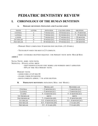
  • 1. 1 PEDIATRIC DENTISTRY REVIEW I. CHRONOLOGY OF THE HUMAN DENTITION A. PRIMARY DENTITION: INITIATION AND CALCIFICATION TOOTH LETTER INITIATION CALCIFICATION ERUPTION CENTRAL A 7 WEEKS (IU) 14 WEEKS (IU) ~ 6 - 9 MONTHS LATERAL B 7 WEEKS (IU) 16 WEEKS (IU) ~ 7 - 10 MONTHS CANINE C 7 WEEKS (IU) 17 WEEKS (IU) ~ 18 MONTHS 1ST MOLAR D 8 WEEKS (IU) 15 WEEKS (IU) ~ 14 MONTHS 2ND MOLAR E 10 WEEKS (IU) 19 WEEKS (IU) ~ 24 MONTHS - PRIMARY ROOT COMPLETION 18 MONTHS POST ERUPTION. (12-18 MOS.) - TEETH ERUPT WHEN THE ROOT IS 2/3 COMPLETE. - MOST FAVORABLE ERUPTION SEQUENCE FOR PRIMARY TEETH BOTH MAX & MAN: ABDCE NATAL TEETH - BORN WITH TEETH. NEONATAL - 30 DAYS AFTER BIRTH - DON'T REMOVE.(UNLESS VERY MOBILE AND WORRIED ABOUT ASPIRATION - 85% OF TIME TRUE PRIMARY TEETH. PRIMARY TEETH: - GERMS FORM AT 6-8 WKS IU - ENAMEL FORMS 4-6 MONTHS - ROOTS COMPLETE APPROX 1 YR AFTER ERUPTION B. PERMANENT DENTITION: INITIATION ( MAX AND MAND ) MAXILLARY MANDIBULAR CENTRAL ~ 5 MONTHS (IU) ~ 5 MONTHS (IU) LATERAL ~ 5 MONTHS (IU) ~ 5 MONTHS (IU) CUSPID 5 - 6 MONTHS (IU) 5 - 6 MONTHS (IU) 1ST PREMOLAR BIRTH BIRTH 2ND PREMOLAR 7 - 8 MONTHS (PP) 7 - 8 MONTHS (PP) 1ST MOLAR 3.5 - 4 MONTHS (IU) 3.5 - 4 MONTHS (IU) 2ND MOLAR 8.5 - 9 MONTHS (PP) 2 YEARS (PP) 3RD MOLAR 3.5 - 4 YEARS (PP) 5 YEARS (PP)
  • 2. 2 C. PERMANENT DENTITION CALCIFICATION: MAXILLARY MANDIBULAR CENTRAL 3 - 4 MONTHS 3 - 4 MONTHS LATERAL 10 - 12 MONTHS 3 - 4 MONTHS CUSPID 4 - 5 MONTHS 10 -12 MONTHS 1ST PREMOLAR 1.5 - 2 YEARS 1.5 - 2 YEARS 2ND PREMOLAR 2 - 2.5 YEARS 2 - 2.5 YEARS 1ST MOLAR BIRTH BIRTH 2ND MOLAR 2.5 - 3 YEARS 2.5 - 3 YEARS 3RD MOLAR 7 - 9 YEARS 8 - 10 YEARS D. PERMANENT DENTITION ERUPTION SEQUENCE: - MAX: 61245378 - MAN: 61234578 EMERGENCE WHEN 2/3 ROOT PRESENT ROOT COMPLETION 3 YEARS POST ERUPTION (2-3 YRS.) ERUPTION SEQUENCE: - MANDIBULAR INCISORS - 1ST PERMANENT MOLARS, UPPER INCISORS - CANINE (MAND) - 1ST PREMOLAR - MAX CANINE - 2ND PREMOLAR ALSO: MAXILLARY: 1ST MOLAR, CI, LI, 1ST PM, 2ND PM, CUSPID, 2ND MOLAR, 3RD MOLAR MANDIBULAR: 1ST MOLAR, CI, LI, CUSPID, 1ST PM, 2ND PM, 2ND MOLAR, 3RD MOLAR PREMATURE ERUPTION: - NOT ENOUGH ROOT FOR STABILITY IN PRIMARY TOOTH - GROSS CARIES PRIMARY MOLARS, LOST EARLY. - ABSCESS CAUSES BONE RESORPTION BENEATH PRIMARY TOOTH RESULTS IN EARLY LOST OF PRIMARY TOOTH AND PREMATURE ERUPTION OF PERMANENT REVIEW OF EMBRYOLOGY 1. INITIATION (BUD STAGE) A. PRIMARY DENTITION: 6-8 WKS IN UTERO
  • 3. 3 B. PERMANENT DENTITION: FROM 5 MOS IN UTERO (ALL ANTERIORS, 1ST BICUSPIDS, 1ST MOLAR) TO 5 YRS OF AGE (THIRD MOLARS) 2. PROLIFERATION (CAP STAGE) 3. MORPHODIFFERENTIATION / HISTODIFFERENTIATION (BELL STAGE) 4. APPOSITION 5. CALCIFICATION VARIABLES THOUGHT TO INFLUENCE PERMANENT TOOTH ERUPTION 1. GENETIC A. FAMILIAL B. RACE (BLACKS SLIGHTLY EARLIER THAN WHITES) C. SEX (FEMALES SLIGHTLY EARLIER THAN MALES) 2. ENVIRONMENTAL A. LOW BIRTH WEIGHT BABIES HAVE DELAYED ERUPTION, PARTICULARLY IN THE PRIMARY DENTITION. B. NUTRITION HAS LITTLE OR NO EFFECT ON ERUPTION 3. SYSTEMIC FACTORS/HERITABLE CONDITIONS THAT AFFECT EXFOLIATION/ERUPTION (ALL TEETH AFFECTED) A. ENDOCRINE - DELAYED EXFOLIATION/ERUPTION - HYPOPITUITARISM - HYPOTHYROIDISM - LOW LEVELS OF HUMAN GROWTH HORMONE B. ENDOCRINE - PREMATURE EXFOLIATION - HYPERPITUITARISM - HYPERTHYROIDISM C. OTHER - PREMATURE EXFOLIATION - CHERUBISM - JUVENILE PERIODONTITIS - PAPILLON LEFEVRE SYNDROME - HISTIOCYTOSIS X - LEUKEMIA - CYCLIC NEUTROPENIA II. ORTHODONTICS AND OCCLUSION REVIEW OCCLUSION CLASSIFICATION THE LOWER FACE IS THE LAST PART OF THE HEAD TO COMPLETE ITS GROWTH.
  • 4. 4 - PRIMARY 2ND MOLAR TERMINAL PLANE RELATIONSHIP ( 75 % OF FIRST MOLAR END TO END RELATIONSHIPS WILL DEVELOP INTO CLASS 1) PRIMARY TERMINAL PLANE 49% MESIAL STEP 37% FTP 14 % DISTAL STEP INITIAL PERMANENT MOLAR RELATIONSHIP 1% CLASS III 27% CLASS I 49% END ON 23% CLASS II FINAL OCCLUSION 3% CLASS III 59% CLASS I 39% CLASS II LEEWAY SPACE: DIFFERENCE BETWEEN THE SUMS OF THE MESIAL - DISTAL WIDTHS OF C, D, AND E AND THE SUCCEEDING PERMANENT TEETH 3, 4 AND 5. - 0.9 MM /QUADRANT IN MAX (PER SIDE) - 1.7 MM /QUADRANT IN MAND (PER SIDE) PRIMATE SPACES: - MESIAL TO PRIMARY MX CANINE AND DISTAL TO PRIMARY MAND CANINES - EARLY SHIFT ( 1ST PERM MOLARS CLOSE POSTERIOR SPACES ) - LATE SHIFT INTO CLASS I HAPPENS WITH LEEWAY SPACE. INCISOR LIABILITY: DIFFERENCE IN SIZE OF PERMANENT INCISORS VS PRIMARY INCISORS - - MAX: 7.6 MM TOTAL - MAND 6.0 MM ARCH CIRCUMFERENCE GREATEST AT AGE THREE CROSS-BITE - ANTERIOR PRIMARY DENTITION, WAIT UNTIL TRANSITION TO TREAT - POSTERIOR IN PRIMARY DENTITION : TREAT IN PRIMARY DENTITION 1. UNILATERAL, FUNCTIONAL ( MIDLINE SHIFT FROM CR TO CO ) 2. BILATERAL, NONFUNCTIONAL: ( NO MIDLINE SHIFT ) - TREATMENT APPLIANCES 1. RPE'S ( 2-4 WEEKS VS SLOW ( 2-6 MONTHS ) 2. SLOW: W-ARCH ( PORTER APPLIANCE ), QUAD HELIX - MID PALATAL SUTURE FUSES IN EARLY TEENS ( 13-16 ) - ANTERIOR CROSSBITE IN PERMANENT DENTITION : ORTHO EMERGENCY SERIAL EXTRACTION - NORMAL EXTRACTION SEQUENCE IS C'S, D'S AND THEN 4'S
  • 5. 5 - TWEED VARIATION: D'S, C'S AND THEN 4'S - INDICATIONS: CLASS I MOLARS AND CANINES SEVERE CROWDING ( GREATER THAN 6 MM PER QUADRANT ) NORMAL OVERBITE AND OVERJET (2 MM EACH) NORMAL ERUPTION SEQUENCE 7-8 YEARS OLD FULL PROFILE - CAN BE INDICATED IN A 7-8 YEAR OLD WHEN FULL FACE BIMAXILLARY PROTRUSION IS FOUND - TREATMENT RESULTS IN INCREASED OVERBITE (GUARD AGAINST LINGUAL DUMPING OF INCISORS) COMPLICATIONS: 1. NOT A CLASS I CASE 2. MISDIAGNOSIS 3. LACK OF FOLLOW UP (COMPLETION OF CARE) ECTOPIC ERUPTION - MOST COMMON: LOWER PERMANENT INCISORS 1. LINGUAL TO PRIMARY INCISORS 2. LATERAL: CAUSES RESORPTION OF PRIMARY CANINES - PERMANENT FIRST MOLARS; INCIDENCE 2-3% ( MOST IN MAXILLA ) 1. 60% SELF CORRECT. 2. IF NOT CORRECTED BY 7-8, ACTIVE TREATMENT IS NEEDED- -EXT 2ND PRIMARY MOLAR, HAWLEY WITH FINGER SPRING. - PUSH 1ST PERMANENT MOLAR BACK. SPACE ANALYSIS: (1) MOYERS: - PREDICTS SPACE FOR BOTH THE MANDIBLE AND MAXILLA - PREDICTS THE SIZES OF THE CUSPIDS AND PREMOLARS - MEASURE WIDTH OF 4 MAND INCISORS. - PREDICT SPACE NEEDED FOR PERM CANINE, 1ST AND 2ND BICUSPID. - MEASURE EXISTING ARCH CIRCUMFERENCE. - 75 PERCENTILE, PREDICTS CROWDING, 75% HAVE SMALLER TEETH. - IT IS INCORRECT 15% OF THE TIME IF THE 85% PROBABILITY CHART IS USED (2) TINAKA-JOHNSON: - MEASURE MANDIBULAR CENTRAL AND LATERAL (ONE SIDE). - MAND ADD 10.5 MM TO GET SPACE NEEDED FOR 345. - MAX ADD 11.5 MM. (3) OTHERS:
  • 6. 6 NANCE. HIXON & OLDFATHER. CAREY. FOSTER. MORE OF SPACE DISCREPANCY MAND THAN MAX: - GOOD, GETS 1ST MOLARS FORWARD A LITTLE INTO CLASS I. SKELETAL AGE ASSESSMENT CARPAL RADIOGRAPH: GROWTH SPURT: - PRESENCE OF RADIAL SESAMOID BONE (SHORT BONE). - CURVING OF EPIPHYSIAL LINES AT END OF LONG BONES. GROWTH AND DEVELOPMENT CLEFT LIP AND PALATE: - PRIMARY PALATE FORMS 4-6 WEEKS IU. - SECONDARY PALATE FORMS 8-12 WEEKS IU. GROWTH SPURT HAS A GREAT DEAL OF INDIVIDUAL VARIATION, BUT TENDS TO OCCUR NEARLY 2 YEARS EARLIER ON THE AVERAGE IN GIRLS THAN BOYS. PUBERTY IN BOYS STARTS LATER AND EXTENDS OVER A LONGER PERIOD OF TIME, ABOUT 5 YEARS AS COMPARED TO 3.5 YEARS FOR GIRLS. INTERCANINE WIDTH DOES NOT INCREASE IN GIRLS AFTER THE AGE OF 9. SPACE MAINTENANCE (1) BAND AND LOOP. (2) LINGUAL ARCH WIRE: - 1ST MOLAR TO 1ST MOLAR. - PUT SPUR DISTAL TO CUSPID, PREVENTS DISTALIZING CUSPID AS PERM INCISORS ERUPT. (3) GOOD CLASS I MOLAR RELATIONSHIP MAY NOT NEED SPACE MAINT. (4) TAKE OUT PRIMARY CANINES: - PUT IN LING ARCH WIRE. - IF LING ERUPTION OF INCISORS REPLACE ARCH WIRE. (5) DISTAL SHOE: - IMPORTANT SPACE MAINTENANCE APPLIANCE. - WHEN 1ST MOLAR ERUPTS BREAK OFF TONGUE AND USE AS BAND AND LOOP.
  • 7. 7 - CONTRAINDICATED IF PATIENT REQUIRES SBE PROPHYLAXIS MISSING TEETH - PRIMARY TOOTH MISSING HAVE INCREASED INCIDENCE OF MISSING PERMANENT TOOTH. - MOST COMMON IS MAND BICUSPID, NEXT MAX LAT, MAX 2ND BI, MAND LAT. - RETAINED PRIMARY MOLAR WILL LAST INTO 40'S. III. FLUORIDES DIETARY FLUORIDE SUPPLEMENTATION SCHEDULE American Academy of Pediatric Dentistry & American Dental Association's Council on Dental Therapeutics Age Less than 0.3 ppm F 0.3 - 0.6 ppm F More than 0.6 ppm F Birth - 6 mos 0 0 0 6 mos - 3 yrs 0.25 mg 0 0 3 yrs - 6 yrs 0.50 mg 0.25 mg 0 6 yrs up to at least 16yr 1.0 mg 0.50 mg 0 • Evaluate patient's fluoride exposure before prescribing appropriate fluoride therapy. Document the patient's fluoride exposure in the record and review at periodic visits. Have a water analysis done prior to issuing supplemental fluoride. • Point-of-use water conditioning/filtration systems significantly reduce fluoride content. Activated carbon filters reduce fluoride concentration by 81%. Bottled water can have variable fluoride concentrations. • Preschool children require supervision during tooth brushing. No more than a pea size amount of toothpaste should be used, avoid swallowing the paste and rinse after brushing. • Fluoride mouth rinses containing 0.05% or 0.2% should not be used by children under the age of six years or those who are unable to adequately expectorate. • Supervise professionally applied topical fluoride treatments. Use 5 - 10 ml of fluoride gel in well fitting trays, have the patient lean forward in the dental chair, admonish not to swallow, and use a saliva ejector positioned between the trays. Fluoride Toxicity:
  • 8. 8 1. Chronic (results in fluorosis) - a. Dean's Classification: 0-4 scale 0- (None)59% of population at optimal fluoride level will exhibit no fluorosis. 1- (Very mild) 7.4% of population at optimal dose 2- (Mild) Greater than 25%, less than 50% of surface with opaque defects greater than 1.2 ppm 3- (Moderate) Brown stains present ( greater than 2 ppm) 4- (Severe) Pitting and attrition problems ( reater than 2-3 ppm ) b. Fluorosis occurs during maturation process. For incisors, age 2-3 years is the critical period. 2. Acute a. CLD ( certain lethal dose ): 32-64 mg/kg in one source, 71-140 mg/kg in another source. A death has been reported at 17 mg/kg. b. PTD ( Probable toxic dose ) 5mg/kg i.e. 50 mg for the average 10 kg 2 year old. c. STD ( Safely tolerated dose ) 8-16 mg/kg d. Toxicity at low doses is due to gastric irritation. Hypocalcemia and hyperkalemia at high doses leads to cardiovascular collapse and death. e. Treatment for overdose: - less than 5mg/kg: oral calcium ( milk) and observe - More than 5 but less than 15 mg/kg: induce vomiting, oral calcium (milk, calcium gluconate, or calcium lactate), admit to hospital - More than 15 mg/kg: admit to hospital immediately, induce vomiting, monitor cardiac function, 10% calcium gluconate solution IV, monitor electrolytes. F. - COMMON SIGNS AND SYMPTOMS OF ACUTE FLUORIDE TOXICITY: LOW DOSAGES HIGH DOSAGES NAUSEA 3-C'S VOMITING CONVULSIONS HYPERSALIVATION CARDIAC ARRHYTHMIA ABDOMINAL PAIN COMATOSE DIARRHEA - PT BECOMES HYPOCALCEMIC DUE TO CA BINDING WITH F. CALCULATING MG F IN COMPOUNDS: 1ST STEP: FIND % F IN COMPOUND. NAME MOLECULAR WEIGHT RATIO APF-ACIDULATED PHOSPHATE FLUORIDE 1
  • 9. 9 NAF-SODIUM FLUORIDE 1/2.2 OR 1/2 SNF2-STANNOUS FLUORIDE 1/4.1 OR 1/4 MFP-SODIUM MONOFLUOROPHOSPHATE 1/7.6 OR 1/8 MULTIPLY % COMPOUND BY MOLECULAR WEIGHT RATIO = % F. 2ND STEP: EXPRESS % F IN COMPOUND IN MG F/ML. BY DEFINITION % SOLUTION IS GRAMS/100ML. GRAMS/100ML = 1000 MG/100ML = 10 MG/ML. MULTIPLY STEP 1 BY 10 = MGF/ML. 3RD STEP: MULTIPLY STEP 2 BY ML INGESTED TO GET MG F INGESTED. FLUORIDES : - 1.23% APF X 1 = 1.23% F X 10 = 12.3 MGF/ML. - .2% NAF X 1/2 = .1% F X 10 = 1 MGF/ML. - 2% NAF X 1/2 = 1% F X 10 = 10 MGF/ML. - .4% SNF2 X 1/4 = .1% F X 10 = 1 MGF/ML. - 10% SNF2 X 1/4 = 2.5% F X 10 = 25 MGF/ML. - COMPARE WITH CLD AND STD FOR WEIGHT OF INDIVIDUAL. WATER FLUORIDATION: - DECREASE IN CARIES: PRIMARY = 40%, SECONDARY = 50-60% MECHANISM OF ACTION OF FLUORIDES: - DECREASES ENAMEL SOLUBILITY. - IMPROVES CRYSTALINITY. - PROMOTES REMINERALIZATION. - DECREASES FREE SURFACE ENERGY OF BACTERIA SO IT CANNOT STICK ON TOOTH. - BACTERIOCIDAL OR STATIC. - CAUSES DEVELOPING CRYSTAL TO GET BIGGER, LESS SOLUBLE FLUORIDE PROTOCOL: - HOME USE IN TRAYS, TOPICAL. - H2O SUPPLY. - SYSTEMIC ALSO TOPICAL WHEN CHEWING TABLET. - HIGH FREQUENCY LOW DOSE IS EFFECTIVE
  • 10. 10 PEDIATRIC DENTAL EMERGENCIES TYPES - PAIN, SWELLING/INFECTION, TRAUMA BEFORE YOU DO ANYTHING 1. GOOD MEDICAL HISTORY A. CONGENITAL HEART PROBLEMS (SBE PROPHYLAXIS) B. CHRONIC ILLNESS (HX ORGAN TRANSPLANT, LEUKEMIA, HEMOPHILIA, ETC.) C. MEDICATIONS/ALLERGIES 2. RULE OUT CHILD ABUSE 3. DOCUMENT EVERYTHING EXTREMELY WELL, PARTICULARLY TRAUMA. DIAGNOSIS - WHICH TOOTH IS IT? 1. WHAT NOT TO DO A. VITALOMETER B. HEAT/ICE TESTS 2. WHAT TO DO A. MUST RELY ON CLINICAL SIGNS AND SYMPTOMS - NO GOOD TESTS AVAILABLE B. DO GET A PA RADIOGRAPH - DEEP CARIES APPROACHING PULP - INTERNAL/EXTERNAL RESORPTION - APICAL OR FURCAL RADIOLUCENCIES C. CHECK FOR MOBILITY. ALMOST ALL PULPALLY INVOLVED TEETH ARE MORE MOBILE THAN THE UNAFFECTED TOOTH ON THE OTHER SIDE. D. PERCUSSION SENSITIVE SOMETIMES CARIES IN THE PRIMARY DENTITION 1. GENERAL INFORMATION A. OCCLUSAL SURFACES OF PRIMARY MOLARS ARE GENERALLY MORE RESISTANT TO DECAY THAN PERMANENT MOLARS B. PRIMARY TEETH HAVE PROPORTIONALLY MORE PULP AND LESS ENAMEL AND DENTIN C. PRIMARY TEETH HAVE SIGNIFICANTLY LESS ENAMEL AND DENTIN PROTECTING THE PULP D. PRIMARY DENTINAL TUBULES ARE LARGER, RELATIVELY MORE POROUS THAN THOSE IN PERMANENT DENTIN E. PROXIMAL CARIES PROGRESSES MORE RAPIDLY THAN OCCLUSAL CARIES AND CAUSES A HIGHER PERCENTAGE OF PULPAL EXPOSURES. F. IF THE PROXIMAL DECAY HAS PROGRESSED (RADIOGRAPHICALLY) 1/2 WAY TO THE DEJ OR FURTHER, THE TOOTH MUST BE RESTORED (UNLESS EXFOLIATION IS IMMINENT) G. USUALLY, IF THERE IS PAIN IT WILL ALMOST ALWAYS BE A PRIMARY MOLAR - ANTERIOR TEETH ABSCESS W/O PAINFUL SYMPTOMS H. RADIOGRAPHIC EVIDENCE OF PULPAL INVOLVEMENT ON PRIMARY MOLAR WILL OCCUR
  • 11. 11 IN THE FURCA AREA, APICALLY ON ANTERIOR TEETH 2. BABY BOTTLE TOOTH DECAY (BBTD) - ETIOLOGY A. CHILD PLACED IN BED AT NIGHT AND/OR NAP TIME WITH BOTTLE OF MILK OR SWEETENED LIQUID B. THE CHILD DOES NOT SWALLOW THE LAST MOUTHFUL OF LIQUID BEFORE HE/SHE FALLS ASLEEP AND THE LIQUID POOLS AROUND THE MAXILLARY TEETH C. THE SWEETENED LIQUID PROVIDES AN EXCELLENT CULTURE FOR ACIDOGENIC BACTERIA D. THE TONGUE PROTECTS THE MAND INCISORS = KEY TO DIAGNOSIS: MAND INCISORS ARE CARIES FREE CHARACTERISTICS OF INFECTIONS IN CHILDREN 1. CAN SPREAD RAPIDLY THROUGH BONE DUE TO LARGE MARROW SPACES 2. CAN SPREAD BEYOND POINTS OF MUSCLE INSERTIONS TO DEEPER AREAS 3. DIFFUSE CELLULITIS IS THE NORM - RARELY MUCH PUS PRODUCTION - USUALLY NOTHING TO I & D 4. MAXILLARY TEETH - MAXILLARY CANINES AND MOLARS CAN INVOLVE THE PERIORBITAL AREA - THIS CAN LEAD TO CAVERNOUS SINUS THROMBOSIS, BRAIN ABSCESSES, SEPTICEMIA, ETC., (ALL POTENTIALLY FATAL COMPLICATIONS) 5. MANDIBULAR TEETH - 1ST AND 2ND PRIMARY MOLAR CAN SEE VESTIBULAR SWELLING OR BUCCAL SPACE INFECTION - RARELY SEE SUBMANDIBULAR INFECTIONS * IF YOU CANNOT FEEL THE INFERIOR BORDER OF THE MANDIBLE, YOU ARE IN TROUBLE * IF THE CHILD COMPLAINS OF DYSPHAGIA, YOU ARE IN TROUBLE ( INFECTION HAS SPREAD BELOW THE INSERTION OF THE MYLOHYOID. MYLOHYOID ELEVATES WHEN YOU SWALLOW, THUS THE DYSPHAGIA. 6. TREATMENT - ANTIBIOTICS - PCN STILL DRUG OF CHOICE (FAIRLY NARROW SPECTRUM, LOW TOXICITY, BACTERICIDAL, PROVEN USE, CHEAP) - ERYTHROMYCIN POOR SECOND CHOICE FOR PCN ALLERGIC TRAUMA 1. THREE PEAK PERIODS FOR INJURIES A. 1 1/2 - 2 1/2 YRS OF AGE B. 5 -6 YRS OF AGE C. 8 - 9 YRS OF AGE 2. ACCURATE HISTORY OF INJURY
  • 12. 12 A. WHERE DID IT OCCUR (CLEAN VS DIRTY) (LAST TETANUS ?) B. WHEN (SOME INJURIES ARE TIME SENSITIVE) C. HOW (MAKE SURE HISTORY AND EXPLANATION MATCH UP) 3. EXAMINATION A. EXAMINE TEETH LAST B. CHECK FOR SIGNS OF CNS DAMAGE (I.E. SUBDURAL HEMATOMA, SKULL FRACTURE, SEVERE CONCUSSION) - AMNESIA - LOSS OF CONSCIOUSNESS - NOT ALERT, NOT ORIENTED TO TIME AND PLACE, EXTREME LETHARGY - IRRITABILITY, DRAMATIC CHANGE IN PERSONALITY - MUSCLE WEAKNESS OF ONE SIDE OR ONE ARM, ONE LEG ETC. - PUPIL DIFFERENCES - NAUSEA AND VOMITING C. IF YOU SEE ANY OF THESE , IT USUALLY WARRANTS IMMEDIATE REFERRAL TO MEDICAL FACILITY - TAKES PRECEDENCE OVER DENTAL TRAUMA D. EXAMINE FOR FACIAL FRACTURES - RARE IN CHILDREN DUE TO LARGE MARROW SPACES, BONE IS LESS DENSE, MUCH LESS BRITTLE - MOSTLY GREENSTICK FRACTURES - ORDER OF FREQUENCY - NASAL, MANDIBULAR (INCLUDING CONDYLES), ZYGOMAXILLARY COMPLEX E. MAXILLARY FRACTURES - ALMOST NEVER SEE TRUE LAFORTE FRACTURES IN CHILDREN DUE TO SMALL SINUSES UNTIL AGE 10 12 F. MANDIBULAR FRACTURES - SIGNS AND SYMPTOMS SAME AS ADULT - PAIN, MALOCCLUSION, SUBLINGUAL HEMATOMA, GINGIVAL TEARS, SWELLING - PALPATE ALL AREAS FOR “STEPS” - 66% IN CONDYLAR AREA - 34% IN BODY - CONDYLAR FRACTURES CAN BE ASSOC WITH FXS OF THE BODY OF THE MANDIBLE ON THE CONTRALATERAL SIDE G. CHIN TRAUMA - CLASSIC TRIAD WITH CHIN TRAUMA * SYMPHYSEAL FRACTURE * POSTERIOR CROWN FRACTURES * CONDYLAR FRACTURE - POSSIBILITY OF CERVICAL VERTEBRAL FX ALSO H. SIGNS AND SYMPTOMS OF CONDYLAR FRACTURE - PAIN - DEVIATION TO AFFECTED SIDE - LIMITATION OF OPENING
  • 13. 13 SOFT TISSUE INJURIES 1. IF DIRTY WOUND. ASK ABOUT TETANUS STATUS A. CLEAN = ANY WOUND THAT CAN BE MECHANICALLY DEBRIDED AND SCRUBBED CLEAN; I.E. SURFACE ABRASIONS, SUPERFICIAL LACERATIONS B. DIRTY = DEEP LACERATIONS, PUNCTURE WOUNDS, FALLING IN MANURE, REIMPLANTING TEETH THAT HAVE BEEN ON THE GROUND, ETC. 2. LACERATIONS A. LOCAL ANESTHESIA AS NECESSARY B. CLEANSE WOUND C. TO R/O FOREIGN BODY IN WOUND (PARTICULARLY IF THERE ARE ROOT FRAGMENTS THAT ARE UNACCOUNTED FOR) PALPATE THEN TAKE RADIOGRAPH OF LACERATED AREA D. ACHIEVE HEMOSTASIS W/ RESORBABLE SUTURES (4-0 OR 5-0 VICRYL) INTRA ORALLY (CAN USE SILK); NYLON ON SKIN, BURY KNOTS INTRAORALLY WHEN POSSIBLE E. FRENUM TEARS - VERY COMMON IN TODDLERS - MOST DO NOT REQUIRE SUTURES UNLESS YOU CANNOT OBTAIN HEMOSTASIS - IN CHILDREN NOT YET WALKING OR CRAWLING, STRONG POSSIBILITY OF FORCED FEEDING, OTHER INAPPROPRIATE TREATMENT = CHILD ABUSE F. TONGUE LACERATIONS - EVERYONE’S WORST NIGHTMARE - NO EASY WAY TO TREAT IF SUTURES ARE REQUIRED AND THEY USUALLY ARE - DIRECT INJECTION INTO TONGUE AROUND WOUND ARE USUALLY BEST - “HOLD ‘N GO” G. DEGLOVING INJURIES - SUTURE BACK INTO POSITION IF ACCORDION TYPE INJURY - IF TISSUE IS LOST, AREA WILL GRANULATE IN BUT THESE CAN GET INFECTED - CONSIDER AB COVERAGE FOR 7 - 10 DAYS H. ELECTRICAL BURNS - TISSUE DAMAGE IS ENORMOUS; MUCH MORE THEN WHAT IS VISIBLE - INJURIES INVOLVE MUCOSA, SUBMUCOSA, MUSCLE, NERVE AND VASCULAR TISSUE - ALL NECROTIC TISSUE, NO PAIN - NECROTIC TISSUE WILL SLOUGH IN 10 - 21 DAYS - CAN LEAD TO LABIAL ARTERY BLEED - TREMENDOUS SCAR FORMATION; UNTREATED WILL RESULT IN SEVERE MICROSTOMIA - APPLIANCE PLACED AFTER TISSUE SLOUGHS WILL ALLOW SCAR TO FORM AROUND A MATRIX; CAN SIGNIFICANTLY DECREASE THE AMOUNT OF DISFIGURATION. MAINTAINS COMMISSURAL REGIONS EQUIDISTANT TO MIDLINE - APPLIANCES MUST BE WORN 24 HRS/DAY FOR 6 - 18 MONTHS DEPENDING ON SEVERITY OF INJURY IV. GUIDELINES FOR PRESCRIBING DENTAL RADIOGRAPHS (NO PREVIOUS RADIOGRAPHS)
  • 14. 14 AGE 3-5 6-7 8-9 10-12 CONSIDERATIONS NO APPARENT ABNORMALITIES (OPEN CONTACTS) NO APPARENT ABNORMALITIES (CLOSED CONTACTS) EXTENSIVE CARIES DEEP CARIES NO APPARENT ABNORMALITIES EXTENSIVE OR DEEP CARIES NO APPARENT ABNORMALITIES OR DEEP CARIES NO APPARENT ABNORMALITIES OR DEEP CARIES RADIOGRAPHS NONE 2 BW'S (SIZE 0) 4 FILM SURVEY SELECTED PA'S AND THE 4 FILM SURVEY 8 FILM SURVEY SELECTED PA'S AND 8 FILM SURVEY 12 FILM SURVEY 12 OR 16 FILM SURVEY
  • 15. 15 THE ABOVE RECOMMENDATION IS FROM MCDONALD'S 5TH EDITION PAGE 7 ACCORDING TO INTERORGANIZATIONAL CONFERENCE, INCLUDING THE ADA AND THE AMERICAN ACADEMY OF PEDIATRIC DENTISTRY IN 1992-93: PATIENT CATEGORY PRIMARY DENTITION (PRIOR TO 1ST PERM MOLAR) NO CLINICAL CARIES POSTERIOR BW EXAMINATION AT 12-24 MONTH INTERVALS IF PROXIMAL SURFACES OF PRIMARY TEETH CANNOT BE VISUALIZED OR PROBED TRANSITIONAL DENTITION (FOLLOWING ERUPTION OF 1ST PERM MOLAR POSTERIOR BW EXAMINATION AT 12 - 24 MONTH INTERVALS ADOLESCENT, PERMANENT TEETH (PRIOR TO THIRDS) POSTERIOR BW EXAMINATION AT 24 - 36 MONTH INTERVALS CLINICAL CARIES OR POSTERIOR BW EXAMINATION AT 6 MONTHS INTERVALS OR UNTIL NO HIGH RISK FACTORS CARIOUS LESIONS ARE EVIDENT IN PRIMARY AND TRANSITIONAL FOR CARIES DENTITIONS. POSTERIOR BW EXAMINATION AT 6 - 12 MONTH INTERVALS FOR PERMANENT TEETH IN ADOLESCENTS AND 12 - 18 MONTHS IN ADULTS OR UNTIL NO CARIOUS LESIONS ARE EVIDENT V. PEDIATRIC PATHOLOGY A. INCLUSION CYSTS - ALL SHED SPONTANEOUSLY KERATIN-FILLED NODULES: (1) EPSTEIN'S PEARLS - TRAPPED EPITHELIUM IN MIDPALATINE RAPHE. - 100% HAVE CLINICAL OR SUBCLINICAL EPSTEIN'S PEARLS. - NOT RARE. (2) BOHN'S NODULES - TRAPPED MUCOUS GLAND TISSUE ON BUCCAL AND LINGUAL OF RIDGE - LABIAL/LINGUAL OF ALVEOLAR RIDGE. (3) DENTAL LAMINA CYST - REMNANTS OF DENTAL LAMINA ON CREST OF RIDGE
  • 16. 16 B. LYMPHANGIOMAS OF ALVEOLAR RIDGE - 1-3% BLACKS. - BLUE DOMED, FLUID FILLED LESION ON THE ALVEOLAR RIDGE - TX: OBSERVATION FOR SPONTANEOUS REGRESSION. EXCISION AFTER SIX MONTHS. C. CONGENITAL EPULIS - PEDUNCULATED LESION ( MAY BE SEVERAL CM IN DIAMETER ) ON RIDGE IN AREA OF MAX INCISORS. - SPONTANEOUS REGRESSION WITHIN FIRST YEAR IS COMMON. OBSERVATION IS INDICATED. - IF SURGERY IS INDICATED DUE TO FEEDING PROBLEM , WAIT UNTIL AFTER 8-10 DAYS OF AGE D. MEDIAN ALVEOLAR NOTCH -NOTCH IN THE MX ALVEOLUS, NOT A TRUE CLEFT ( CONNECTIVE TISSUE DEFECT ) - MORE FREQUENT IN BLACKS - MAY RESULT IN DIASTEMA E. NEUROECTODERMAL TUMOR OF INFANCY - HIGHLY PIGMENTED ( MELANOTIC ) BENIGN TUMOR APPEARING WITHIN THE FIRST YEAR OF LIFE, USUALLY IN THE ANTERIOR MAXILLA. - DESTRUCTIVE RADIOLUCENT LESION: FLOATING TEETH TX; CONSERVATIVE EXCISION - HIGH URINARY LEVELS OF VANILMANDELIC ACID ( COMMON WITH NEURAL CREST TUMORS ) F. NATAL( AT BIRTH ) AND NEONATAL ( WITHIN 30 DAYS OF BIRTH ) - USUALLY LOWER PRIMARY INCISORS ( 85% ) RARELY SUPERNUMERARIES - TX: NONE IF TOOTH NOT LIKELY TO EXFOLIATE OR CAUSE ULCERATION - RIGA-FEDA DISEASE: ULCERATION OF VENTRAL SURFACE OF TONGUE DUE TO IRRITATION OF LOWER INCISORS WHEN NURSING. - NO SURGERY UNTIL AFTER 10 DAYS DUE TO HYPERPROTHROMBINEMIA. G. AMELOGENESIS IMPERFECTA (RADIOGRAPHICALLY PULP LOOKS NORMAL) 3 TYPES: (1) HYPOPLASTIC: - MATRIX DID NOT FORM PROPERLY. - THIN ENAMEL, SHOVEL SHAPED, PITS AND DEFECTS. (2) HYPOCALCIFIED: - MATRIX OK, CALCIFICATION NOT PROPER. - SHEARS, BREAKS.
  • 17. 17 - MOTH EATEN X-RAY APPEARANCE. (3) HYPOMATURE:(HYPOMINERALIZED) - QUALITATIVE DEFECT IN ENTIRE CALCIFIED AREA. - MORE RADIOLUCENT. H.. DENTINOGENESIS IMPERFECTA (PULP CHAMBER OBLITERATED RADIOGRAPHICALLY) THREE TYPES: (1) SHIELDS TYPE I: - MOST COMPREHENSIVE, SYSTEMIC PROBLEMS, OSTEOGENESIS IMPERFECTA. - BRITTLE BONE DISEASE. - LATERAL SKULL FILMS SHOW RADIOPACITY. - PRIMARY TEETH WORSE THAN THE PERMANENT (2) SHIELDS TYPE II: - DOES NOT HAVE SYSTEMIC INVOLVEMENT. - BOTH DENTITIONS EQUALLY AFFECTED (3) SHIELDS TYPE III:(BRANDYWINE)(RACIAL ISOLATE IN MARYLAND) - BELL SHAPED CROWNS. - SHELL TEETH. - MULTIPLE PULP EXPOSURES IN PRIMARY TEETH I. PERICORONITIS: - CAN HAVE ON 6 YEAR MOLAR. - IF AN ERUPTION CYST FORMS DISTAL TO THE SECOND PRIMARY MOLAR, THE INITIAL TREATMENT OF CHOICE IN MOST CASES IS TO WAIT AND JUST OBSERVE IT. IN MOST CASES NORMAL ERUPTION WILL TAKE PLACE. IF IT DOES NOT, THAN IT CAN BE INCISED AND THE FIRST MOLAR EXPOSED. J. ERUPTION SEQUESTRUM: - NOT ALL BONE OVER TOOTH RESORBED. K. TURNER TOOTH - HYPOPLASIA OF PERMANENT TOOTH DUE TO INFECTION OR TRAUMA TO THE PRIMARY PREDECESSOR - ETIOLOGY IS A DISTURBANCE TO THE ENAMEL ORGAN DURING FORMATION OF THE PERMANENT CROWN - FACIAL ASPECT OF A MAXILLARY INCISOR IS USUALLY THE AFFECTED AREA L. HIV IN CHILDREN -ETIOLOGY- MOSTLY PERINATAL OTHER: SEXUAL, DRUG ABUSE BREAST MILK RARE
  • 18. 18 -INFANTS USUALLY CONVERT IN 15 MONTHS - MOTHERS TAKING AZT 25% OF NEWBORNS CONVERT, NO AZT 30-50% CONVERT - CLINICAL MANIFESTATIONS ARE FAILURE TO THRIVE, FEVER CHRONIC DIARRHEA, LYMPHADENOPATHY, ORAL CANDIDA, PERSISTENT DIFFUSE PAROTITIS OCCASIONALLY AND ORAL VIRAL LESIONS VI. STAGES OF TOOTH DEVELOPMENT -INITIATION, PROLIFERATION, HISTODIFFERENTIATION, MORPHODIFFERENTIATION, APPOSITION (CALCIFICATION), MATURATION VII ANOMALIES OF TEETH A. INITIATION PHASE -SUPERNUMERARY TEETH: - MORE OFTEN IN PERM DENTITION, MESIODENS IS MOST COMMON, CLEIDOCRANIAL DYSPLASIA - MULTIPLE SUPERNUMERARY TEETH - MISSING TEETH: RARE IN PRIMARY DENTITION -IN PERM DENT (OTHER THAN 3RDS) MAND 2ND BI > MAX 2ND BI > MAX LI, ( SOME DISAGREEMENT ABOUT WHETHER MAX 2ND BI OR MAX LI ) -ECTODERMAL DYSPLASIA - ANODONTIA OR HYPODONTIA B. TWINNING ANOMALIES (PROLIFERATION PHASE) (1) GEMINATION: DIVISION OF SINGLE GERM - BIFID CROWN, SINGLE ROOT (2) FUSION: TWO BUDS, TWO TEETH, SEPARATE PULP CANALS - DENTIN CONFLUENCE, COMMONLY MISSING ONE OF THE SUCCESSORS (3) CONCRESCENCE: CEMENTAL UNION, TWO BUDS, TWO TEETH C. SIZE AND SHAPE - MACRODONTIA, MICRODONTIA, PEG LATERAL, DENS-IN-DENTE - TURNER'S TOOTH: HYPOPLASTIC DEFECT D. ANKYLOSIS OF 1ST PRIMARY MOLAR - TAKE OUT OR NOT? - HOW FAR ERUPTED OR INTRUDED. - STAGE OF ERUPTION. - COOPERATION OF CHILD. - SUBMERGED, TAKE OUT. - DISTAL TIPPING CANINE.
  • 19. 19 - MESIAL TIPPING 2ND MOLAR CAUSES ANKLYLOSED TOOTH TO BE IMPACTED. - CALCIFICATION OF BICUSPID, LOOK AT X-RAY. E. MULTIPLE SUPERNUMERARY TEETH - CLEIDOCRANIAL DYSOSTOSIS. - GARDENERS SYNDROME VIII. PULP THERAPY FOR PRIMARY TEETH RADIOGRAPHIC EVIDENCE OF PULPAL NECROSIS IN PRIMARY POSTERIOR TEETH IS USUALLY OBSERVED IN THE BIFURCATION AREA. A. PULP CAPPING - INDIRECT PULP CAPS: SAME INDICATIONS AND TECHNIQUE IN BOTH PRIMARY AND PERM TEETH. - MINIMUM REENTRY TIME 6-8 WEEKS ( REENTRY IS NOT REQUIRED ) - DIRECT PULP CAPS: GENERALLY DISCOURAGED FOR POSTERIOR TEETH - FOR SMALL NONCARIOUS EXPOSURES IN PRIMARY TEETH - CAOH IS THE MEDICAMENT OF CHOICE ALSO: 1. INDICATIONS A. ASYMPOTMATIC TOOTH B. SMALL, MECHANICAL EXPOSURES C. NORMAL PULPAL RESPONSE - NO PROLONGED HEMORRHAGE D. PERIPHERY OF EXPOSURE CONSISTS OF SOUND DENTIN 2. CONTRAINDICATIONS A. RARELY INDICATED ON PRIMARY TEETH - PULPOTOMY HAS HIGHER SUCCESS RATE B. RADIOGRAPHIC CHANGES C. HEMORRHAGE OR PUS AT EXPOSURE SITE D. CARIOUS EXPOSURE B. PULP TESTING OF PRIMARY TEETH IS UNRELIABLE C. PULPOTOMY: - REMOVAL OF CORONAL PORTION OF EXPOSED PULP AND TX OF EXPOSED SURFACES OF APICAL PULP TISSUE - INDICATIONS: A. SYMPTOMATIC OR ASYMPTOMATIC POSTERIOR PRIMARY TEETH B. INFLAMMATION IS CONFINED TO THE CORONAL PULP NOT EXTENDING INTO THE ROOT CANALS
  • 20. 20 C. NO EVIDENCE OF PATHOLOGIC CHANGES RADIOGRAPHICALLY -CONTRAINDICATIONS: A. EVIDENCE OF FURCAL OR PERIAPICAL PATHOLOGY B. HX OF SPONTANEOUS OR UNPROVOKED PAIN C. INABILITY TO CONTROL HEMORRHAGE FROM PULPAL STUMPS D. LESS THAN 2/3 ROOT REMAINS (TOOTH NEAR EXFOLIATION) E. TOOTH NON-RESTORABLE F. INTERNAL RESORPTION G. PULP WITH SEROUS OR PURULENT HEMORRHAGE H. FISTULA TECHNIQUE: 1. REMOVE ALL CARIOUS DENTIN PRIOR TO ENTERING THE PULP CHAMBER 2. UNROOF THE PULP CHAMBER AND WIDEN ACCESS TO ALLOW GOOD VISUALIZATION OF THE ENTIRE PULP CHAMBER 3. REMOVE THE CORONAL PULP TISSUE DOWN TO THE ORIFICES OF THE CANALS WITH A SHARP SPOON EXCAVATOR OR STERILE ROUND BUR. 4. PRESS DAMP STERILE COTTON PELLETS AGAINST PULP STUMPS TO CONTROL HEMORRHAGE 5. PLACE NEARLY DRY FORMOCRESOL PELLET AGAINST PULP STUMPS FOR 5 MINUTES 6. AFTER REMOVING THE FORMOCRESOL PELLET, HEMORRHAGE SHOULD BE COMPLETELY CONTROLLED. PULP STUMPS SHOULD APPEAR GRAYISH AND DRY. 7. MIX ZINC-OXIDE AND EUGENOL TO MAKE A THICK PASTE AND PLACE OVER PULPAL STUMPS 8. PLACE PERMANENT RESTORATION: RESTORATION OF CHOICE IS SSC - CONCERNS: - CLINICAL SUCCESS 90+ % VS X-RAY AND HISTO SUCCESS - SYSTEMIC ABSORPTION OF FC WITH TISSUE BINDING AND IMMUNE RESPONSE, BUCKLEY'S FC IS 19% - SUGGESTED MODIFICATIONS - USE 1:5 CONCENTRATION OF FC, ( DILUTE WITH GLYCERIN AND WATER ) 3 PARTS GLYCERIN + 1 PART WATER + 1 PART BUCKLEY’S FORMOCRESOL - SHORTER FIXATION TIME - CONSIDER GLUTERALDEHYDE INSTEAD OF FC FORMOCRESOL VS. GLUTARALDEHYDE (1) 1:5 FORMOCRESOL VS GLUTARALDEHYDE (2) WHY NOT FORMOCRESOL? - POSSIBLE EFFECTS ON PERMANENT TEETH. - MAY CAUSE AUTO ANTIBODY FORMATION TO FIXED TISSUE. (3) STUDIES SHOW: - GLUTARALDEHYDE HAS < PERFUSION TO APEX. - GLUTARALDEHYDE HAS > SUPPRESSION OF RESPIRATORY ENZYME (LACTIC DEHYDROGENASE).
  • 21. 21 (4) ADVANTAGES OF GLUTARALDEHYDE: - LESS APICAL DAMAGE OF TISSUE. - LESS NECROSIS. - FIBROBLASTIC PROLIFERATION TISSUE IMMEDIATELY ADJACENT. D. PULPECTOMY/OBTURATION: - THIS IS A VIABLE TREATMENT OPTION FOR PRIMARY TEETH (50-80% SUCCESS) - COMPLICATING FACTORS: - ANATOMY OF CANAL SYSTEM IN PRIMARY TEETH - THINNESS OF ROOT WALLS AND PULPAL FLOOR( EASY PERFS ) - COOPERATION OF CHILD - FILLING MATERIALS - NO SUCCEEDING TOOTH- USE GUTTA PERCHA - WITH SUCCEEDING TOOTH, USE ZOE ( PURE ), NOT IRM OR OTHER REINFORCED CEMENT - CONTRAINDICATED FOR PATIENTS AT RISK FOR ENDOCARDITIS - MUST HAVE 100% APICAL CLOSURE OF ROOT IN ORDER TO ATTEMPT - THERE IS AN INCREASED INCIDENCE OF HYPOPLASIA IN THE PERMANENT SUCCESSOR PARTIAL PULPECTOMY: THE REMOVAL OF ALL ACCESSIBLE PULP TISSUE IN A TOOTH WITH A VITAL BUT IRREVERSIBLE PULP. NO ATTEMPT IS MADE TO PENETRATE THE APICES AND ONE APPOINTMENT TECHNIQUE MAY BE USED. COMPLETE PULPECTOMY: THE REMOVAL OF THE CONTENTS OF THE CORONAL AND RADICULAR PULP IN A TOOTH WITH A NECROTIC PULP. SEVERAL APPOINTMENTS MAY BE REQUIRED AND APICES ARE USUALLY PENETRATED. THE CANALS ARE FILLED WITH A RESORBABLE PASTE. E. APEXIFICATION/APEXOGENESIS: FOR PERMANENT TEETH WITH OPEN APICES - APEXIFICATION- FRANK TECHNIQUE (FOR NON-VITAL TEETH) - APEXOGENESIS - CVEK TECHNIQUE (FOR VITAL TEETH) IX BEHAVIOR MANAGEMENT AGE 1: - COMMUNICATION DIFFICULT. - IN/OUT FAST. AGE 2: - NO IS FAVORITE WORD.
  • 22. 22 - KEEP PROCEDURES SHORT. AGE 3: - PRAISE FOR ROUTINE BEHAVIOR. AGE 4: - FIRMNESS NEEDED. AGE 5: - GOOD AGE. - PRAISE, FIRMNESS, COMPLIMENTS. AGE 6-7: - NEEDS UNDERSTANDING. - SHOW, TELL, DO. AGE 8: - INTELLECTUAL LEVEL. - FIRMNESS. - CLASSIFICATION OF BEHAVIOR - CLINICAL CLASSIFICATION: -COOPERATIVE....PRECOOPERATIVE....POTENTIALLY COOPERATIVE - FRANKL BEHAVIOR RATING SCALE: 1. (--) DEFINITELY NEGATIVE ...REFUSES TREATMENT 2. (-) NEGATIVE.....RELUCTANT, SULLEN ,WITHDRAWN, WHINING 3. (+) POSITIVE ....ACCEPTANCE OF TX BUT RESERVED 4. (++) DEFINITELY POSITIVE ....GOOD RAPPORT WITH DENTIST, INTERESTED IN TX. - INFORMED CONSENT - ALL TREATMENT REQUIRES CONSENT - ALL CONSENT MUST BE INFORMED -PHARMACOLOGICAL APPROACH: (1) INDICATIONS: - VERY YOUNG PATIENT. - EMOTIONALLY DISTURBED OR MENTALLY HANDICAPPED. - HX TRAUMATIC PREVIOUS MED/DENT EXPERIENCE. (2) LIGHT CONSCIOUS SEDATION: - N20 AND O2 (3) MODERATE CONSCIOUS SEDATION: - CHLORAL HYDRATE: - 25-50 MG/KG MAX OF 1GM - 30 MIN ONSET, DURATION 5 HOURS, PEAK EFFECT IN 1 HOUR - NO REVERSAL AGENT AVAILABLE - HIGH DEGREE OF GASTRIC IRRITATION - DEMEROL - MOST COMMON NARCOTIC AGENT
  • 23. 23 - DOSE: 1-2.2 MG/KG NOT TO EXCEED 100 MG ALONE IN COMBO 1 MG/KG - ACTION REVERSED BY NARCAN - ANTIEMETICS AND ANTIHISTAMINES - BENADRYL 1MG/KG WITH NARCOTIC - ATARAX 1-2MG/KG WITH NARCOTIC - PHENERGAN 1MG/KG WITH NARCOTIC (4) MONITORING MINIMAL EQUIPMENT NEEDED IS PRECORDIAL STETHOSCOPE, BPCUFF, PULSE OX. DYNAMAP AND PULSE OX ARE STD OF CARE. (5) CHILDREN ARE NOT LITTLE ADULTS: THEY HAVE LARGE HEADS, SHORT NECKS, LARGE TONGUES MORE INTERNAL FATTY TISSUE NARROW NASAL PASSAGES AND LONG STIFF EPIGLOTTIS. THEY HAVE FEWER LUNG ALVEOLI AND A HIGHER METABOLIC RATE. THE FUNCTIONAL RESIDUAL CAPACITY IS MUCH LOWER IN PEDIATRIC PATIENTS. THEY MUST BE MONITORED CLOSELY. NITROUS OXIDE AND OXYGEN CONSCIOUS SEDATION - ALERT, EASILY AROUSED, PROTECTIVE REFLEXES INTACT DEEP SEDATION - DIFFICULT TO AROUSE, PROTECTIVE REFLEXES DEMINISHED GENERAL SEDATION - UNCONSCIOUS, NO REFLEXES ASA CLASSIFICATION - BASIC CNS MONITORING - ALERT, VERBAL, PAIN, UNRESPONSIVE PROTOCOL FOR THE APPREHENSIVE PATIENT 1. PATIENT/ DOCTOR TRUST 2. TX PLAN 3. PREAPPOINTMENT SEDATION 4. AM APPOINTMENT 5. INTRAOPERATIVE SEDATION 6. INTRAOPERATIVE PAIN CONTROL
  • 24. 24 7. POST-OPERATIVE PAIN CONTROL 8. POST -OPER CONTACT PROPERTIES OF NITROUS OXIDE 1. SWEET SMELLING, COLORLESS, NONIRRITATING 2. NONFLAMMABLE AND NONEXPLOSIVE 3. SPECIFIC GRAVITY OF 1.53 VS 1.0 FOR AIR (WILL SETTLE TO FLOOR) 4. NONALLERGENIC 5. ONLY INORGANIC GAS USED FOR ANESTHESIA CO2 AND N2O ARE THE ONLY INORGANIC COMPOUNDS THAT HAVE CNS DEPRESSANT PROPERTIES POTENCY 1. LEAST POTENT OF ANESTHETIC GASES 2. SECOND STAGE ANESTHESIA RARELY REACHED 3. ANALGESIC EFFECT AT 35% IS SUPPOSEDLY EQUAL TO 10-15 MG MORPHINE 4. CONSCIOUS SEDATION VS DEEP SEDATION PHARMACOLOGY 1. ABSORPTION - RAPIDLY ABSORBED BECAUSE OF A LOW BLOOD/GAS PARTITION COEFFICIENT AND LARGE CONCENTRATION GRADIENT A. RELATIVELY INSOLUBLE IN BLOOD - CARRIED IN PHYSICAL SOLUTION - DOES NOT COMBINE WITH HEMOGLOBIN - 100 TIMES MORE SOLUBLE THAN O2 - REPLACES NITROGEN IN THE BLOOD - N2O IS ABOUT 35 TIMES MORE SOLUBLE THAN NITROGEN - N2O CAN ENTER AIR SPACES MORE QUICKLY THAN NITROGEN LEAVES. THIS CAN CAUSE DISTENTION IN THE MIDDLE EAR, BOWEL, STOMACH, AND PLEURAL SPACES B. FACTORS INVOLVED WITH ABSORPTION CONCENTRATION OF THE GAS IN THE INHALED MIXTURE. THE CONCENTRATION OF THE ANESTHETIC GAS IS PROPORTIONAL TO THE PARTIAL PRESSURE OF THAT GAS. PRESSURE GRADIENT. FLICKS LAW OF MIGRATION OF MOLECULES - A GAS OR VAPOR MIGRATES FROM AN AREA OF HIGH PRESSURE TO ONE OF LESSER PRESSURE AND CONTINUES TO DO SO UNTIL THE PRESSURES ARE EQUALIZED. E.G. OXYGEN EXHIBITS THE FOLLOWING PARTIAL PRESSURES WHICH DRIVE ITS MIGRATION. AIR 152 MMHG, ALVEOLI 105 MMHG, ARTERIAL BLOOD 90 MMHG, TISSUES 0 MMHG SOLUBILITY IN BLOOD. THIS IS THE MOST IMPORTANT SINGLE FACTOR IN DETERMINING
  • 25. 25 RATE OF UPTAKE AND EXCRETION OF VOLATILE ANESTHETICS. - BLOOD/GAS PARTITION COEFFICIENT = OSTWALD COEFFICIENT - DEFINED AS THE RATIO OF DISTRIBUTION OF A GAS OR VAPOR IN IDENTICAL VOLUMES OF A GASEOUS AND LIQUID PHASE EQUILIBRATED AT A GIVEN TEMPERATURE AT ONE ATMOSPHERE ANESTHETIC BLOOD/GAS NITROUS OXIDE 0.47 HALOTHANE 2.4 ETHYL ETHER 12.0 - COEFFICIENT = VOLUME % IN BLOOD / VOLUME % IN ALVEOLI - AGENTS WITH LOW BLOOD/GAS COEFFICIENTS ARE LESS SOLUBLE IN BLOOD - EFFECT OF BLOOD/GAS COEFFICIENT ON INDUCTION AND RECOVERY TIMES LOW COEFFICIENT = RAPID INDUCTION AND RECOVERY HIGH COEFFICIENT = SLOW INDUCTION AND RECOVERY - UPTAKE ABOUT 1L/MIN AT 75% NITROUS OXIDE 2. DISTRIBUTION A. FACTORS ALTERING DISTRIBUTION OF NITROUS OXIDE SOLUBILITY IN TISSUES - TISSUE/BLOOD PARTITION COEFFICIENTS FOR MOST ANESTHETICS ARE SIMILAR - COMPARED TO OTHER ANESTHETICS NITROUS OXIDE IS THE LEAST SOLUBLE IN FAT LOW LIPID SOLUBILITY CONTRIBUTES TO RAPID RECOVERY B. BLOOD SUPPLY TO TISSUES DETERMINES THE RELATIVE EXPOSURE OF THE TISSUES TO THE BLOOD BORNE ANESTHETIC - THE VESSEL RICH GROUP (VRG) INCLUDES THE BRAIN, HEART, KIDNEY AND LIVER - RAPID EQUILIBRATION IN THE VESSEL RICH GROUP OF TISSUES BLOOD EQUILIBRATES IN 3-5 MIN BRAIN EQUILIBRATES IN 8-10 MIN C. NO TRANSFORMATION IN THE BODY 3. ELIMINATION A. RAPID AND COMPLETE RECOVERY. MAJOR PORTION OF GAS ELIMINATED IN 3-5 MIN B. HIGHER GAS CONCENTRATION AND LONG EXPOSURE CAN TEMPORARILY AFFECT FINE MOTOR SKILLS C. DIFFUSION HYPOXIA - HEADACHE, NAUSEA, LETHARGY - WHEN N2O IS TURNED OFF, THE RAPID DIFFUSION OF N2O FROM THE BLOOD INTO THE LUNGS GAS RUSHES FROM THE LUNGS DILUTES THE AVAILABLE OXYGEN.
  • 26. 26 THE N2O GAS RUSHES FROM THE LUNGS TAKING WITH IT A MIXTURE OF THE OTHER GASES (O2, CO2, N) WITH IT. MORE CO2 IS REMOVED THAN NORMAL. THE REDUCED CO2 OR DILUTION OF THE OXYGEN IN THE ALVEOLI MAY DIMINISH THE RESPIRATORY DRIVE AND THE DECREASE IN LUNG OXYGEN CONCENTRATION MAY LEAD TO A TRANSIENT HYPOXIA (DIFFUSION HYPOXIA) UNTIL THE N2O IS ELIMINATED. - TO AVOID A DROP IN THE OXYGEN CONCENTRATION, 100% OXYGEN IS CONTINUED FOR 4 MINUTES FOLLOWING THE TERMINATION OF THE N2O. METABOLISM - NITROUS OXIDE IS NOT METABOLIZED - MOST IS EXHALED IN 3-5 MINUTES WHEN THE GAS IS TERMINATED - ABOUT 1% IS ELIMINATED MORE SLOWLY OVER 24 HOURS THROUGH THE SKIN, BOWEL, AND LUNG MECHANISM OF ACTION OF NITROUS OXIDE IS UNKNOWN EFFECTS ON SYSTEMS 1. CNS - PRIMARY SITE OF ACTION A. CORTICAL DEPRESSION AFFECTING ALL SENSATIONS INCLUDING SIGHT, TOUCH, PAIN. B. POSTREMA (VOMITING CENTER) - AFFECTED ONLY IN PRESENCE OF NITROUS OVERDOSE, HYPOXIA, OR ANOXIA. C. SHORT TERM MEMORY, ABILITY TO PERFORM CALCULATIONS, ABILITY TO CONCENTRATE AFFECTED. 2. SIGNS IN RESPONSE TO NITROUS OXIDE AND OXYGEN CONSCIOUS SEDATION CONCENTRATION OF N2O RESPONSE 10 - 20% BODY WARMTH TINGLING OF HANDS AND FEET 20 - 30% CIRCUMORAL NUMBNESS NUMBNESS OF THIGHS 20 - 40% NUMBNESS OF TONGUE NUMBNESS OF HANDS AND FEET DRONING SOUNDS PRESENT HEARING DISTINCT BUT DISTANT DISSOCIATION BEGINS AND REACHES PEAK MILD SLEEPINESS ANALGESIA (MAX AT 30 - 35%) EUPHORIA FEELING OF HEAVINESS OR LIGHTNESS OF BODY 30 - 50% SWEATING NAUSEA
  • 27. 27 AMNESIA INCREASED SLEEPINESS 40 - 60% DREAMING, LAUGHING, GIDDINESS FURTHER INCREASED SLEEPINESS, TENDING TOWARD UNCONSCIOUSNESS 50% AND OVER UNCONSCIOUSNESS AND LIGHT GENERAL ANESTHESIA 3. CARDIOVASCULAR SYSTEM A. AT NORMAL DOSE (20 - 50%) FOR CONSCIOUS SEDATION, THERE IS NO SIGNIFICANT EFFECT. - NO SIGNIFICANT EFFECT ON BLOOD PRESSURE OTHER THAN SLIGHT DECREASE AS RELAXATION INCREASES - CUTANEOUS VASODILATATION IS OBSERVED. FLUSHING AND PERSPIRATION. B. AT 80% YOU GET A SLIGHT DECREASE IN MYOCARDIAL CONTRACTILITY. 4. RESPIRATORY SYSTEM A. SLIGHT DECREASE IN THE RESTING RESPIRATORY VOLUME B. NO CHANGE IN CO2 RESPONSE C. REDUCED GAG REFLEX 5. NO OTHER SYSTEMS EFFECTED. DOSAGE OPTIMAL CONCENTRATION TO PRODUCE MAXIMAL DEGREE OF ANALGESIA AND YET MAINTAIN THE COOPERATION OF THE PATIENT IS APPROX. 30 - 40% THE CONCENTRATION MAY BE MODIFIED BY: - MOUTH BREATHING DILUTES THE N2O. THIS GIVES THE PATIENT CONTROL OVER THE DEGREE OF DESIRED ANALGESIA. - LEVEL OF ANXIETY - TYPE OF PROCEDURE - PATIENT PAIN THRESHOLD - SYNERGISM WITH OTHER DRUGS -- POTENTIATES COMPLICATIONS OF OTHER DRUGS INDICATIONS - ANXIETY - PAIN CONTROL - EXTENDED APPOINTMENT - MANAGE MEDICALLY COMPROMISED - MI, CVD, RESP, SEIZURES, DIABETIC, GAGGING CONTRAINDICATIONS TO N2O
  • 28. 28 1. UPPER RESPIRATORY INFECTIONS 2. ACTIVE TB OR ANY PULMONARY DISEASE A. EMPHYSEMA - HAVE LOWERED CO2 DRIVE. IF YOU INCREASE THE % OXYGEN, YOU WILL DEPRESS THE DRIVE FOR BREATHING B. ATELECTASIS - AIRLESS STATE OF THE LUNGS DUE TO PRESSURE OF A PLEURAL EFFUSION BLOCKING THE SMALLER BRONCHIAL TUBES WITH MUCOUS. 3. ABNORMAL AIR-FILLED BODY SPACES - PEUMOTHORAX, PNEUMOCARDIUM, COMPLETE BOWEL OBSTRUCTION. 4. PSYCHOTIC - PERSONALITY DISORDERS, PERSONS TAKING MOOD ALTERING ANTIDEPRESSANTS, PERSONS WHO HAVE A TENUOUS GRIP ON REALITY. 5. FIRST TRIMESTER OF PREGNANCY 6. SICKLE CELL ANEMIA 7. MYESTHESIA GRAVIS 8. BLEOMYCIN THERAPY - CANNOT TOLERATE ELEVATED OXYGEN 9. UNCOOPERATIVE PATIENT - MOUTH BREATHER, UNABLE TO ACCEPT MASK (VERY YOUNG OR CLAUSTROPHOBIC) ADVANTAGES OF NITROUS OXIDE SEDATION 1. NONFLAMMABLE 2. NON-IRRITATING 3. NO DELETERIOUS EFFECT ON ORGAN SYSTEMS 4. INEXPENSIVE 5. POST-ANALGESIC NAUSEA AND VOMITING UNUSUAL 6. EXCELLENT ANTI-ANXIETY AND SEDATIVE AGENT 7. RAPID ONSET AND RECOVERY 8. EASILY TITRATED AND PATIENT CAN SELF CONTROL LEVEL OF SEDATION EQUIPMENT 1. TANKS A. NITROUS OXIDE - MARKETED AS A COMPRESSED LIQUID WHICH BECOMES A GAS AT ATMOSPHERIC PRESSURE - BLUE CYLINDERS REGISTER 750 PSI. - ABOUT 30% (WHEN FULL) OF TANK IS IN LIQUID FORM - STABLE UNDER PRESSURE - U.S.P. STATES N2O MUST BE 97% PURE (CO AND NO ARE POSSIBLE IMPURITIES) - NITROUS OXIDE METER WILL READ 750 PSI UNTIL TANK IS NEARLY EMPTY B. OXYGEN - COMPRESSED TO 2000 PSI IN TANK IN A GAS STATE - METER REFLECTS THE QUANTITY OF REMAINING GAS - GREEN TANKS C. SIZE - PORTABLE - SIZE E - FIXED - SIZE G (OXYGEN), SIZE H (N2O)
  • 29. 29 D. USE APPROX. 2.5 OXYGEN TO 1 NITROUS TANK E. HOSE CONNECTIONS ARE DIS OR OHIO - DIS = DIAMETER INDEX SYSTEM - OHIO CONNECTORS ARE A PIN TYPE SYSTEM SCAVENGING OF N2O REDUCES ANESTHETIC EXPOSURE IN THE OPERATORY A. PROBLEMS - UNCOMPLIANT PATIENT - MOUTHBREATHING, TALKING, CRYING - LOOSE NASAL MASK - LEAKS!! - LEAKS - TANKS LOOSE OR HOSES LOOSE WHEN MOVING UNIT - LEAKS - RUBBER GOODS CRACKED - RECIRCULATING VENTILATION SYSTEM - SMALL CLOSED OPERATORIES - SUCTION IS INADEQUATE B. BREATHING ZONE VS DISTANT AREA C. RECOMMENDED SAFE LEVEL OF NITROUS OXIDE - BUMED - 50 PPM - NIOSH - 25 PPM - INDUSTRIAL HYGIENISTS - 50 PPM D. ADJUNCT OPTIONS - ADDITIONAL SUCTIONING, FAN MONITORS A. BASIC REQUIREMENTS = BP, HEART RATE, RESPIRATORY RATE. B. PROCHORDIAL STETHOSCOPE C. AUTOMATED MONITORS - PULSEOXIMETER - MEASURES ARTERIAL SATURATION OF OXYGEN CONTINUOUSLY - NON - INVASIVE - NORMAL VALUE = 95 - 100% - MILD DESATURATION = 90 - 95% - SEVERE DESATURATION <70% (CYANOSIS) -AUTOMATED BP MACHINE - SET LIMITS ADMINISTRATION A. EQUIPMENT CHECK B. HQR - CONGESTION? C. BATHROOM? D. TRIEGER TEST AND PREOPER VITAL RECORDS E. ADMINISTER N2O - RECLINE IN CHAIR - 100% OXYGEN AT 5-6 LPM AND ADJUST MASK - CHECK FLOW RATE - TITRATE N2O AT 20% AND INCREASE AT APPROX. 5 - 10% JUMPS EVERY 60 - 90 SECONDS
  • 30. 30 - COMPLETE PLANNED PROCEDURE - ADMINISTER 100% OXYGEN FOR 4 MINUTES - EVALUATE RECOVERY - VERBAL - SAME AS IF NOTHING HAPPENED - VITAL SIGNS SIMILAR TO PREOPER - TRIEGER TEST THE SAME RECORDS A. JUSTIFICATION FOR SEDATION B. HEALTH HISTORY REVIEW C. CONSENT D. RECORD OF SEDATION (SF 517) E. POST-OP NOTE - NITROUS OXIDE USED FOR 30 MIN AT 40%. GOOD SEDATION. 100% OXYGEN FOR 4 MINUTES TO COMPLETE RECOVERY AT ALERT STATE. POI WRITTEN. SEE 517 FOR MONITORING AND EQUIPMENT CHECK. COMPLICATIONS 1. INADEQUATE SEDATION 2. OVERDOSE - MAY OCCUR MORE THAN WE THINK - BEHAVIOR - TALKING, LAUGHING, SLEEPING 3. NAUSEA AND VOMITING - MOST COMMON COMPLICATION A. REDUCE OCCURRENCE BY: - NPO - NO FOOD OR DRINK - SHORT EXPOSURE < 45 MIN - LOWER CONCENTRATION < 50% - CONSIDER PREMED 4. SWEATING - INDICATES OVERDOSE A. EXCESSIVE PERSPIRATION - ALONE WITH NO OTHER SIGN DECREASE N2O B. EXCESSIVE PERSPIRATION - WITH LOSS OF COLOR, BP, HR - GIVE 100% OXYGEN AND RECHECK 5. SHIVERING - SYMPTOMATIC 6. EXPECTORATING - HAVE ADEQUATE SUCTION 7. PSYCHOTIC REACTION - PATIENT WITH PREVIOUS PSYCHIATRIC PROBLEMS - PATIENT LOSS OF SELF-CONTROL CURRENT ISSUES 1. SEXUAL PHENOMENA 2. TOXICITY A. PATIENT - SHORT EXPOSURE PRESENTS NO TOXIC EFFECT B. MEDICAL/DENTAL STAFF EXPOSED TO AMBIENT N2O PRESENT AN INCREASED RISK OF SPONTANEOUS ABORTION AND REDUCED FERTILITY. OTHER POSSIBLE RISKS INCLUDE: - CONGENITAL ABNORMALITIES - HEPATIC AND RENAL DISEASE - NEUROLOGIC DISEASE
  • 31. 31 - BONE MARROW DEPRESSION 3. RECREATIONAL ABUSE X. CHILDHOOD DISEASES A. HERPES: - 1-3 YEARS PRIMARY. - MOST SUBCLINICAL. - CREEPING VESICLES, FEVER, LYMPHADENOPATHY. VIRAL INFECTION (PRIMARY HERPETIC GINGIVOSTOMATITIS) 1. DIAGNOSIS A. 2-3 DAYS OF MALAISE, ANOREXIA, FATIGUE, FEVER, LYMPHADENOPATHY B. MULTIPLE SHALLOW ULCERATIONS, INTRA AND EXTRA-ORALLY C. FIERY RED GINGIVA D. SYMPTOMS RANGE FROM A SUB-CLINICAL, NON-RECOGNIZED INFECTION TO SEVERE ILLNESSES REQUIRING HOSPITALIZATION FOR DEHYDRATION 2. TREATMENT A. SYMPTOMATIC SUPPORT B. TYLENOL OR MOTRIN C. FORCE FLUIDS D. COLD MILK PRODUCTS, POPSCICLES, ICE CHIPS E. TOPICAL ANESTHETICS FOR OLDER CHILDREN 3. WARNINGS TO PARENT A. CHILDREN ARE HIGHLY CONTAGIOUS B. NO SCHOOL, DAY CARE ETC. C. STERILIZE EATING AND DRINKING UTENSILS D. DX IS SELF-LIMITING; 10-14 DAYS IN DURATION B. HERPANGINA: COXSACKIE A VIRUS C. CHICKEN POX: VARICELLA VIRUS D. MEASLES: RUBEOLA, KOPLIKS SPOTS E. APHTHOUS ULCERS: UNATTACHED MUCOSA. F. WHEN TO HOSPITALIZE: - AIRWAY PROBLEM. - CANINE SPACE INFECTION. XI. TRAUMA - TAKE X-RAY.
  • 32. 32 - INCOMPLETE ROOT FORMATION MORE FAVORABLE PROGNOSIS. -DO NOT REIMPLANT PRIMARY TEETH. IF THE TOOTH IS INTRUDED, WATCH FOR REERUPTION IN 3-4 WEEKS. IF NO MOVEMENT, EXTRACT. IF THE TOOTH IS DISPLACED OUT OF THE BONE, EXTRACT. IF THERE IS A CROWN ROOT FRACTURE, EXTRACT THE CROWN IF NOT EASILY RETRIEVED, THE ROOT SHOULD RESORB. OBSERVE CLOSELY! A. AVULSED TOOTH: - PROBABLY DID NOT HURT THE ALVEOLUS. B. SUBLUXATED MAY NOT NEED TR. C. LUXATED: - REPOSITION AND STABILIZE 7-10 DAYS. D. INTRUDED: - CRUSHED ALVEOLUS. - PASSIVELY RE-ERUPT. - PULPECTOMY. - IF PRIMARY TOOTH EXTRACT. E. ROOT FX: - STABILIZE 2-3 MONTHS. - RIGID SPLINT. - HEALING: - FIBROUS UNION. - CALLOUS FORMATION. - OSSEOUS, PDL BTW SEGMENTS, WIDE SEPARATION. - GRANULATION TISSUE, WORST KIND HEALING. F. ASK PARENT WHAT TOOTH LOOKED LIKE BEFORE TRAUMA: - LOOKS LIKE EXTRUSIVE LUXATION MAY BE SUBLUXATED. - INTRUDED PRIMARY TOOTH ALLOW TO RE-ERUPT BY ITSELF: - IF LING INTRUSION TOWARD PERM TOOTH EXTRACT. G. ELECTRICAL BURNS: - EDEMA, SPONTANEOUS BLEEDING. - NEED TO RETRACT COMMISSURES SO THEY DON'T HEAL CONTRACTED XII CARIES (3 THEORIES) A. ACIDOGENIC THEORY: - BACTERIA ATTACK MINERALS OF TOOTH. - CAUSE IT TO DISSOLVE.
  • 33. 33 B. PROTEOLYTIC THEORY: - BACTERIA ATTACK PROTEIN MATRIX. - AS MATRIX BREAKS DECALCIFIED AREAS BREAK OFF WITH IT. C. CHELATION THEORY: - CA++ BIND WITH BACTERIA AND IS REMOVED. D. BACTERIA: (1) LACTOBACILLUS - PIT AND FISSURES. (2) STREP MUTANS: - PITS AND FISSURES - SMOOTH SURFACE. - BABY BOTTLE CARIES HAVE HIGH STREP MUTANS. - NEW BORNS DO NOT HAVE STREP MUTANS. - PRODUCES GLYCANS WHICH ARE STICKY AND MORE BACTERIA ATTACH. - METABOLIZES SUCROSE WHICH IS FRUCTOSE AND GLUCOSE. (3) ACTINOMYCOSIS VISCOSES(?)- ROOT SURFACE E. BACTERIA ATTACHMENT: (1) STICKY STUFF: - SALIVARY GLYCOPROTEINS. - STREP MUTANS PRODUCES GLYCANS WHICH ARE STICKY. (2) CELL MEMBRANE: - NEG CHARGE OF PELLICLE. - POSITIVE CHARGE OF BACTERIA CELL MEMBRANE. (3) PIT AND FISSURES: - MECHANICAL ATTRACTION. (4) FRUCTOSE: - MORE INTERCELLULAR ENERGY STORES. - PRODUCE OWN ENERGY SOURCE AND STICKY STUFF. (5) STREP MUTANS: - PRODUCE ACID INTERCELLULAR POLYSACCHARIDE. - IS ITS OWN INTERNAL FOOD SOURCE. - PRODUCE EXTRACELLULAR POLYSACCHARIDE. - GLYCANS ARE STICKY. F. INCIPIENT CARIES: - FLUORIDE CAN REMINERALIZE. - SURFACE ZONE IS INTACT. - SUBSURFACE ZONE STARTS TO DEMINERALIZE. - SURFACE IS POROUS. - WHEN SURFACE CRACKS AND BREAKS DOWN REMINERALIZATION NO LONGER POSSIBLE. G. SALIVA:
  • 34. 34 - PROTECTS TEETH PHYSICALLY. - WASHES AWAY BACTERIAL COMPONENTS FROM TOOTH. - LUBRICATES, CLEANS. H. NURSING BOTTLE CARIES: - MAXILLARY ANTERIOR TEETH. - DO NOT USE BOTTLE AS PACIFIER. - AT AGE 12 MONTHS GIVE JUICE IN A CUP. XIII. OPERATIVE A. MAXIMUM DOSE OF LOCAL ANESTHETIC - XYLOCAINE: (36 MG PER CARPULE) - 2 MG/LB = <2 CARPULES FOR 30 LB CHILD. - XYLO/MEPIVICAINE MAX = 4MG/KG B. MORPHOLOGIC DIFFERENCES BETWEEN PRIMARY AND PERMANENT DENTITION 1. CERVICAL RIDGE INCISORS MORE PROMINENT LABIALLY LINGUALLY IN PRIMARY. 2. CROWNS AND ROOTS PRIMARY MOLARS MORE SLENDER MESIODISTALLY IN CERVICAL 1/3. 3. BUCCAL CERVICAL RIDGE IN PRIMARY MOLARS MORE PROMINENT. 4. ROOTS PRIMARY MOLARS RELATIVELY LONGER AND MORE SLENDER. 5. BUCCAL AND LINGUAL SURFACES ABOVE CERVICAL CURVATURE FLATTER IN PRIMARY MAKING OCCLUSAL NARROWER. 6. PRIMARY LIGHTER IN COLOR. 7. PULP CHAMBERS LARGE JUST AFTER ERUPTION. - FOLLOWS OUTLINE OF CROWN. - EXAMINE BTW X-RAY BEFORE OPERATIVE PROCEDURES. - PULP HORNS HIGHER AND CLOSER TO SURFACE. C. RESTORATIVE OPTIONS 1. AMALGAM: - CLASS I OR SMALL CLASS II - MINIMAL CARIES RISK - ROUNDED LINE ANGLES, BEVELED GROOVED AXIAL PULPAL LINE ANGLE AND FLAT GINGIVAL FLOOR - HIGHEST FAILURE IN CLASS II PRIOR TO AGE FOUR 2. SS CROWN: - EXTENSIVE CARIES. - > 2 SURFACES. - PULPAL THERAPY.
  • 35. 35 - PATIENT DOES NOT HAVE GOOD ACCESS TO DENTIST. - CLASS II LESIONS ON YOUNG PATIENTS - HIGH CARIES RISK - DURING PREPARATION, USE MINIMAL REDUCTION. COMMON ERROR IN PREP IS LEDGING. RETENTION MORE DEPENDENT ON CEMENT THAN PREP. 3. COMPOSITE/GLASS IONOMER -INDICATIONS- ANTERIOR CARIES, ESTHETIC CONCERNS - CLASS I POSTERIOR RESTORATIONS - STRIP CROWNS - PRR COMPOSITE/ SEALANT COMBINATION PREVENTION AND INFANT ORAL HEALTH GUIDELINES FOR FIRST EXAM - AAPD AND ADA = AGE 1 YEAR - AAP = AGE 3 YEARS INFANT EXAM - FAMILY DENTAL HISTORY - MATERNAL POST-PARTUM PATTERNS OF SUGAR CONSUMPTION WERE ASSOCIATED WITH CARIES RATE IN CHILDREN - MOTHERS AND INFANTS HAVE SIMILAR SUGAR CONSUMPTION - MOTHERS WITH HIGH STREP MUTANS COUNTS HAVE HIGHLY INFECTED CHILDREN - DIE T - MOST PARENTS STILL THINK IN TERMS OF “GOOD FOOD/BAD FOOD” - NUMBER OF ACIDOGENIC EXPOSURES/DAY IS MORE IMPORTANT - WEAN CHILD BY AGE 12 - 14 MOS FROM BOTTLE (USE SIPPER CUP) - NEVER PUT A BOTTLE IN BED WITH THE CHILD - DON’T USE THE BOTTLE AS A PACIFIER ALL DAY - ORAL HYGIENE (CLEAN FOLLOWING ALL MEALS) - PARENTS MUST TAKE RESPONSIBILITY FOR CHILD’S OH UNTIL THE CHILD IS AGE 6 OR 7. - DIETARY FLUORIDE - TRAUMA AND SAFETY A Review of Pediatric Dentistry
  • 36. 36 PREVENTIVE DENTISTRY I. Exam Schedules: A. First exam 1. ADA, AAPD, & ASDC (by 12 months of age) a. Why so early? 2. NIDR: 2 years 3. AAP: 3 years II. Caries Experience/Prevalence: A. 1971-74 NCHS Survey: 28% caries free perm teeth B. 1979-80 NIDR Survey: 37% caries free perm teeth between 5-17 yrs, of which 84% pit/fissures perm teeth 16% smooth surfaces perm teeth C. 1986-87 NIDR Survey: 50% Caries free perm teeth DMFT: 3.76 dmft: 3.91 D. 1988-91 NHNE Survey: 55% of school children have caries free perm teeth 40% 10 yr olds have primary caries 80 % of caries in 20% of children E. Head Start children have high caries risk (30-89% - high S. mutans levels) F F. Crall (PD12:233) Caries is multifactoral: Single parent (+), fluoride (), G insurance (-), gender (0), mom at home (0). H Effect on caries rate (+)=increased, (-)=decreased, (0)=no change G. Four factors that influence caries development: Time, microorganisms, teeth, carbohydrates (Venn diagram). Saliva is a contributing factor. III. Nursing Bottle Caries Syndrome (NBCS, BBTD, ECC, Bottle Mouth): A. Diagnosis/caries pattern: smooth surfaces, max incisors B. Etiology: frequency, intensity, and duration of exposure to fermentable carbos C. Caries model: Fermentable carbo + plaque + susceptible tooth surface + time D. Age of infectivity: 19-28 months of age, maternal in origin (mother’s saliva Strep mutans- vertical transmission - justifies first exam by 1 year). IV. Fluoride: A. Relative Basis for Efficacy: 1. Enhances remineralization/post eruption enamel maturation 2. Inhibits acid production 3. Decreases acid solubility (demineralization -16%) 4. Bactericidal activity with high concentrations 5. Improves surface morphology (minimal effect) B. Effectiveness of Fluorides: Mainly for smooth surface protection 1. Water fluoridation: 60% 2. School fluoridation: 60% 3. Tablets: 40% 4. Rinses: 30% 5. Toothpaste: 30% 6. Fluoride varnishes: 50% C. Systemic Therapy: (0.05 mg/kg/day where 1ppm = 1 mgF/L) 1. Water fluoridation: optimum levels dependent on annual mean maximum daily temp Water conditioners: may decrease fluoride concentration below optimal levels 2. Supplementation schedule in mg of F per day (where water F level is < 1ppm) Have water assayed prior to Rx F Bottled water may have varied amount of fluoride concentrations 3. Exclusively breast or prepared formula-fed infant (> 6 mo.) will require F
  • 37. 37 supplements 4. Food and milk decrease the F supplement uptake MOST CURRENT FLUORIDE SCHEDULE: The Dietary Fluoride Supplement Workshop of January 31 - 1 February 1994 was co-sponsored by the ADA, the American Academy of Pediatrics, and The American Academy of Pediatric Dentistry. The recommended fluoride supplement schedule is as follows: AGE < 0.3 ppm F 0.3 - 0.6 ppm F > 0.6 ppm F Birth - 6 months 0 0 0 6 mo. - 3 yrs. 0.25 0 0 3 yrs. to 6 yrs. 0.5 mg 0.25 mg 0 6 yrs. to16 yrs. 1.0 mg 0.50 mg 0 D. Topical Fluoride Therapy: 1. Efficacy depends on potency, frequency, duration a. Low doses at high frequency is currently believed to be the best method 2. Routine office applications: high potency & low frequency a. 2% NaF: 4 applications over 2 weeks at 2-3 yr intervals (10 mgF/ml) b. 8% SnF2 for 4 min Gel or Powder (20 mgF/ml) c. 1.23% APF for 4 min (thixotropic gels) (12.3 mgF/ml) d. Foam vs Gel (0.6-0.8 mg/tray vs 3-4 mg/tray) 3. Home or school programs: high frequency & low potency a. 0.2% NaF weekly 1 min rinse (1 mgF/ml)-most rinses are with 10 ml b. 0.05% NaF daily 1 min rinse (0.25 mgF/ml) high alcohol concentration (+20%) 0.2 mgF/ml bid offers better protection c. 0.4% SnF2 gel, daily in trays or brush (1.0 mgF/ml =1000 ppm) d. Toothpaste (1000ppm is 0.1% F): one 9oz tube has 270 mg F E. Toxicity - National Academy of Sciences (ADA News, Sep 6, 1993) - no link to health problems 1. Chronic (may result in fluorosis) - EPA has a ceiling of 4ppm F in drinking water a. Dean's classification (0-4 scale) - None [0] (59% of population at optimal fluoride will exhibit no fluorosis) - Questionable [0.5] - Very mild [1] - 7.4 % of population at optimal dose - Mild [2] >25%, <50% of surface with opaque defects - (> 1.2 ppm) - Moderate [3] Brown stains present - (2 ppm) - Severe[4] pitting & attrition problems - (> 2-3 ppm) b. No decrease in dmfs or DMFS with levels greater than 0.7 ppm c. May occur to primary dentition mainly to molars - Less severe and prevalent than in perm dentition - Due to postnatal exposure d. Fluorosis occurs during the maturation process - For perm incisors, age 18-30 mo is the critical period. 2. Acute a. CLD (certain lethal dose) estimated: 32-64mg/kg or 71-140 mg/kg (different sources)
  • 38. 38 b. PTD (probable toxic dose): 5 mg/kg; 50 mg F for the average 10 kg (2 yr old) c. Toxicity at low doses leads to gastric irritation (nausea, vomiting) - Hypocalcemia & hyperkalemia at high doses leads to convulsions, cardiovascular arrhythmia, comatose, death d. Treatment for fluoride overdose: - Less than 5 mg/kg: oral Ipecac syrup, calcium (milk) and observe - More than 5 but less than 15 mg/kg: induce vomiting; oral soluble calcium (milk, calcium gluconate, or calcium lactate) & admit to hospital - More than 15 mg/kg: admit to hospital immediately, induce vomiting, monitor cardiac function, 10% calcium gluconate solution IV, monitor electrolytes e. Packaging restricts fluoride products to have < 250 mg NaF. V. Sealants: A. Efficacy due to physical obstruction of pits and fissures B. May be used over sites with incipient, active caries C. Effective for primary teeth D. Fluoride releasing sealants I E. Sealant failure occurs during first year, check closely and repair F. Dentin Bonding Agent improves sealant bond in wet environment G. Timing: Increase failure (54%) with occlusal gingival tags VI. Antiplaque mouthrinses: A. Used for those who are at a high caries risk B. Active ingredients: (Sanquinarine, chlorhexadine, and a phenolic compound) VII. Child Abuse: 2000 deaths/year; 1 million reported cases, 625,000 substantiated yearly. A. Legal requirement to monitor and refer/report (child abuse screening exam) B. Diagnosis: 1. Torn frenum in non ambulatory child under 12 months 2. Long bone fx (especially twisting) 3. Failure to thrive 4. Subdural hematoma 5. Inconsistent or unreasonable history of injury 6. Multiple burns 7. Bruises in varying stages of healing 8. Bite marks C. Considerations: Inappropriate behavior (parent/child), Inappropriate clothing D. Investigate if punishment resulted in bruising, required medical tx, used fist or kicked, repeated blows, delivered before child learns to walk, administered >3x day, vigorous shaking E. Dental Neglect GROWTH & DEVELOPMENT/INTERCEPTIVE ORTHODONTICS I. Tooth Identification Systems: A. Universal system: 1-32 and a-t (A-T), placing a “d” after the primary tooth number B. Palmer system: 1-8 and a-t (A-T), midline back by quadrant used to describe groups of teeth. (Commonly used by orthodontists) C. International system: a. Permanent: quadrants 1-4 (clockwise) and teeth 1-8 (front to back) b. Deciduous: quadrants 5-8 (clockwise) and teeth 1-5 (front to back) Universal Palmer International #14 6 26 II. Calcification and Eruption:
  • 39. 39 A. Kronfield and Logan "Chronology of the Human Dentition" 1935 B. Lunt and Law-1974 (modification to above chronology) 1. 2-6 weeks earlier for initiation and calcification 2. Modified sequence of calcification: a, d, b, c, e 3. Increased calcification of primary molars by birth 4. Later eruption schedule (+2 months) C. Deciduous 1. Calcification initiated at 4-6 weeks in utero and completed by 1-12 mo pp 2. Root completion is 1.5 years post eruption D. Permanent 1. Calcification initiated: Time Maxillary Mandibular Birth 6's 6's 4 mo 1,3 1,2,3 10-12 mo 2 - 2 yrs 4,5 4,5 3 yrs 7 7 8 yrs 8 8 2. Root completion is 3 years post eruption J 3. Crown calcification completion is approximately half of the age of that tooth’s eruption. E. Eruption sequence: 1. Deciduous Maxilla: ABDCE Mand: ABDCE 2. Permanent: Maxilla: 61245378 Mand: 61234578 F. Eruption starts when root begins to form (PD lig/bone resorption) G. Emergence when ½-2/3 root present III. Occlusion Classification/Terminology & Principles of Transition K A. Primary 2nd molar terminal plane relationship: (Baume-1950) 1. The distal surface of this tooth determines the primary molar relationship L 2. 75% of permanent first molar End to End relationships will develop into Class I Primary Terminal Plane Initial perm molar relationship Final occlusion 49% Mesial step 1 % Class III 3% Class III 37% FTP 27 % Class I 59% Class I 49 % End On 14% Distal step 23 % Class II 39% Class II B. Primate space: 1. Mesial to the max primary canine and distal to the mand canine C. Early (mesial) shift (1’st perm. molar eruption closes posterior (primate) spacing) D. Late (mesial) shift occurs when the second perm molar erupts and causes the first perm molar to shift mesially into the leeway space. E. Leeway space: Difference between the sum of M-D width of C, D, & E and their succeeding, perm teeth. Max: 0.9 mm/side; Mand: 1.7 mm/side F. Incisor liability : The difference between the M-D width from the perm incisors and the primary teeth they replace to include any interproximal spacing. Max: 7.6 mm, Mand: 6.0 mm. G. Arch Changes 6-18 yrs Mandible Maxilla
  • 40. 40 Arch Width Intercanine + 3 mm + 5 mm Intermolar + 2 mm + 4 mm Arch Length - 1 mm < 1 mm decrease Arch Circumference - 4 mm + 1 mm H. How do we arrive at a final molar relationship? 1. Differential shift during transition (late mesial shift) and 2. Differential jaw growth (mainly occurs posteriorly) I. Arch Circumference greatest at age 3 years. IV. Space Requirement Prediction/Mixed Dentition Analysis: A. Moyers (1973) Measure width of 4 lower perm incisors, enter value in table. 1. Original analysis Rx use of 75% level (others Rx use of 65% level) B. Hixon-Oldfather (1958) Most Accurate: (Refined by Bishara and Staley-1984) 1. Sum (widths of lower 1, 2, 4, & 5) (1&2 actual) (4&5 x-ray) used to enter on a predictive table C. Kaplan-Smith: Width of lower central + 4 & 5 (x-ray); enter table D. Curve of Spee: Steeper curve = greater crowding. Allow 1mm for each mm of curve. V. Serial Extraction: A. Allows for the transfer of incisor crowding posteriorly to the premolar extraction sites B. Normal extraction sequence: c's then d's then 4's C. Tweed variation: d's then c's then 4's D. Indications: Class I canines and molars, severe crowding (> 6 mm per quad), normal overbite and overjet (2 mm each), normal eruption sequence, & 7-8 yrs old, full profile. E. Tx results in increased overbite. LLA required to prevent ling dumping of incisors F. Complications: 1. Do not use on a non Class I case 2. Misdiagnosis: Space requirements 3. Lack of Orthodontic follow up VI. Crossbites: A. Anterior in Primary dentition: Treat at this time if behavior is not a factor, otherwise wait until mixed dentition. If the patient is 4.5-5 yrs old, may want to wait for the perm dentition to erupt B. Posterior, in Primary dentition: Treat unless behavior is an issue. Do not treat at 5.5-6 years of age, due to first perm molar eruption; treatment during their eruption, may allow them to erupt into a crossbite relationship. 1. Functional mand shift (appears unilateral clinically): (midline shift from CR to CO). Usually due to maxillary bilateral constriction or an occlusal interference. 2. Bilateral, nonfunctional mand shift: (no midline shift); most likely skeletal in nature. C. Treatment appliances: 1. RPE's (2-4 weeks) vs slow (2-6 months). This appliance causes more palatal expansion. 2. Slow: W-Arch (Porter appliance), Quad helix. Used at a young age(<9 yrs) these appliances may cause palatal expansion. When used at older ages, they mainly lead to dental tipping. D. Mid-palatal suture fuses in early teen years (13-16) E. Anterior, in Permanent dentition-Treatment should be done ASAP. F. Posterior crossbites with mand functional shifts should be treated because they may result in future adult facial asymmetries and possible TMD problems. VII. Ectopic Eruption A. Most common: lower perm incisors 1. Perm incisor erupts lingual to primary incisor
  • 41. 41 2. Lateral incisor eruption may cause resorption of primary canine’s root B. Permanent first molars: incidence: 2-3 % (mostly in maxilla) 1. 66% self-correct 2. If not self corrected by 7-8 yr of age, active treatment indicated 3. Optimum positioning of erupting molar dictates treatment method a. Treatment options: Separator/metal spring mesial to erupting molar, Holterman fixed appliance to distalize the perm. molar VIII. Diastema/Frenum Problems: A. Size of diastema at eruption of centrals is poor indicator of final size B. Best prediction is following eruption of laterals C. 80% of diastemas equal to or less than 2 mm will close after eruption of perm canines (age 12) D. Active tx should follow completion of eruption of max canines E. If surgery is indicated it should follow orthodontic closure SPACE MAINTENANCE (SM) I. Principles A. Passive preservation of M-D space following premature loss of primary tooth to prevent the compromise of the succedaneous tooth eruption. B. Indicated when clinically significant space loss will likely occur C. Contraindicated when inadequate space currently exists or follow-up is uncertain D. May utilize either fixed or removable appliances E. Most space loss occurs within 6 months of tooth loss; it may also be due to interproximal caries F. Perm teeth erupt when approximately ½-2/3 of root is formed G. Primary molar loss before 7 yr of age will retard eruption; after 7 will accelerate eruption (generally) II. Primary incisors (Loss is primarily due to trauma and secondary to caries) A. Concerns: 1. S/M 2. Function 3. Speech 4. Esthetics B. Mand: No SM necessary C. Max: No SM necessary after max canines have erupted III. Primary canines A. Unilateral loss dictates extraction of antemere to prevent midline shift B. Max: No SM necessary C. Mand: Lower lingual arch to prevent incisor lingual dumping IV. Primary First Molars (loss is primarily caused by caries) A. Max and mand: SM is necessary if loss is before stable occlusion of perm. 1st molar B. Band and loop for unilateral loss or LLA for bilateral loss V. Primary Second Molars A. Most critical of all space loss situations B. Always needs SM (unless 2nd premolar eruption is imminent) C. After 1st perm molar eruption 1. LLA is appliance of choice 2. Band and loop if it is unilateral M D. Prior to the eruption of the 1st perm molar N 1. Distal shoe appliance
  • 42. 42 VI. Appliances A. Band & loop (generally E to C) B. LLA (mand): 6 to 6 or E to E C. For max: TPA (transpalatal appliance) or Nance: 6 to 6 or E to E D. Gerber appliances (prefabricated band/crown and loop) 1. Narrow buccal-lingual dimension often prevents eruption of premolar 2. "Tipping" within the space being maintained 3. Advantage is "direct" fabrication method E. Removable appliances: Loss/destruction, and compliance are major concerns F. Distal shoe (guides eruption of 1st perm molar; invasive: contraindicated for patients at risk for endocarditis) RESTORATIVE DENTISTRY I. Amalgams A. Indications: Minimal caries hx or risk; class I or small class II's B. Rounded line angles, beveled-grooved axial pulpal line angle, flat gingival floor O C. Pulpal floor depth 1.5mm, axial wall 0.5 mm into dentin isthmus no more than 1/3 B-L intercuspal width D. Proximity to exfoliation should determine amalgam vs SSC P E. Primary tooth enamel rods run occusal-gingivally 1. Gingival floor should parallel these rods to avoid unsupported enamel rods a. M-D gingival seat width is 1 mm F. Varied failure rate for class II restorations G. The back to back amalgam II. Stainless Steel Crowns: A. Indications: Gross caries/high caries risk hx, pulpotomized/pulpectomized teeth, class II lesions on young patients with high caries risk, 3 surface tooth decay, hypolastic/hereditary anomalies, mentally challenged patients with poor OH, abutments for SM (relative indication) B. Types: Precontoured/festooned vs flat contours C. Prep: Minimal reduction (1-1.5 mm) D. Common error: "ledging" margin must be subgingival and feather edged. E. Retention more dependent on cement than prep; but also depends on the crimping of the gingival portion of the crown. F. No significant difference in retention between PCA, glass ionomer, ZnPO4 cements G. New esthetic "white" crowns - problem: facings break off III. Composite/glass ionomer: A. Indications: anterior caries, esthetic concerns 1. Used in combination with acid etch techniques; moisture control is important 2. Modified class III prep (Doyle-1967): Lingual and/or labial locks B. Class I and II posterior restorations 1. Use conventional conservative preps with bevels 2. Studies with resins show marginal leakage after one year 3. Initial two year clinical study show vitrimer to be equal to amalgam 4. Glass ionomer releases F, has a chemical bond a. Susceptible to fracture and occlusal wear C. Strip crowns: breakage is a clinical problem, do not use in a deep bite anterior occlusion. D. Preventive Resin Restoration (PRR):Composite/sealant combination E. Sandwich restorations (resin bonded or vitremer bonded amalgam) decrease
  • 43. 43 microleakage in vitro. ENDODONTICS/PULP THERAPY FOR PRIMARY TEETH I. History: Q A. Healthy, non medically compromised patient R B. Testing: S 1. Percussion, symptoms (pain), mobility are reliable signs T 2. Pulp testing, thermal sensitivity, radiographic signs are often not reliable U II. Pulp Capping: (inactivates the minimal vital remaining bacteria) A. Indirect pulp caps: same indications and technique in both primary and perm teeth 1. Maintains pulpal vitality; no signs and symptoms of pulpal degeneration 2. Ca(OH)2 and ZOE 3. Re-entry is not advocated or required 4. Require a stable restoration (SSC) B. Direct pulp caps: (for small, non-carious exposures) 1. Ca(OH)2 is medicament of choice a. DBA may be used with varied success 2. Not encouraged for primary teeth, pulpotomy is advocated III. Pulpotomy: A. Removal of affected/infected coronal portion of pulp and tx of exposed healthy radicular pulp tissue B. Indications: exposed, vital pulp with absence of irreversible inflammation in apical pulp. Tooth should be asympotmatic without periapical pathosis or clinical parulis. C. Contraindications: irreversible inflammation in apical pulp 1. Evidence of furcal or apical pathosis 2. Hx of spontaneous pain 3. Swelling, fistula 4. Mobility 5. External/internal root resorption 6. Excessive hemorrhagic from amputated apical pulp tissue 7. Patients at risk for endocarditis 8. < 2/3 of root remains 9. Usually not done with anterior teeth because pulpal chamber floor is difficult to denote clinically. D. Classic Technique: 1. Removal of coronal pulp with round slow speed bur 2. Hemorrhage control of remaining pulp in canals with dampened cotton pellet 3. Fixation of apical pulp tissue: (formocresol (FC){1:5 dilution}-vaporized pellet for 5 minutes) 4. ZOE fill in pulp chamber followed by cementation of a SSC E. Concerns: 1. Clinical success (70-97%). FC mummifies pulpal tissue, even though inflammation persists. 2. Systemic absorption of FC with tissue binding/necrosis/immunologic/mutagenic response F. Suggested modifications/alternatives: 1. Use of 1:5 concentration of FC (Buckley's solution)- 3 parts glycerin and 1 part water combo then add 1 part Buckley’s to make a 5:1 dilution. Has equal efficacy to full strength and less toxic risk 2. Shorter fixation period (5 min) 3. Use of gluteraldehyde
  • 44. 44 a. Mild Fixative (optimal time and concentration is still unknown) b. Low antigenic c. Less penetration, less tissue binding. d. Less necrosis e. Not toxic (eliminated by urine and conversion to CO2 gas) f. Gluteraldhyde not available from dental suppliers. Must be mixed fresh at V this point. The recommended dilution is yet to be established and FDA W approval is questionable. X g. 18-42% failure rate 4. Partial pulpotomy with Ca(OH)2 pulp cap (Swedish technique) 83% success Reported 53% - success Ca(OH)2 with classic amputation (high rate of internal resorption) 5. Ferric sulfate, electrocautery, and lasers have not been used as much clinically but may serve as treatment options as research is demonstrating. a. Ferric sulfate is a hemostatic agent and has less success rate than that of FC IV. Pulpectomy/Obturation: (for chronic radicular pulpal inflammation or necrosis) A. Goal is to maintain primary teeth that would otherwise be lost 1. Is a viable treatment option for primary teeth (50-80% success) B. Contraindications: 1. Medical compromise (endocarditis) 2. Gross root structure is absent 3. Advanced internal/external root resorption C. Complicating factors: 1. Anatomy of canal system in primary teeth 2. Thinness of root walls and pulpal floor, easy to perforate 3. Curved roots of molars 4. Cooperation/behavior of patient D. Filling materials: 1. No succeeding tooth: gutta purcha 2. With succeeding tooth: ZOE, not IRM or other reinforced cement 3. Iodoform paste, Calcium hydroxide (?) E. Must have 100% root and apical closure. F. Increased incidence of hypoplasia in permanent successor? Y G. Reserved for important space management cases. i.e. for a second primary molar prior to the eruption of the six year molar. V. Apexification/Apexogenesis (for permanent teeth with open apicies) A. Apexification: To induce an apical stop for non-vital teeth 1. Termed “Frank technique” 2. Clinical and radiographic signs of periapical reaction 3. Ca(OH)2 is replaced every 6-12 mo until root closure is achieved 4. Require conventional RCT post apical stop formation Z B. Apexogenesis: (induces continued root length development with direct contact of Ca(OH)2) AA 1. (Cvek technique-vial pulpotomy): For vital teeth BB 2. Affected coronal pulp tissue is removed with a round diamond bur CC 3. Ca(OH)2 – i.e. Pulpdent is placed on the vital pulpal tissue DD 4. Conventional endo with gutta percha is done after root development is complete BEHAVIOR MANAGEMENT I. Classification of Behavior: A. Clinical classification 1. Cooperative 2. Non cooperative 3. Potentially cooperative
  • 45. 45 4. Precooperative B. Frankl Behavioral Rating Scale 1. (--) Definitely negative: Refusal of Tx, overt/forceful negativism 2. (-) Negative Reluctant, sullen, withdrawn, whining (tx is able to be accomplished) 3. (+) Positive Acceptance of Tx but reserved & cautious 4. (++) Definitely positive Good rapport with DDS. Interested in Tx II. Techniques - consented (written and verbal) by parent A. Pharmacological 1. Oral/inhalation sedation 2. General anesthesia B. Non-pharmacological 1. Tell-show-do 2. Positive reinforcement 3. Voice control 4. Distraction 5. Non verbal communication 6. Physical restraint 7. Hand over mouth (HOM) III. Informed Consent (IC) A. Legal issue that protects and informs the patient; the right that a mentally competent adult receives to consent to treatment that is to be done. B. Risks of tx, risks of no tx, and tx alternatives C. All treatment requires consent D. All consent must be informed E. Implied vs expressed consent 1. Implied: determined by the patient’s willful behavior 2. Expressed: written as well as oral F. Oral vs written consent 1. Written IC - most substantial; protects from litigation IV. Conscious Sedation A. Patient is able to maintain a patent airway spontaneously and is able to respond to verbal and physical Stimuli B. Indications: 1. Age 2. Mental/physical challenge 3. Medical problems 4. Treatment requirements C. AAPD guidelines for conscious sedation (requires proper training, personnel, facilities, equipment) D. Major problem is respiratory depression leading to CV problems E. Medications: 1. Chloral hydrate (most common sedative/hypnotic used in Pediatric Dentistry) a. Non-barbiturate: PO dose: 25-40 mg/kg; Max - 1 gm b. Onset: P.O.- 30 min. Duration: 5 hours. Peak effect: 1 hour c. Sedative/depressive effects are additive to other agents including local anesthesia. d. No reversal agent available; overdose requires direct physical support e. Metabolite (therapeutic agent) is trichloroethanol f. High degree of gastric irritation g. Most often used in combination with other drugs (#3 below) h. Relaxes the tongue which may drop back against enlarged tonsils and cause airway blockage 2. Meperidine (Demerol) a. Most common narcotic agent used
  • 46. 46 b. May cause repiratory depression, apnea, hypoxia c. Dose: 1-2.2 mg/kg (1 mg /lb) not to exceed 100 mg (alone) d. Action reversed by naloxone (Narcan) e. Potentiates actions of (1 and 3) 3. Antiemetic-antihistaminic sedatives (antianxiety and sedative/hypnotic properties) a. Diphenhydramine (Benadryl) 1 mg/kg with narcotic b. Hydroxyzine (Atarax, Vistaril) 1-2 mg/kg with narcotic EE c. Promethazine (Phenergan) 1 mg/kg (0.5 mg/lb) with narcotic F. Nitrous Oxide 1. Properties: a. Heavier than room air b. Can be titrated c. Reduces the amount of other drugs needed d. Sweet odor e. Colorless gas f. Nonallergenic g. Inorganic h. Nonflammable 2. Pharmacology: a. Low blood/gas partitian coefficient b. Low solubility c. Not metabolized d. No effect on organ systems e. High concentration gradient drives rapid uptake and exit f. Toxicity: - Staff * Spontaneous abortion, congenital anomalies, decrease in pregnancy rate - Patient * None; transient effect on DNA synthesis, avoid in 1st and 2nd trimester of pregnancy 3. Diffusion hypoxia: Occurs when N20 is withdrawn without giving 100% oxygen. The N20 diffuses from the bloodstream into the lungs and dilutes the oxygen content there. As the N20 is breathed off, it carries with it CO2 and O2 and this depresses the respiration drive, leading to hypoxia. To avoid this, the patient should be given100% for 5 min post N20 usage. 4. Complications: Nausea, vomiting, shivering, psychotic reaction, sexual dreaming, abuse G. Monitoring 1. Minimal equipment for oral conscious sedation: precordial stethoscope, BP cuff , pulse oximeter 2. Periodic: BP, respirations, and pulse 3. Current standard of Care: Dynamap and pulse oximeter 4. For deep sedation, a dedicated individual must monitor patient vital signs. A capnograpgh is recommended 5. For N20 run at less than or equal to 50%, no monitors are required H. Kids are not just little adults: FF 1. Large heads, short necks, large tongue, more internal fatty soft tissue, narrow nasal passages. 2. Larynx is more cephalad and anterior. Glottis at C3-4 (adult C4-5) GG 3. Epiglottis is long and stiff, protrudes posteriorly at 45 degrees. HH 4. Narrowest portion of airway is at cricoid ring which is above cords, therefore kids are more prone to obstruction from swelling after intubation 5. Angles of main stem bronchi from trachea are equal. 6. Fewer lung alveoli: 20 million at birth -300 million at age 8 yrs. 7. Higher metabolic rate results in greater alveolar ventilation than adults. 8. Functional residual capacity (FRC) much lower in the pediatric patient. More prone to arterial hypoxemia. Supine position decreases FRC 20 - 30 %. 9. Metabolic demand for oxygen 60% greater in children, hypoxemia develops
  • 47. 47 rapidly. Bradycardia initial response to hypoxemia. Treat with 100% oxygen. PATHOLOGY I. Down Syndrome A. 3 types: Trisomy 21 (95%); dislocation of Chrom 21 (4%); Mosaic (1%) B. Physical findings: simian crease (palm crease); cardiac defects: 25-35%; flat nasal bridge; epicanthal folds; upward slanting palpebral fissures C. Oral-facial: 1. Deficient maxilla resulting in pseudo Class III 2. Relative macroglossia, scrotal tongue 3. Low caries rate 4. High incidence of periodontal disease 5. Delayed eruption 6. Missing teeth 7. Misshapen, small, hypoplastic teeth D. Mentally challenged II. Premature Exfoliation: Cherubism, Acrodynia, Hypophosphatasia, Pseudohypophosphatasia, Cyclic Neutropenia, Hyperpituitariam, Juvenile Diabetes, Progeria, Histiocytosis III. Delayed Eruption: Down syndrome, Cleidocranial Dysostosis, Hypothyroidism, Hypopituitarism, Achrondroplastic Dwarfism, Fibromatosis, Ellis Van Creveld, Gardner's syndrome, Ricketts IV. Supernumerary teeth A. Mesodens 1. Most common supernumerary tooth 2. Anterior maxilla 2. Incidence (1-3%) (2:1 for males) 3. Extraction is recommended. Timing of extraction is determined by degree of interference with eruption and maturity of perm. teeth in area. A dentigerous cyst, pericoronal space ossification, or permanent crown resorption may occur if mesodens not removed. 4. Upright vs inverted a. Inverted grow towards the nose (nosey tooth) b. Most are located palatal to the centrals B. Found in Apert, Gardener, Down, Crouzon, Oral-Facial-Digital syndromes, cleidocrainal dysplasia, Sturge-Webber disease V. Eruption Hematoma: A. Enlargement of follicular space around crown of erupting primary or permanent tooth 1. Tooth secondary to accumulation of tissue fluid or blood 2. Usually deep blue color B. Tx: none; Will rupture spontaneously VI. Infant Dental Abnormalities: A. Natal (at birth) and Neonatal (within 30 days of birth) teeth 1. Usually lower primary incisors (85%) (rarely supernumerary tooth) 2. Tx: none if tooth is not likely to exfoliate (aspiration) or cause ulceration 3. Riga-Fede: Ulceration of ventral surface of tongue due to irritation of incisors while nursing 4. Natal:Neonatal (3:1) B. Inclusion Cysts 1. Epstein's Pearls: trapped epithelium in midpalatal raphe
  • 48. 48 2. Bohn's Nodules: trapped mucous gland tissue on buccal and lingual of ridge 3. Dental Lamina cyst: Remnants dental lamina on crest of ridge 4. No tx required for any of above: Spontaneous resolution C. Congenital Epulis of the Newborn 1. Pedunculated lesion (may be several cm in diameter) on ridge in area of max incisors 2. Spontaneous regression within first year is common. Observation indicated. 3. If surgery is indicated due to a feeding problem, wait after 8-10 days of age. D. Alveolar Lymphangioma 1. Blue domed, fluid filled lesion on the alveolar ridge 2. Incidence: 3.7% in black children 3. Tx: observation for spontaneous regression. Excise after 6 months. E. Median Alveolar Notch 1. Notch in the max alveolus, not a true cleft (connective tissue defect) 2. More frequent in blacks 3. May result in a diastema F. Neuroectodermal Tumor of Infancy 1. Melanotic pigmented benign tumor appearing within first year of life usually in anterior maxilla 2. Destructive, radiolucent lesion: (floating teeth) Tx: Conservative excision. 3. High urinary levels of Vanilmandelic acid (common with neural crest tumors) 4. 90% in females 5. Low to no recurrence VII. Tooth Development Abnormalities A. Conjoined Teeth 1. Gemination: one bud, Bifid crown, single root (incidence 0.5%) 2. Twinning: one bud, two separate teeth 3. Fusion: two buds, two teeth, confluent dentin, separate canals, commonly missing one of the Successors (incidence 0.5%) 4. Concresence: two buds, two teeth, junction limited to cementum a. Etiology: trauma or adjacent tooth malposition 5. 1 & 3 above may lead to retarded eruption of perm teeth and are more common in primary teeth B. Dentinogenesis Imperfecta (Hereditary Oplaescent Dentin) 1. Shields type I: with Osteogenesis Imperfecta (AD trait) a. Primary teeth worse than perm dentition b. Increase risk of long bone fracture at a young age (also think of child abuse) c. Both dentitions can have a reddish brown to gray opalescent color d. Slender roots with bulbous crowns e. Small or absent pulp chamber with ribbon-like pulp canals 2. Shields type II: Independent condition a. Both dentitions equally effected as above 3. Shields type III: Brandywine type (found in southern MD) a. "Shell teeth" b. Multiple pulp exposures to primary teeth. C. Amelogenesis Imperfecta (3 major types) 1. Hypoplastic (AD and AR trait) 2. Hypomaturation (XLR, AR, AD trait) 3. Hypocalcification (AD, XLD trait) D. Turner’s tooth: hypoplasia of permanent tooth due to infection or trauma to the primary predecessor E. Environmental hypoplasia II 1. Systemic: due to elevated fever, infection, medications JJ 2. Local: due to trauma, localized infection, iatrogenic incident
  • 49. 49 MISCELLANEOUS I. Surgery Considerations: A. Complications during extraction of primary molars due to proximity of premolar tooth bud, curvature and thinness of primary roots, and caries that may weaken the clinical crown and lead to fracture during exodontia. II. Local anesthesia A. Mandibular foramen is lower for children B. Maximum doses: Mepivacaine and Lidocaine: 4 mg/kg (2mg/lb) 1. Should always calculate for a sedation patient treatment C. Electronic dental anesthesia - 60% effective for class I restoration into dentin III. Dental trauma A. Primary teeth 1. Do not re-implant avulsed primary teeth 2. Intrusions: Watch for re-eruption in 3-4 weeks. If no movement: extract KK a. If the tooth is displaced out of the bone, extract. 3. Cown/root fracture: Extract if mobile crown, root should resorb if not easily retrieved a. Observe and follow-up. 4. Sequela to Trauma: a. Pulpal hyperemia b. Internal hemorrhage c. Calcific pulpal obliteration - Calcific metamorphosis (tooth appears yellow clinically) d. Internal Resorption e. External Resorption f. Pulpal Necrosis g. Ankylosis h. Damage to the adjacent permanent tooth B. Permanent teeth 1. Root fractures – best prognosis if at apical 1/3 of root a. Tx: Reposition, firm immobilization with splint for 2-3 mo, RCT prn 2. Pulpal canal obliteration – RCT prn 3. Inflammatory resorption (internal and external) a. Tx: Pulpectomy, CaOH2 for 6-24 mo then RCT 4. Ankylosis- roots can be completely resorbed in 2-3 yrs a. Tx: Consider RCT 5. Luxation-Better prognosis with immature, open apicies teeth (goal is to maintain a vital PDL) a. Lateral luxation-Reposition, splint for 3-8 wks, RCT post splinting if apicies are closed b. Subluxation-Follow-up with radiographs x 1 yr, no splinting, RCT prn c. Intrusion-Worst prognosis, orthodontic repositioning with light forces, pulpectomy within 2 wks with CaOH2 fill and follow-up for one yr, RCT. Only surgically reposition loose permanent intruded teeth. d. Extrusion-Repositon, splint for 2-3 wks. RCT if apicies are closed e. Concussion-Good prognosis, follow-up, take tooth out of occlusion prn f. Avulsion-Reimplant ASAP, do not scrub PDL fibers - Non formed roots-functional splint for 2 wks, RCT (CaOH2 therapy), prn - Completely formed roots-functional splint for 7-10 days, pulpectomy in 1 wk with CaOH2 fill and gutta percha fill 1 mo later.
  • 50. 50 IV. Ankylosis: A. Incidence (7-14%) higher among Caucasian than African-American children 1. 50% of patients having one ankylosed tooth have another one as well 2. Primary Md 1st molar > prim Md 2nd molar > prim Mx 1st molar > prim Mx 2nd molar B. Appears submerged clinically 1. May not exfoliate on its own 2. Let parents know of this condition 3. Observe for mesial perm molar tipping with submerged second primary molars and possible super-eruption of opposing teeth. C. Usually does not delay eruption of perm. teeth. D. Extract if: 1. Over retained, becoming surgical hazard, below height of contact V. HIV in Children: A. Etiology. Mostly perinatal. Others: transfusion, sexual, drug abuse, breast milk rare. 1. Infants usually convert within 15 months. Hypergammaglobulinemia common before a decrease in helper T cells. 20% transmission rate from mother to infant; Zidovudine cuts this down to 10%. 2. 50% of mothers not drug abusers or homosexual. Fathers were infected. 3. Mothers taking AZT 25% of newborns infected. Without AZT, 30-50% infected children. B. Clinical Manifestations: (50% of HIV infected children, oral manifestations are often the first signs) 1. Failure to thrive 2. Fever 3. Chronic diarrhea 4. Cervical lymphadenopathy 5. Oral pseudomembranous (PO) canadidiasis a. Most common oral symptomatic manifestation in children 6. Persistent diffuse parotitis sometimes seen 7. Oral viral lesions common C. Different than in the adult: increased susceptibility to bacterial infections and more severe reaction. Particularly polysaccharide-encapsulated organisms. 1. Otitis media 2. Haemophilus influenzae pneumonia 3. Sepsis and meningitis are common 4. Oral neoplasms are rare. Higher parotic swelling but less xerostomia. Less aphthous. D. Susceptible to most opportunistic infections as seen in adults with AIDS, although this is a late complication. E. Pulmonary disease is the most common cause of death. Survival is longer in children infected after 2 yrs of life. Median survival time after diagnosis of HIV is 38 months. F. Susceptibility to infection is not always correlated with severe immunologic abnormalities. G. Oral findings: 1. Parotid swelling 2. Erythematous gingivitis 3. Candadial infections a. Oral Canadida: LL - Nystatin. 500,000 units swabbed QID by parent on mucosa. P-O patients resolve after 1-4weeks. P-2 patients - variable success with ketaconozole 5-10mg/kg/day in one PO dose for one to three weeks. Ketaconozole can be hepatotoxic. MM Note: Increased decay in HIV children may be associated with the
  • 51. 51 use of zidovudine (retrovir) and nystatin which both have a high sucrose content. Many children on high carb nutritional supplement. - Ensure parents are performing oral hygiene after meds are administered VI. Odontogenic Infections: NN A. Common, rapidly spreading through bone, tx: pulpectomy or most effective- extraction of tooth OO B. Serious complications PP 1. Cavernous sinous thrombosis QQ 2. Brain abscess RR 3. Airway obstruction SS 4. Mediastinal infection spread TT C. Clinical signs UU 1. Elevated temp (102-4 ) VV 2. Difficulty swallowing, breathing WW 3. Nausea, fatigue, sweating XX D. Antibiotic tx: YY 1. PCN ZZ a. Over 60 lb (500 mg q 6 h x 10 days) AAA b. Under 60 lb (250 mg q 6 h x 10 days) BBB 2. SBE prophylaxis regimen CCC a. Amoxicillin 50 mg/kg 1 hr prior dental tx DDD b. PCN allergy: Clinamycin 20 mg/kg 1 hr prior to dental tx VII. Habits: A. Digit, pacifier, lip biting, tongue thrust, grinding of teeth, mouth breathing B. Dental effects 1. Anterior open bite tongue thrust 2. Flared max incisors 3. Lingual version of mand incisors 4. Enamel/dentinal wear C. Treatment modalities 1. Positive reinforcement techniques 2. Appliance therapy VIII. Lab values: A. Kids generally have higher values than adults IX. Adolescent Tobacco Cessation Counseling A. Smoking and oral nicotine products X. Supplemental Handouts: A. Guidelines for prescribing dental radiographs B. Occlusal relationships & apexification/genesis information C. Operative materials and amalgam prep design D. Conscious sedation & oral HIV manifestations information E. Definition of conscious & inhalation sedation XI. References: A. Journal of the American Academy of Pediatric Dentistry; Reference Manual 1999-00, Vol 21, No 5 B. Pediatric Dentistry, Infancy Through Adolescence; Pinkham, 3rd Edition 1999 D. Dentistry for the Child and Adolescent; McDonald, 6th Edition, 1994 -Dental awareness: -teething signs: irritability, increased salivation, decreased appetite, sleep disturbances (not fever or diarrhea).
  • 52. 52 -fluoride toxicity: 5mg/kg (b/c fluoride can bind to calcium in calcium channel). -digit sucking and pacifier use can cause: flaring of max ant teeth or max constriction leading to a posterior cross-bite. I. Child maltreatment -trauma: patient with an electric lip burn should be hospitalized for 3 weeks after injury due to possible spontaneous labial artery bleeding. -dating bruises: -0-2 days: swollen, tender -0-5 days: red, blue, purple -5-7 days: green -7-10 days: yellow -10-14 days: brown -2-4 weeks: cleared -causes of death from trauma: neglect (42%) and abuse (54%). -maltreatment: acts or omissions that harm or threaten harm. -4 risk factors for abuse: 1. less than 3 years 2. impaired child/maternal bonding 3. congenital abnormalities/chronic illnesses 4. adopted children -4 Types of maltreatment: 1. physical abuse 2. emotional/mental abuse 3. sexual abuse 4. neglect -physical abuse: non-accidental act of commission by a person resulting in observable physical injury and demonstrating unnecessary force or repetition. -child sexual abuse: the involvement of a child in any sexual act or situation in which the purpose is to provide sexual gratification or financial benefit to the perpetrator. -child neglect: deprivation of necessity when means exist to provide for the child. -physical neglect: failure of the parent or guardian to seek treatment for known or visually untreated caries, oral infection, pain; or failure to follow through with treatment once informed of its significance. -emotional neglect: failure to provide the support or affection necessary to a child’s psychological and social development. -educational neglect: neglect or refusal to provide their child with proper education necessary for their health. -Reporting child maltreatment: -Dentists have a professional responsibility. -For military, contact family advocacy II. Fluoride
  • 53. 53 -Fluoride helps prevent tooth decay systemically when ingested during tooth development and topically when applied to erupted teeth. -The most cost effective way to deliver fluoride systemically and topically is the delivery through fluoridated water. -Fluoride contents in bottled water range from 0.04 ppm to 1.40ppm. -A water softening system will not likely effect -Fluoride, because it will ionize to form a negatively charged ion. -Reverse osmosis systems and distillation units remove significant amounts of fluoride from the water supply. However, water softeners and water softening processes cause no significant change in fluoride levels. A. Topical Fluoride 1. Dentifrices: NaF , NaMFP, SnF2, NaF/CaPO4 combinations 2. Mouth rinses: NaF, SnF2, APF/SnF2 combinations 3. Fluoride gels: NaF, APF, SnF2 4. Varnishes: NaF 5. Professionally applied products: concentrated Fl rinses (APF/SnF2 combinations), in-office Fl gels (paint on full arch trays-1.23% APF or 12,300 ppm, 8% SnF2 or 20,000 ppm, & 2% NaF or 9,000ppm). 6. Patient applied products: toothpaste (1,00-1,100 ppm), extra strength Aim (1,500ppm), PreviDent 5000 (5,000ppm or 1.1% NaF), Rinses (0.05% daily OTC rinses---ACT or Fluorigard 7. The optimum daily intake of Fl is approx. 0.05-0.07mg/kg body wt. B. Systemic/Topical Fluoride -The cariostatic benefits from Fl are achieved primarily by topical incorporation of fluoride into the enamel and dentin or teeth post-eruptively. -Although systemic ingestion of Fl and its subsequent incorporation into tooth structure during development many not be as significant in caries prevention as originally thought, it can be linked to the formation of fluorosis. B. Mechanism of action -As bacteria ferment the CHO; acid is produced as a by-product. -The diffusion of the lactic acid into tooth enamel and dentin dissolves the carbonated hydroxyapatite mineral. The acidogenic lactic acid producing bacteria are mutans streptococci and lactobacilli. -The protective capacity of saliva includes the presence of calcium, phosphate and proteins. -Topically applied fluoride has been shown to prevent and arrest caries by inhibiting bacterial metabolism, inhibiting demineralization, and enhancing/accelerating remineralization of dental hard tissue. -How does Fl inhibit bacterial metabolism? In the ionized form, fluoride cannot move through the bacterial cell wall and membrane. However in the HF form, it is able to. As the pH of plaque is rapidly lowered by bacterial metabolism of fermentable CHOs
  • 54. 54 (sucrose, glucose, & fructose), and lactic acid is formed, some of the ionized fluoride in the plaque solution can combine with the H ion to convert to HF. This can diffuse into the cell wall, and once inside, it dissociates into H+ ion and F- ion, which acidifies the bacterial cell. The F- ion can now interfere with bacterial enzyme function by inhibiting enolose (an enzyme that bacteria need to metabolize CHO). -Inhibiting demineralization of enamel, dentin, and cementum: Carbonated hydroxyapatite is the mineral of our teeth. During demineralization, the carbonate is lost. During remineralization, it is not included within the newly formed mineral. Fluorapatite has F- ion replacing the OH- ion in hydroxyapatite, which results in a mineral that is highly resistant to acid attack and mineral dissolution. C. The role of saliva -Saliva provides calcium, phosphate, and proteins that maintain the supersaturation of calcium in the plaque. -As the saliva flows over plaque, it acts to neutralize the pH, which allows for the halting and reversal of the demineralized process. -Remineralization occurs by replacing the mineral in the partially demineralized regions of the carious lesion. Remineralization is enhanced by fluoride. E. Complications: Toxicity and Fluorosis -Fluoride toxicity can occur with ingestion of extreme amounts of fluoride over a short period of time. -Symptoms of acute fluoride overdose: GI symptoms (nausea, vomiting, diarrhea, and abdominal pain). -Symptoms of chronic fluoride overdose may include skeletal fluorosis, dental fluorosis, and possible kidney damage. -Serum hypocalcemia can occur leading to paresthesia, paresis, muscle fibrillation, tetany, convulsions, decreased myocardial contractility, and CV collapse. Serum hyperkalemia can occur leading to ventricular arrhythmia and cardiac arrest. -Mild GI symptoms may occur at doses up to 5mg F/kg body wt. The estimated “certainly lethal dose” (CLD) is approximately 32-64mg F/kg body wt. The “safely tolerated dose” (STD) is ¼ the CLD or approximately 8-16mg F/kg body wt. -Since the child cannot effectively expectorate until the age 3 or 4, a Fl dentifrice is not recommended until age 4. -Emergency txmnt for Fl overdose depends on the amount ingested per kg body wt. If less than 5mg F/kg body wt, then give Ca orally (milk, ice cream). If greater than 5mg F/kg body wt, empty the stomach (induce vomiting with an emetic) and admit to hospital. If greater than 15mg F/kg, admit to hospital immediately. D. The Dietary Fluoride Supplementation Schedule (Fl in drinking water) Age <0.3ppmF 0.3-0.6ppmF >0.6ppm 0-6mo 0 0 0 6mo-3yr 0.25mg/day 0 0 3yrs-6yrs 0.50mg/day 0.25mg/day 0 6yrs-16yrs 1.0mg/day 0.50mg/day 0
  • 55. 55 F. Fluorosis -Def: hypomineralization of tooth enamel or dentin by the long continued ingestion of excessive amounts of fluoride during tooth development. -Clinical appearance: It can affect entire enamel surface at varying depths. It’s characterized by a spectrum of changes than can range from slightly perceptible accentuation of the perikymata pattern to disfiguration and nearly complete loss of the external portions of the enamel. Pitting is not a characteristic. -Fluorosis was originally described as affecting the secretory phase of amelogenesis, but is now known to also influence the maturation phase---which means that the developing enamel is susceptible for a longer period than originally thought. -Distribution pattern of fluorosis from high to low: premolars, 2nd molars, max incisors, canines, 1st molars, and lower incisors. E. Calculating the % of Fluoride in a Product 1. NaF is approx. ½ its molecular wt conversion ratio (MWCR) 2. SnF is approx. ¼ its MWCR 3. NaMFP is approx. 1/8 its MWCR 4. APF is given in its % ion conc and therefore 1.23% = 1.23%F Ex: 2% NaF would be divided by 2 to yield 1%F ion conc. 8% SnF would be divided by 4 to yiedl2%F ion conc. 5. Conversions: -1%F = 10mg/ml = 10,000ppm -0.1%F = 1mg/ml = 1,000ppm -.0001%F = 1ppm -so, to convert % to mg/ml, multiply by a factor of 10 (1% = 10mg/ml, 2% = 20mg/ml, & 3% = 30mg/ml) 6. To calculate the amount of mg of F ingested by a child, you have to multiply the # mg/ml by the amount ingested. Ex: a child has ingested 10mg/ml of 0.20% NaF rinse a. Calc %F ion conc: .20% divided by 2 = .10%F ion. b. To calc mg/ml, multiply by 10: 0.10 x 10 = 1mg/ml c. Calc mg ingested, multiply by the # of ml ingested: 10 x 1 = 10mg. III. Restorative Dentistry for Primary Teeth A. Morphologic differences b/w primary and permanent teeth: 1. Primary tooth enamel is thinner (about 1mm thinner than perm enamel) 2. Primary teeth pulps are larger in relation to crown size. 3. Pulp chambers in primary teeth generally conform to the tooth surface contour and are closer to its outer surface. Pulp horns are usually located below cusp tips (the MB
  • 56. 56 cusp horn is the most prominent). 4. Enamel rods in the gingival portion of the primary teeth extend from the DEJ in an occlusal direction. The rods in the perm dentition extend in an apical direction at the gingival portion. 5. Primary teeth have broad, flat proximal contacts. 6. Primary teeth have a relatively narrow occlusal surface and have wider crowns mesiodistally. 7. Primary teeth are more constricted at the CEJ B. Why restore primary teeth? 1. To prevent disease and infection 2. To guide eruption 3. To prevent space loss 4. For function 5. For speech 6. For appearance C. Local anesthetic -Maximum dose is 2mg/lb or 4.4mg/kg. To calculate this in a child, divide by 36mg/cartridge for 2% xylocaine---this gives the maximum # of cartridges you can give. D. Control of Moisture (Rubber Dam) -Use slit dam technique---helps keep dam down interproximally. -Use winged clamp to keep dam down. -Mouth props: McKesson bite block or molt mouth prop (place this one outside of rubber dam) -High speed handpiece rotates 100,000-300,000 rpm and a slow speed rotates 500- 15,000 rpm. E. Restoring Primary anterior teeth 1. Indication: 1-2 surface caries. 2. Ideally, the axial wall is 0.5mm into dentin. 3. For Class V restorations, pulpal wall should be parallel to outer enamel surface with lateral walls slightly flared. 4. Full coronal restorations for primary anterior teeth: a. resin strip crown (celluloid crown): reduce incisal edge 1.5mm and proximal surfaces 1.0mm with a 169 bur. Reduce labial 1mm and lingual 0.5mm. Composite lines this crown and it’s cured. b. SSC w/ or w/out resin facing: use for extensive caries or developmental defects. Select crown according to tooth’s incisal edge width. Prep is same for resin strip crown, except no undercut is placed for retention. For facing, cut out a facial window leaving a 1.5-2.0mm