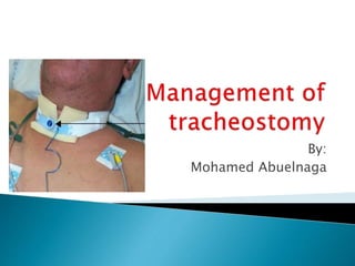
Management of tracheostomy
•Download as PPTX, PDF•
14 likes•9,118 views

how to deal with tracheostomy in critically ill patients

Recommended
More Related Content
What's hot
What's hot (20)
Viewers also liked
Similar to Management of tracheostomy
Similar to Management of tracheostomy (20)
More from mohamed abuelnaga
More from mohamed abuelnaga (10)
Recently uploaded
Recently uploaded (20)
Management of tracheostomy
- 2. 50 years female patient admitted to ICU with DCL due to spont. ICH Tracheostomy was done after 7 days with MPDT technique After 4 days ,with patient care ,the outer part of the traceostomy tube becomes longer with no explanation Then the patient shows surgical emphysema and hypoxia. umbo ventilation was done with worsening of the condition ,then patient arrested. Icu resident tries to do oro-tracheal intubation ,he found air way edema ,small sized ETT was inserted ,CPR started but the patient died What is your explanation?
- 3. Definition. Indications: #facilitate weaning from prolonged ventilation # bypass an obstruction of the URT #prevent aspiration from the pharynx or GIT #provide pulmonary toilet and facilitate removal of secretions by aspiration #as part of another procedure, as head and neck surgery
- 4. Absolute contraindication for tracheotomy is skin infection ,and prior major neck surgery which completely obscures the anatomy Technique: Surgical tracheotomy Percutaneous dilatational tracheotomy .
- 5. Effects of Tracheostomy: -The larynx is bypassed and so the patient is unable to phonate -decreased anatomical and respiratory dead space, decreasing the work of breathing -There is loss of the humidification and filtration function of the nasal mucosa
- 6. -There is an increased risk of respiratory tract infection -There is a redundant area above the tracheal opening and below the larynx in which mucus can accumulate and fall back into the lungs. -A foreign body reaction can occur causing local inflammation
- 7. 1-Improvement of respiratory mechanics: facilitate weaning due to reduced (WOB) because of a decrease in flow resistance 2-Reduced laryngeal ulceration: Endotracheal intubation can result in severe injury of the upper airway which can be largely prevented by early tracheotomy
- 8. 3-Improved nutrition, enhanced mobility and speech. 4-Improved patient comfort. 5-Patient can be nursed outside intensive care unit (ICU). 6-Clearance of secretion
- 10. Tracheostomy tubes can be metal or plastic. Metal tubes are constructed of silver or stainless steel. Plastic tubes are most commonly used and can be made from polyvinylchloride or silicone. Polyvinyl chloride softens at body temperature (thermolabile), conforming to patient anatomy and centering the distal tip in the trachea.
- 11. Silicone is naturally soft and unaffected by temperature. Some plastic tracheostomy tubes are packaged with a tracheal wedge . The tracheal wedge facilitates removal of the ventilator circuit while minimizing the risk of dislodgement of the tracheostomy tube
- 12. The dimensions of tracheostomy tubes are given by their ID, OD, length, and curvature. Single cannula tracheostomy tubes use the International Standards Organization method of sizing, determined by the ID of the outer cannula at its smallest dimension. Dual cannula tracheostomy tubes also use the International Standards Organization method. If an inner cannula is required for connection to the ventilator, the published ID is the ID of the inner cannula. The OD is the largest diameter of the outer cannula
- 13. When selecting a tracheostomy tube, the ID and OD must be considered. If the ID is too small, it will increase the resistance through the tube, make airway clearance more difficult, and increase the cuff pressure required to create a seal in the trachea. A tube with a larger OD will be more difficult to pass through the stoma.
- 14. Tracheostomy tubes can be angled or curved Because the trachea is essentially straight, the curved tube may not conform to the shape of the trachea, potentially allowing for compression of the membranous part of the trachea, while the tip may traumatize the anterior portion. If the curved tube is too short, it can obstruct against the posterior tracheal wall, which can be remedied by using either a larger tube, an angled tube, a tube with a flexible shaft, or a tube with extra length
- 15. Angled tracheostomy tubes have a curved portion and a straight portion. They enter the trachea at a less acute angle and may cause less pressure at the stoma.
- 16. Tracheostomy tubes are available in standard length or extra-length. Extra-length tubes are constructed with extra- proximal length (horizontal extra length) or with extra-distal length (vertical extra length). In the case of one manufacturer, extra-distal length is achieved by a double cuff design. This design also allows the cuffs to be alternatively inflated and deflated, which may reduce the risk of tracheal-wall injury, although this has never been subjected to appropriate clinical study
- 17. Differences in length exist between tubes of the same ID. Extra-proximal length facilitates tracheostomy tube placement in patients with a large neck (eg, obese patients). Extra -distal length facilitates placement in patients with trachealmalacia or tracheal anomalies. Care must be taken to avoid inappropriate use of these tubes, which may induce distal obstruction of the tube
- 19. Cuff: Tracheostomy tubes can be cuffed or uncuffed. The cuff reduces aspiration and leakage of air during anaesthesia and positive pressure ventilation. Specific types of cuffs used on tracheostomy tubes include high-volume low-pressure cuffs,tight-to-shaft cuffs (low-volume high- pressure), and foam cuffs.
- 20. Inner cannula: Some tracheostomy tubes are designed to be used with an inner cannula, and these are called dual-cannula tracheostomy tubes. It has the safety advantage of being easily and quickly removed to relieve life threatening obstruction due to blood clots or secretions.
- 21. Fenestration: Fenestrations may be single or multiple and are sited at the site of maximum curvature of the tracheostomy tube. With the inner cannula removed, the cuff deflated, and the normal air passage occluded, the patient can inhale and exhale through the fenestration and around the tube . This allows for assessment of the patient’s ability to breathe through the normal oral/nasal route (preparing the patient for decannulation) and permits air to pass by the vocal cords (allowing phonation)
- 23. Flexibility and adjustable flange: Several tube designs have a spiral wire reinforced flexible design. These also have an adjustable flange design to allow bedside adjustments to meet extra- length tracheostomy tube needs. This is useful in obese patients or those with local tissue swelling, where the soft tissue depth is increased.
- 25. Subglottic suction: Some newer tracheostomy tubes include a subglottic suction port, the aim of which is to try and reduce the incidence of VAP. Speaking valve: Speaking valves are one way valves that are designed to be used with fenestrated tracheostomy tubes or unfenestrated tubes (with the cuff deflated). They allow inspiration but not expiration. Hence the expired air is forced through the larynx allowing the patient to phonate
- 26. Mini-Tracheostomy Tubes: The mini-tracheostomy tube is a small bore cannula (4.0mm ID) inserted into the trachea through the cricothyroid membrane or the tracheal stoma after decannulation. It can be used for oxygen administration. However, it is used primarily for patients with airway clearance issues because it allows bronchial lavage and suctioning with a 10 French suction. It is uncuffed and generally unsuitable for provision of positive pressure ventilation
- 27. Nursing care: the tracheotomy cannula is secured in place and the tracheotomy is left to heal for 5-7 days, to allow for development of a stable and patent cutaneo-endotracheal tract. The tracheotomy wound has to be kept clean and dry to prevent post-incisional wound infection.
- 28. Changing tracheostomy tubes: When a dual cannula is used, there is usually no need to change the outer cannula. The inner cannula is changed daily or more frequently if necessary. The first change should not occur within 72 hours of the tracheostomy being sited and ideally not for 7 days after a percutaneous insertion. This is to allow for the formation of a more reliable ‘track’ for the new tube to pass through. The only indication to change the tube is when the cuff has been damaged or when a tracheotomy tube of different size or shape is found to be necessary
- 29. Emergency airway equipment, including a smaller tracheostomy tube, and emergency drugs should be immediately available during the change. The tracheostomy tube may be changed over a soft suction or airway exchange catheter or soft tipped Ryle’s tube. The use of a rigid gum-elastic bougie for this purpose may increase the risk of creating a false passage (i.e. the new tracheostomy comes to lie next to rather than within the trachea)..
- 30. If a soft tipped Ryle’s tube or similar is used, it may be reassuring to see fogging within that tube with respiration. Alternatively, the track may be gently dilated with a gloved little finger. There should be a low threshold for suspicion of erroneous placement if it is difficult to ventilate the patient. If difficulty is encountered in replacing the tracheostomy tube, the clinical need for a tube must be re-assessed. If in doubt, re-intubation with an oral endotracheal tube may be required
- 31. Tube cuff pressure: Tracheal capillary perfusion pressure is normally 25–35 mm Hg so it is generally agreed that 25 mm Hg (34 cm H2O) is the maximum acceptable intra-cuff pressure. If the cuff pressure is too low (below 18 mmHg) may cause the cuff to develop longitudinal folds, promote microaspiration of secretions collected above the cuff and increase the risk for nosocomial pneumonia. Tracheotomy tube cuffs require monitoring to maintain pressures in a range of 20-25 mmHg.
- 32. Cuff pressures should be monitored with calibrated devices and recorded at least once every nursing shift and after manipulation of the tracheotomy tubes . It has been suggested to deflate tracheotomy cuffs on a regular schedule to allow mucosal perfusion at the site of the cuff and to improve clearance of secretions accumulated around the cuff, but too little evidence is available to advocate this practice.
- 33. To minimize damage to the tracheal wall by the distal end of the tracheotomy tube, the tube has to be maintained in a central position, avoiding angling and contact between tracheal mucosa and tube.
- 34. Humidification: Cold and unfiltered air is an irritant when inhaled and can lead to mucosal damage, loss of mucociliary transport increased production and viscosity of secretions. This can be uncomfortable for the patient as well as causing tracheal mucosal keratinisation. The increasingly viscous secretions will be difficult to clear, causing sputum retention, increase the risk for lower respiratory tract infection , atelectasis, impaired gas exchange and even life threatening blockage of the tracheostomy tube.
- 35. It is therefore essential that inhaled oxygen is appropriately humidified and heated using conventional techniques such as heat and moisture exchange (HME) filters or heated water baths. Clearance of secretions: Because suctioning is uncomfortable and is associated with ventilatory complications such as airway collapse and alveolar derecruitment, it should be performed only when indicated and not at a fixed frequency.
- 36. Frequency of airway suctioning should hence be determined on a patient level, taking into account factors such as viscosity and quantity of mucus, neurological and muscular performance and presence of active cough reflexes and efforts. The upper airway should also be suctioned periodically to remove oral secretions and to minimise stasis of pooled secretions above the tracheotomy cuff with subsequent potential aspiration into the lower airways.
- 37. Shallow suctioning, involving the insertion of the aspiration catheter to a premeasured depth not beyond the distal end of the tracheotomy tube, is preferred to deep suctioning, in which the aspiration tube is inserted beyond the tracheotomy tube until resistance is met. Deep suctioning should be avoided since it is associated with airway mucosal damage and inflammation and may induce bronchial bleeding with subsequent risk of airway occlusion.
- 38. Speech: Ventilator-dependent patients, who require low minute ventilations may accomplish whispered speech during periods of partial deflation of the tracheotomy tube cuff, provided a good swallowing function is present and stasis of secretions above the cuff is limited. After removal from mechanical ventilation, the inner cannula is removed in awake and adequate patients, allowing expiratory airflow through the larynx when the external end of the tracheotomy tube is occluded transiently.
- 39. Application of a one-way valve permits inspiratory airflow through the tracheotomy tube during inspiration but closes during expiration, promoting airflow around a deflated cuff. Some centres use a fenestrated tube promoting airflow through the tube fenestrations during expiration. Nutrition It is conventional to feed intubated, ventilated patients enterally unless there is a good reason not to. This is usually via a nasogastric or nasojejunal tube but it may be possible for patients with tracheostomies to be fed orally
- 40. However, swallowing is still adversely affected by the presence of a tracheostomy tube, which has a tendency to limit normal movement of the larynx. In addition, the inflated cuff causes a sense of pressure in the upper oesophagus and the difficulty that occurs with swallowing may result in an increased risk of aspiration of food into the lungs. Between 20 and 70% of patients with a chronic tracheotomy experience at least one episode of aspiration every 48 h.
- 41. Patients may be fed orally, with the cuff inflated or partially deflated, but staff must be alert to signs of aspiration, such as coughing, increased secretions and impaired gas exchange. To decrease risks for nosocomial pneumonia caused by aspiration, patients with tracheotomy are kept with their heads elevated to 45o during periods of tube feeding. It is prudent to commence with sips of water and some form of swallowing assessment by a fiber optic.
- 42. There are many ways of assessing adequate breathing around the tracheostomy tube. Patients can be trialed with increasing periods of cuff deflation. This allows patients to become re-accustomed to swallowing more normally and to having to clear their own secretions. Alternatively, an occlusion cap may be used which completely blocks the tracheostomy tube.
- 43. Of course this must be used with a fenestrated tube or an unfenestrated tube with the cuff deflated, and this greatly increases the work of breathing due to the increased airway resistance. It will be harder for patients to breathe in this situation than without the tracheostomy in place and this must be taken into account when interpreting the success or failure of such a trial.
- 44. Decannulation can be carried out when: • The patient is not dependent on ventilatory support and has an adequate respiratory reserve (dead space will be increased without the tracheostomy tube) • The patient is able to cough and swallow effectively and manage their own secretions whilst being able to protect their own airway • The patient is able to cough and clear his tracheal secretions •Patient can tolerate cuff deflation or capping of the tracheostomy tube.
- 45. Criteria for tracheotomy decannulation: #Stable arterial blood gases. #Absence of distress. #Haemodynamic stability. #Absence of fever or active infection. #PaCO2 < 60 mmHg. #Absence of delirium or psychiatric disorder. #Normal endoscopic examination or revealing stenotic lesion occupying <30% of the airway. #Adequate swallowing. #Able to expectorate.
- 46. Technique of decannulation: Decannulation itself can be performed in the morning, with a rested patient and daylight hours in which to review their progress. After deflation of the tracheotomy cuff, a gloved finger occludes the opening of the tube and one observes the patient for objective signs of respiratory distress. In case of problems, one promptly returns the patient to breathing through the tracheotomy tube and performs an endoscopic examination to check for upper airway obstruction.
- 47. If no lesions are present, one should consider whether the tube is not too large and try again after changing the tube. The tube can be removed and the stoma is covered with a semi- permeable dressing. The patient is encouraged to gently press over this defect with whilst speaking or coughing. They should be subsequently be monitored for signs of respiratory distress. Equipment and expertise to re-secure the airway, either via the stoma or via oral intubation, should be available.