
Recommended
Recommended
More Related Content
What's hot
What's hot (20)
Similar to Anatomical landmarks for edentulous patients and facial landmarks
Similar to Anatomical landmarks for edentulous patients and facial landmarks (20)
Recently uploaded
Recently uploaded (20)
Anatomical landmarks for edentulous patients and facial landmarks
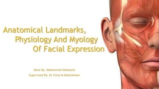
- 1. Anatomical Landmarks, Physiology And Myology Of Facial Expression Done By: Mohammed Abdulaziz Supervised By: Dr Faiza M AbdulAmeer
- 2. Contents… Introduction Extra-Oral Anatomical Landmarks Intra-Oral Anatomical Landmarks -Classification Of Oral Mucosa -Landmarks Of Maxillary Edentulous Arch -Landmarks of Mandibular Edentulous Arch Myology Of Face And Oral Cavity Physiology Of Oral Cavity Conclusion
- 3. Introduction… The good knowledge of anatomy and physiology is a keystone in getting the best result during the fabrication of any prosthesis, this best result is which restores the missing oral parts and preserves what is remaining.
- 4. Facial Anatomical Landmarks Effects Of Edentulism on face: 1- 2-
- 5. Eye
- 6. Ear
- 7. Nose
- 8. Lips
- 10. Naso Labial Angel Indication of lip support by upper anterior teeth. Used In jaw-relation clinic to correct the position of wax rim anteriorly 90-110 in average.
- 11. Interpupillary line + Ala Tragus Line
- 14. Oral Mucosa Epithelial Layer Connective Tissue layer Sub-Mucosa
- 15. Types Of Oral Mucosa Masticatory Mucosa Lining Mucosa Specialized Mucosa
- 16. Masticatory Mucosa Free, attached gingiva and hard palate comes in primary contact with food during mastication and is keratinized.
- 17. Lining Mucosa the lips cheeks, vestibule, floor of the mouth, ventral surface of the tongue and soft palate. It does not function in mastication. It is soft, pliable and non-keratinized.
- 18. Specialized Mucosa SPECIALIZED MUCOSA: on the dorsal surface (dorsum) of the tongue. It is covered with cornified epithelial papillae.
- 19. Intra Oral Anatomical Landmarks Maxillary Anatomical Landmarks Mandibular Anatomical Landmarks
- 20. Maxillary Anatomical Landmarks a} Limiting structures - Labial frenum - Labial vestibule - Buccal frenum - Buccal vestibule - Hamular notch - Posterior palatal seal area b} Supporting structures -Hard palate -Residual ridge -Palatal rugae Maxillary Tuberosity c} Relief areas - Incisive papilla - Mid palatine raphe - Canine Eminence - Fovea palatine - Palatal Tori
- 21. Limiting Structures Labial Frenum It contains no muscle and has no action on its own. It should be properly relieved.
- 22. Limiting Structures Labial Vestibule The labial vestibule is divided into a left and right labial vestibule by the labial frenum and extends upto the buccal frenum on either side. The main muscle of the lip, which forms the outer surface of the labial vestibule, is the orbicularis oris.
- 23. Limiting Structures Buccal Frenum Consist of one or more bands. Influenced by 3 muscles Orbicularis oris (forward) Buccinator (backward) Levator anguli oris (position)
- 24. Limiting Structures Buccal Vestibule Buccal frenum to hamular notch
- 25. Limiting Structures Hamular Notch Situated between the tuberosity and hamulus of the medial pterygoid plate. Distal extension of denture. If the denture extends too far into the hamular notch, the mucous membrane covering the raphe will be traumatized. Anterior Vs Posterior Vibrating lines
- 26. Limiting Structures Posterior Palatal Seal Area The soft tissue area at or beyond the junction of the hard and soft palates on which pressure,within physiologic limits, can be applied by a denture to aid in its retention”. (GPT-8). Posterior palatal seal has several advantages.
- 28. Supporting Structures Hard Palate The horizontal part of palate is considered as primary stress bearing area.
- 29. Supporting Structures Residual Ridge “The portion of the alveolar ridge and its soft tissue covering, which remains following the removal of teeth”.(GPT-8).
- 30. Supporting Structures Palatal Rugae This area contributes to the stress- bearing role (secondary stress bearing area) as well as to retention although in a secondary capacity.
- 31. Supporting Structures Maxillary tuberosity It is considered as secondary stress bearing area, if it is firm it can withstand more forces. Sometimes it is oversized either with soft tissue or with bone.
- 33. Relief areas Incisive Papilla This covers the incisive foramen and is located in the midline immediately behind and between central incisors. Care should be taken that the denture base does not impinge on them and hence should be relieved.
- 34. Relief areas Mid Palatine Raphe Adequate relief should be provided in this area as, mucosa covering the raphe is extremely thin and is traumatized easily.
- 38. Mandibular Anatomical Landmarks Limiting structures - Labial frenum -Labial vestibule -Buccal frenum -Buccal vestibule -Lingual frenum -Alveololingual sulcus -Retromolar pad Supporting structures -buccal shelf area. -residual alveolar ridge. Relief areas -mental foramen. -genial tubercle. -Mylohyoid ridge. -Torus mandibularis.
- 46. Supporting Structures Buccal Shelf Area It is considered as primary stress bearing area. It is horizontal and made up of dense cortical bone.
- 47. Supporting Structures Residual Alveolar Ridge It is considered as secondary stress bearing area (lateral slopes). The crest of the ridge should be relieved.
- 50. Relief Areas Mylohyoid Ridge Soft tissue usually hides the sharpness of mylohyoid ridge. The mucous membrane overlying the sharp or irregular mylohyoid ridge needs to be relieved.
- 52. Is it time for rest Any energy remained
- 53. Myology Of Face And Oral Cavity -Physiology Of Muscles -Muscles Of Facial Expression -Sopra-Hyoid Muscles -Infra-Hyoid Muscles -Muscles Of Mastication -Muscles Of The Tongue -Muscles O Soft-Palate
- 54. Muscles Of Facial Expression
- 55. Modiolus
- 56. Modiolus
- 57. Muscles Of Facial Expression Buccinator Is a thin quadrilateral muscle, occupying the interval between the maxilla and the mandible at the side of the face. It forms the anterior part of the cheek or the lateral wall of the oral cavity.
- 58. Muscles Of Facial Expression Buccinator
- 59. Muscles Of Facial Expression Orbicularis Oris “The orbicularis oris muscle is a complex of muscles in the lips that encircles the mouth. Until recently, it was misinterpreted as a sphincter, or circular muscle, but it is actually composed of four independent quadrants that interlace and give only an appearance of circularity.” Saladin, "Anatomy & Physiology: The Unity of Form and Function". 5th edition. McGraw Hill. Page 330
- 60. Muscles Of Facial Expression Mentalis Muscle When contracts it can dislodge the denture. It dictates the level of extension of the labial flange of mandibular denture. Reduce the lower labial vestibule when it contracts.
- 61. Supra-Hyoid Muscles Act in elevating the hyoid bone and larynx and depression of the mandible.
- 62. The mylohyiod and geniohyiod may influence border of mandibular denture. The mylohyoid constitutes the floor of the mouth.
- 63. Infra-Hyoid Muscles actions of these muscles are important to the prosthodontist, for they are a part of the kinetic chain of the mandibular movement.
- 64. Muscles Of Mastication These muscles have masticatory and non-masticatory movements, but in concern of complete denture, the non-masticatory movements have more influence on denture borders. Also these muscles especially the temporalis aid in obtaining centric relation.
- 65. Muscles Of Mastication Temporalis Muscle ACTIONS OF TEMPORALIS - Elevates the mandible, this movement requires both the upward pull of anterior fibers and backward pull of the posterior fibers. - Posterior fibers draw the mandible backwards after it has been protruded.
- 66. Muscles Of Mastication Masseter Muscle
- 67. ACTIONS OF MASSETER: Elevates the mandible to close the mouth and to occlude the teeth in mastication. It has a small effect in side-to- side movement, protraction and retraction.
- 68. Muscles Of Mastication Medial And Lateral Pterygoids The fibers of the lateral pterygoid, pull the mandible forwards (protrusion) and medially. The fibers of the medial pterygoid also perform the same actions in addition elevate the mandible.
- 69. Muscles Of The Tongue Intrinsic muscles Extrinsic muscles Styloglossus Palatoglossus Genioglossus hyoglossus
- 70. Importance of Tongue In Prosthodontics After loss of teeth, tongue expands into the space created by lost teeth, this enlargement make the impression more difficult, and reduce the stability of denture. The level of occlusal table in relation to tongue level is so important because high occlusal level reduces the retention of denture, while low occlusal level contributes in tongue biting during function.
- 71. Muscles Of Soft Palate 1. Tensor veli palatini : It influences the denture border in the hamular notch 2. Levator veli palatini: it helps in determining the vibrating line. 3. Palatoglossus: it exerts pressure on the lingual extension of the lower denture mainly when the tongue is moved toward the cheek. 4. Palatopharyngeous 5. The uvula: is unpaired
- 72. Classification of soft palate: House’s classification Class I: the soft palate is almost horizontal curving gently downwards Class II: the soft palate turns downward at about 45 angle from the hard palte Class III: the palate turns downward sharply at about 70 angle to the hard palate.
- 73. Physiology Of Oral Cavity Physiology Of Muscles Physiology Of Bone Saliva
- 77. Physiology Of Bone The reduction of bone may occur due to: -Anatomical factor -Prosthodontic factor (pressure mediated resorption ) -Functional Factor -Metabolic and systemic factor: 1-Osteoporosis 2-Hyperthyroidism
- 79. Prosthodontic factor Intensive denture wear Unstable occlusal contact Immediate denture treatment
- 80. Functional Factor Loss of stimulation is essential in accelerating the bone resorption.
- 81. Metabolic and systemic factors: Osteoporosis Hyperthyroidism
- 82. Saliva Saliva is a watery substance formed in the mouth. Human saliva comprises 99.5% mostly water, plus electrolytes, mucus, white blood cells, epithelial cells (which can be used to extract DNA), glycoproteins, enzymes, antimicrobial agents such as secretory IgA and lysozyme
- 83. Roles Of Saliva Lubrication Digestion Role In Taste Immune Other ( In prosthodontics )
- 84. Major Salivary Glands Parotid Submandibular Sublingual They produce about 90 % of total salivary production.
- 85. Minor Salivary Glands Labial, buccal and lingual mucosa Soft palate Lateral parts of hard palate Floor of the mouth The produce about 10 % of saliva.
- 86. Sialorrhea Excessive production of saliva (hypersalivation). Make impression procedure more difficult. May occur temporarily after insertion of denture.
- 87. Xerostomia And Hyposalivation Injury due to losing lubricant abilities of saliva. Loss of retention Causes Treatment
- 88. Conclusion
- 89. Thank You For Listening