Contrast media & reaction
•Download as PPTX, PDF•
81 likes•28,534 views

Contrast media & reaction

Recommended
More Related Content
What's hot
What's hot (20)
Similar to Contrast media & reaction
Similar to Contrast media & reaction (20)
More from Dr. Mohit Goel
More from Dr. Mohit Goel (20)
Recently uploaded
Recently uploaded (20)
Contrast media & reaction
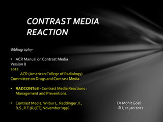
- 1. CONTRAST MEDIA REACTION Dr Mohit Goel JR I, 21 jan 2012 Bibliography- • ACR Manual on Contrast Media Version 8 2012 ACR (American College of Radiology) Committee on Drugs and Contrast Media • RADCONT08 - Contrast Media Reactions : Management and Preventions. • Contrast Media,Wilbur L. Reddinger Jr., B.S.,R.T.(R)(CT),November 1996.
- 2. Categories of Reactions Mild: nausea, vomiting, cough, sneezing, warmth, headache, dizziness, shaking, altered taste, itching, pallor, flushing, chills, sweats, rash, hives, nasal stuffiness, anxiety, sneezing, swelling – eyes, face. Moderate: tachycardia / bradycardia, hypertension / hypotension, pronounced cutaneous reaction, dyspnea, bronchospasm / wheezing. Severe: laryngeal edema, convulsions, profound hypotension, clinically manifest arrhythmias, unresponsiveness, cardiopulmonary arrest.
- 3. Types of reactions: 1. Anaphylactoid 2. Non-anaphylactoid a. chemotoxic b. vasovagal c. idiopathic 3. Combined (1 and 2)
- 4. Anaphylactoid reactions • Anaphylactoid reactions occur unexpectedly and the specific cause is uncertain.Therefore, anaphylactoid reactions are often referred to as “idiosyncratic”. • Other reactions relating to osmotic, chemotoxic, direct organ toxicity, or vasomotor effects are more predictable and better understood.These reactions do not have the characteristics of an anaphylactoid reaction and are therefore referred to as nonanaphylactoid. • In some patients, both types of reactions occur, and the result is a combined reaction.
- 5. Chemotoxic Reactions : • Chemotoxic side effects include neurotoxicity, cardiac depression, arrhythmia, electrocardiogram changes, and renal tubular or vascular injury. • Some chemotoxic side effects appear to relate to the ionic nature and content of contrast media that dissolved in solution. Nonionic contrast media are associated with fewer chemotoxic side effects.
- 6. Vasovagal Reactions : • Vagal reactions occur as a result of increased vagal tone on the heart and blood vessels.The result is bradycardia and decreased blood pressure and may be accompanied by apprehension, confusion, sweating, unresponsiveness, and loss of bowel or bladder control signals. • Some vagal reactions may not be caused by the contrast media but instead may be the result of coincident events related to the examination (e.g., needle puncture, or abdominal compression).
- 7. Combined Reactions: • Anaphylactoid reactions and nonanaphylactoid reactions can occur or appear to occur simultaneously.The end result may be a complex, life-threatening situation with a patient in shock. • Careful attention to the specific signs and symptoms of a reaction should help in identifying the exact causes of the reaction. • A careful history of any medications ingested prior to the exam can aid in identifying possible contributory effects of the medications.
- 8. The adverse reactions are commonly related to the following:
- 9. 1.The physical properties of the contrast media include • the ions or particles associated with the chemical breakdown of the contrast media when it enters a solution, • the number and size of the iodine molecules, and • the number and size of the molecules of any chemical additive.
- 10. The chemical composition of ionic and non-ionic contrast media contains iodine. • One of the primary differences between ionic and non-ionic contrast media is that an ionic compound dissociates or dissolves into charged particles when it enters into blood. • Ionic media breakdown into cations and anions. • For every three iodine molecules present in an ionic media, one cation and one anion are produced when it enters blood. • Ionic contrast media are generally referred to as 3:2 compounds.
- 11. • Non-ionic contrast media do not dissolve into charged particles when it enters a solution. • For every three iodine molecules in a non-ionic solution, one neutral molecule is produced. • Non-ionic contrast media are referred to as 3:1 compounds. The resultant ion charges from an ionic compound have the potential to disrupt the electrical charges associated with the brain as well as the heart. Neurotoxicity is the term that describes the potential that the ionic charges have for disrupting the electrical charges of the brain.
- 12. The radiographic significance of the osmolality value of contrast media is that it is higher than the osmolality value of blood plasma. Contrast media is primarily divided into two categories, high osmolar contrast media (HOCM) and low osmolar contrast media (LOCM). • Most non-ionic agents are placed into the LOCM category • while all ionic agents are in the HOCM category. 1. An injection of ionic HOCM results in a big increase in the number of particles contained in the vascular system. 2. The introduction of contrast media into the vascular system causes water from body tissue (cells ) to move into the vascular system in an attempt to equalize concentrations (osmosis). 3. The blood vessels dilate (Hypervolemia) in an attempt to compensate from the increased fluid volume.
- 13. • The rapid fluid movement throughout the vascular system is believed to contribute to pain associated with vessel dilation,flushing, damage to the vascular endothelium, red cell changes, nausea, vomiting, and dehydration. The osmotic effect can cause the arteries of the kidneys to expand. When the arteries expand vasoconstrictors are released to compensate for the artery expansion. The vasoconstrictors constrict the arteries resulting in diminished blood supply to kidneys which can lead to total shut down of the kidneys. If the kidneys are non-functional, the fluid is forced to seek other avenues of escape causing fluid overload to occur in other body systems. One of the major results of this event is pulmonary edema.
- 14. The iodine concentration The iodine concentration of an individual contrast agent determines how radiopaque the agent will be. The higher the iodine concentration, the better the chance that more x-ray photons will be absorbed therefore, that particular contrast agent maybe more radiopaque than a comparative low iodine concentrated agent. The iodine concentration has an effect on the severity of an adverse reaction.The higher the iodine concentration, the greater the risk of an adverse reaction.
- 15. The total volume or dose is dependent upon several factors. 1. Iodine concentration of the contrast media, 2. type of contrast media being injected (ionic or non-ionic), 3. body weight, 4. anatomical structures or regions, 5. speed of the injection and 6. age or disease process that could increase the risk of an adverse reaction. Increasing the injected volume increases the possibility of an adverse reaction occurring.An injection of a large amount of contrast media usually is accompanied by the use of a lower iodine concentrated agent.
- 16. Increasing the speed or rate of injectionmay increase the risk of an adverse reaction occurring. Contrast medias with a higher viscosity values will have to be injected at a slower rate. Heating the contrast media , usually to body temperature, reduces viscosity. All power injection devices are equipped with a "warming pad“ covering a portion of the syringe. Iodine concentration, viscosity, temperature of the contrast media, catheter inner diameter, catheter length, and the number of catheter holes are all factors that influence contrast media flow.
- 17. Incidence of Adverse Effects • Use of low osmolality ionic and nonionic contrast media is associated with a lower overall incidence of adverse effects, particularly serious ones. • A large-scale (337,647 cases), nationwide comparative clinical study in Japan on adverse drug reactions (ADRs) to high- osmolar ionic contrast media and low-osmolar nonionic contrast media was performed prospectively. --- The overall prevalence ofADRs was 12.66% in the ionic contrast media group and 3.13% in the nonionic contrast media group. Severe ADRs occurred in 0.22% of the ionic and 0.04% of the nonionic contrast media examinations.
- 18. Patient Selection & Preparation Strategies The approach to patients has two general aims: 1. to prevent a reaction from occurring and 2. to be fully prepared to treat a reaction should one occur. • History should focus on the factors that may indicate either a contraindication to contrast media use or an increased likelihood of a reaction. • Hemodynamic, neurologic, and general nutritional status should be assessed. In regard to specific risk factors. • True concern should be focused on patients with significant allergies, such as prior anaphylactic response to one or more allergens.A history of asthma indicates an increased likelihood of a contrast reaction. • Patients with any know allergies have a four fold chance of a reaction to contrast media.
- 19. Administration of contrast medium to breast feeding mothers: • Less than 1 % of the administered dose of contrast iodinated or gadolinium based is excreted in the breast milk. • Less than 1% of the contrast in breast milk taken in by and infant is absorbed by the GI tract. • Literature and data published by the ACR suggest that it is safe to continue to breast feed after receiving contrast iodinated or gadolinium. Contrast is nearly 100% excreted from the body within 24 hours after receiving contrast. • The practice standard should follow the manufacturers guidelines and substitute bottle feedings for breast feedings for the 24 hours following the injection of contrast media.
- 20. Contrast Reactions In Children Children have a lower frequency of contrast reactions that adults • They tend to have allergic-type reactions rather that cardiac problems. • In addition to fewer reactions, nonionic contrast media has the added benefit of decreased nausea and vomiting and the possibility of diminished morbidity from extravasation into soft tissue. NOTE: Children’s airways are smaller and more easily compromised that those in adults. It is important to have pediatric emergency equipment, contrast kit and protocols available in the radiology department.
- 21. Iodinated Gastrointestinal Contrast Media • Iodinated /Aqueous (water soluable) contrast agents are absorbed rapidly from the interstitial spaces and peritoneal cavity, a feature that makes them uniquely useful in examining patients with a suspected perforation of a hollow viscous. • The current use of iodinated contrast media is primarily limited to those situations in which the administration of barium sulfate is contraindicated: Suspected or potential intestinal perforation. Administration before surgical or endoscopic procedures involving the bowel. Confirmation of the position of percutaneously placed bowel catheters. • Barium sulfate contrast media continue to be the preferred agents for opacification of the gastro-intestinal tract.
- 22. Treatment It is most important to target premedication to those who, in the past, have had moderately severe or severe reactions requiring treatment. Two frequently used regimens are: 1. Prednisone – 50 mg by mouth at 13 hours, 7 hours, and 1 hour before contrast media injection, plus Diphenhydramine (Benadryl®) – 50 mg intravenously, intramuscularly, or by mouth 1 hour before contrast medium. or 2. Methylprednisolone (Medrol®) – 32 mg by mouth 12 hours and 2 hours before contrast media injection.An anti-histamine (as in option 1) can also be added to this regimen injection. If the patient is unable to take oral medication, 200 mg of hydrocortisone intravenously may be substituted for oral prednisone.(Greenberger protocol) • Premedication strategies (ACR Manual on Contrast Media ,Version 8 , 2012)
- 23. In evaluating a patient for a potential contrast reaction, five important immediate assessments should be made: • How does the patient look? • Can the patient speak? How does the patient’s voice sound? • How is the patient’s breathing? • What is the patient’s pulse strength and rate? • What is the patient’s blood pressure? The level of consciousness, the appearance of the skin, quality of phonation, lung auscultation, blood pressure and heart rate assessment will allow the responding physician to quickly determine the severity of a reaction. These findings also allow for the proper diagnosis of the reaction including urticaria, facial or laryngeal edema, bronchospasm, hemodynamic instability, vagal reaction, seizures, and pulmonary edema.
- 24. Management of acute reactions in adults Urticaria 1. Discontinue injection if not completed 2. No treatment needed in most cases 3. Give H1-receptor blocker: diphenhydramine (Benadryl®) PO/IM/IV 25–50 mg. If severe or widely disseminated: give alpha-agonist (arteriolar and venous constriction): epinephrine SC (1:1,000) 0.1–0.3 ml (= 0.1–0.3 mg) (if no cardiac contraindications).
- 25. Facial or Laryngeal Edema 1. Give O2 6–10 liters/min (via mask). 2. Give alpha agonist (arteriolar and venous constriction): epinephrine SC or IM (1:1,000) 0.1–0.3 ml (= 0.1–0.3 mg) or, especially if hypotension evident, epinephrine (1:10,000) slowly IV –3 ml (= 0.1–0.3 mg). Repeat as needed up to a maximum of 1 mg. If not responsive to therapy or if there is obvious acute laryngeal edema, seek appropriate assistance (e.g., cardiopulmonary arrest response team).
- 26. Hypotension withTachycardia 1. Legs elevated 60 degree or more (preferred). 2. Monitor: electrocardiogram, pulse oximeter, blood pressure. 3. Give O2 6–10 liters/min (via mask). 4. Rapid intravenous administration of large volumes of Ringer’s lactate or normal saline. If poorly responsive: epinephrine (1:10,000) slowly IV 1 ml (= 0.1 mg) Repeat as needed up to a maximum of 1 mg.If still poorly responsive seek appropriate assistance (e.g., cardiopulmonary arrest response team)
- 27. Hypotension with Bradycardia (Vagal Reaction) 1 Secure airway: giveO2 6–10 liters/min (via mask) 2. Monitor vital signs. 3. Elevate legs. 4. Secure IV access: rapid administration of Ringer’s lactate or normal saline. 5. Give atropine 0.6–1 mg IV slowly if patient does not respond quickly to steps 2–4. Repeat atropine up to a total dose of 0.04 mg/kg (2–3 mg) in adult. 6. Ensure complete resolution of hypotension and bradycardia prior to discharge.
- 28. Hypertension, Severe 1. Give O2 6–10 liters/min (via mask). 2. Monitor electrocardiogram, pulse oximeter, blood pressure. 3. Give nitroglycerine 0.4-mg tablet, sublingual (may repeat × 3); or, topical 2% ointment, apply 1-inch strip. 4. If no response, consider labetalol 20 mg IV, then 20 to 80 mg IV every 10 minutes up to 300 mg. Transfer to intensive care unit or emergency department.
- 29. Seizures or Convulsions 1. Give O2 6–10 liters/min (via mask). 2. Consider diazepam (Valium®) 5 mg IV (or more, as appropriate) or midazolam (Versed®) 0.5 to 1 mg IV. 3. If longer effect needed, obtain consultation; consider phenytoin (Dilantin®) infusion — 15–18 mg/kg at 50 mg/min. 4. Careful monitoring of vital signs required, particularly of pO2because of risk to respiratory depression with benzodiazepine administration.
- 30. Proper dilution of Epinephrine for IV use in patient with anaphylaxis
- 32. First-line emergency drugs and instruments that should be in the room where contrast medium is injected • Oxygen • Adrenaline 1:1000 • Antihistamine H1 – suitable for injection • Atropine • B2 agonist metered dose inhaler • Intravenous fluids-normal saline or ringer’s solution • Anti convulsive drugs (diazepam) • Sphygmomanometer • One-way mouth “breathing” apparatus
- 33. Thank you