SlideShare a Scribd company logo
1 of 36
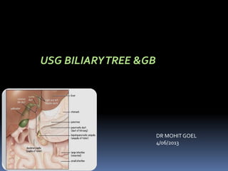
USG BILIARYTREE &GB
DR MOHIT GOEL
4/06/2013
GB
Gallstone Disease
Sonography is highly sensitive in the
detection of stones within the
Gallbladder.
The large difference in the acoustic
impedance of stones and adjacent
bile makes them highly reflective,
which results in an echogenic
appearance with strong posterior
acoustic shadowing.
Mobility is a key feature of stones, allowing differentiation from polyps
or other entities.
Various maneuvers may be used to demonstrate mobility of a stone;
scanning with the patient in the right or left lateral decubitus or upright
standing position may allow the stone to roll within the gallbladder.
Biliary Sludge
Biliary sludge, also known as biliary sand or microlithiasis, is defined as a
mixture of particulate matter and bile that occurs when solutes in bile
precipitate.
The sonographic appearance of sludge is that of amorphous, low-level
echoes within the gallbladder in a dependent position, with no acoustic
shadowing.
Acute Cholecystitis
It is caused by gallstones in more than 90% of patients.
Clinically, patients present with a prolonged, constant RUQ or
epigastric pain associated with RUQ tenderness. Fever,
leukocytosis, and increased serum ALP and bilirubin levels
may be present.
Porcelain Gallbladder
Calcification of the gallbladder wall is termed “porcelain” gallbladder.
The degree and pattern of calcification determines the sonographic
appearance.
When the entire gallbladder wall is thickly calcified, a hyperechoic
semilunar line with dense posterior acoustic shadowing is noted.
Mild calcification appears as an echogenic line with variable degrees of
posterior acoustic shadowing.
Chronic Cholecystitis
Chronic cholecystitis is associated with the mere presence of gallstones;
therefore patients are usually asymptomatic and have mild disease. 
More advanced cases involve wall thickening and fibrosis, appearing on
sonographic examination as a thickwalled gallbladder with gallstones.
Differentiation from acute cholecystitis is made by the absence of other
signs, namely, gallbladder distention, Murphy’s sign, and hyperemia in the
wall.
Bouts of acute cholecystitis may complicate chronic cholecystitis.
Adenomyomatosis (Adenomatous Hyperplasia)
Gallbladder adenomyomatosis is a benign condition caused by exaggeration of
the normal invaginations of the luminal epithelium (Rokitansky-Aschoff
sinuses) with associated smooth muscle proliferation.
The great majority of adenomyomatoses are asymptomatic.
Adenomyomatosis may be focal or diffuse.
The most common appearance on sonography is tiny, echogenic foci in the
gallbladder wall that create comet-tail artifacts , presumably caused by either the
cystic space itself or the internal debris.
Prominent mass like focal areas of adenomyomatosis, called adenomyomas,
are the next most common manifestation.
Echogenic foci with ringdown or “twinkling” artifact on Doppler examination
are also typical.
Adenomyomatosis: Focal adenomyomatosis, the most common manifestation.
A, Small focal area of thickening of the anterior fundal wall (arrow) with a bright echogenic
focus with a distal comet-tail artifact.
B, Multiple bright foci (arrow)with distal artifacts.
C, Highly echogenic focal thickening of the gallbladder wall (arrow).Increased echogenicity
is unusual for a malignant tumor that is more likely to appear hypoechoic.
Polypoid Masses of Gallbladder
Cholesterol Polyps
Approximately one half of all polypoid gallbladder lesions are cholesterol
polyps.These represent the focal form of gallbladder cholesterolosis, a
common nonneoplastic condition of unknown etiology.
The sonographic appearance of cholesterol polyps is multiple ovoid,
nonshadowing lesions attached to the gallbladder wall.
Gallbladder Carcinoma
The following three patterns of disease have been described:
1. Mass arising in the gallbladder fossa, obliterating the gallbladder and
invading the adjacent liver (most common pattern)
2. Focal or diffuse, irregular wall thickening
3. Intraluminal polypoid mass
On Doppler interrogation the mass may demonstrate internal arterial and
venous flow.
Diffuse, malignant thickening of the wall differs from other causes in that
the wall is irregular with loss of the normal mural layers.
Polypoid intraluminal masses are differentiated from nonneoplastic
abnormalities by immobility of the mass, larger size (>1 cm), and prominent
internal vascularity.
Gallbladder carcinomas may produce large quantities of mucin,which distends the
gallbladder
Gallbladder cancer:
A and B - Polypoid mass.
B, Examination with high-frequency linear probe shows no invasion beyond the gallbladder
wall.
C, Corresponding coronal MR image shows the enhancing polyp.
Huge mass replacing the gallbladder fossa and invading the liver.
H, Biliary obstruction caused by the invasive mass (arrow). I,Corresponding CT scan.
BiliaryTree
The normal caliber of the CHD/CBD in patients without history of biliary
disease is up to 6 mm.
Although diameters of up to 10 mm have been recorded in an asymptomatic
normal population, the great majority of the diameters are under 7 mm.
Therefore, a ductal diameter of 7 mm or greater should prompt further
investigations, such as correlation with serum levels of cholestatic liver
enzymes.
Choledochal Cysts
Choledochal cysts represent a heterogeneous group of congenital diseases
that may manifest as focal or diffuse cystic dilation of the biliary tree.
Type I. Fusiform dilation of the common bile duct is seen, but no obstructive lesion is
noted.This is the most common type of choledochal cyst.
Caroli’s Disease
Caroli’s disease is a rare congenital disease of the intrahepatic biliary tree that
results from malformation of the ductal plates, the primordial cells that give
rise to the intrahepatic bile ducts.
Caroli’s disease has been associated with cystic renal disease, most often
renal tubular ectasia (medullary sponge kidneys).
Caroli’s disease leads to saccular dilation or less often fusiform dilation of the
intrahepatic biliary tree, resulting in biliary stasis, stone formation, and bouts of
cholangitis and sepsis.
Cholangiocarcinoma develops in 7% of patients with Caroli’s disease.
Primary sclerosing cholangitis.
A, Isoechoic inflammatory tissue causing obliteration of right and left hepatic ducts (arrows)with
proximal dilation.
B, Dilated intrahepatic ducts “rat-tail” as they extend centrally toward the hepatic hilum. Note
the hypoechoic ductal/periductal tissue obstructing the central ducts (arrowheads).
C, Saccular diverticula (arrow)of the duct are occasionally seen.
D, Minute intraductal stones (arrows)are seen in irregular, mildly dilated ducts.
Mirizzi Syndrome
Mirizzi syndrome describes a clinical syndrome of jaundice with pain and fever
resulting from obstruction of the common hepatic duct caused by a stone
impacted in the cystic duct.
It occurs most often when the cystic duct and CHD run a parallel course.
Mirizzi syndrome should be considered on sonography when biliary obstruction
with dilation of the biliary ducts to the CHD level is seen with acute or chronic
cholecystitis.
A stone impacted in the cystic duct with surrounding edema at the level of the
obstruction is confirmatory.
BiliaryTree Infection
Acute (Bacterial) Cholangitis
The clinical presentation is usually that of
(1) fever (~90%),
(2) right upper quadrant (RUQ) pain (~70%), and
(3) jaundice (~60%), the classicCharcot’s triad.
The sonographic findings of bacterial cholangitis include the following :
• Dilation of the biliary tree
• Choledocholithiasis and possibly sludge
• Bile duct wall thickening
• Hepatic abscesses
Cholangiocarcinoma
Cholangiocarcinomas are classified based on the anatomic
location:
1. intrahepatic, also called peripheral (~10%);
2. hilar, also called Klatskin’s (~60%)
3. distal(~30%).
Approximately 90% of cholangiocarcinomas are adenocarcinomas, with
squamous carcinomas the next most common subtype.
Peripheral cholangiocarcinoma.
A, Ultrasound, and B,T2-weighted MR, images depict a solid mass encasing
the right hepatic vein. Differentiation from a metastasis is not possible by imaging.
THANKYOU

More Related Content

What's hot

Retroperitoneal masses radiology
Retroperitoneal masses radiologyRetroperitoneal masses radiology
Retroperitoneal masses radiologyDr. Mohit Goel
 
Presentation1, abdominal ultrasound anatomy.
Presentation1, abdominal ultrasound anatomy.Presentation1, abdominal ultrasound anatomy.
Presentation1, abdominal ultrasound anatomy.Abdellah Nazeer
 
Imaging of the large bowel
Imaging of the large bowelImaging of the large bowel
Imaging of the large bowelArchana Koshy
 
Liver Ultrasound
Liver UltrasoundLiver Ultrasound
Liver UltrasoundSafi. Khan
 
Ultrasound of the gallbladder
Ultrasound of the gallbladderUltrasound of the gallbladder
Ultrasound of the gallbladderSamir Haffar
 
Imaging of Acute Appendicitis
Imaging of Acute Appendicitis Imaging of Acute Appendicitis
Imaging of Acute Appendicitis Sakher Alkhaderi
 
Presentation1.pptx, radiological imaging of esophageal lesions.
Presentation1.pptx, radiological imaging of esophageal lesions.Presentation1.pptx, radiological imaging of esophageal lesions.
Presentation1.pptx, radiological imaging of esophageal lesions.Abdellah Nazeer
 
KEYS OF RADIOLOGY SPOTTERS GIT
KEYS OF RADIOLOGY SPOTTERS GITKEYS OF RADIOLOGY SPOTTERS GIT
KEYS OF RADIOLOGY SPOTTERS GITAnish Choudhary
 
Acute appendicitis - Ultrasound first
Acute appendicitis  - Ultrasound firstAcute appendicitis  - Ultrasound first
Acute appendicitis - Ultrasound firstSamir Haffar
 
Ultrasound of the urinary tract - Renal cysts
Ultrasound of the urinary tract - Renal cystsUltrasound of the urinary tract - Renal cysts
Ultrasound of the urinary tract - Renal cystsSamir Haffar
 
Diagnositc Imaging of the Esophagus
Diagnositc Imaging of the EsophagusDiagnositc Imaging of the Esophagus
Diagnositc Imaging of the EsophagusMohamed M.A. Zaitoun
 
small intestine imaging
small intestine imagingsmall intestine imaging
small intestine imagingSumer Yadav
 
Presentation1 liver ultrasound
Presentation1 liver ultrasoundPresentation1 liver ultrasound
Presentation1 liver ultrasoundAbdallah Bashe
 
Imaging of Bowel Obstruction
Imaging of Bowel ObstructionImaging of Bowel Obstruction
Imaging of Bowel ObstructionRathachai Kaewlai
 
Presentation2.pptx imaging of the biliary system
Presentation2.pptx  imaging of the biliary systemPresentation2.pptx  imaging of the biliary system
Presentation2.pptx imaging of the biliary systemAbdellah Nazeer
 
anatomy and sonography of kidney
anatomy and sonography of kidneyanatomy and sonography of kidney
anatomy and sonography of kidneynoor fatima
 
Imaging Acute Abdomen (Part 1)
Imaging Acute Abdomen (Part 1)Imaging Acute Abdomen (Part 1)
Imaging Acute Abdomen (Part 1)Rathachai Kaewlai
 

What's hot (20)

Retroperitoneal masses radiology
Retroperitoneal masses radiologyRetroperitoneal masses radiology
Retroperitoneal masses radiology
 
Presentation1, abdominal ultrasound anatomy.
Presentation1, abdominal ultrasound anatomy.Presentation1, abdominal ultrasound anatomy.
Presentation1, abdominal ultrasound anatomy.
 
Imaging of the large bowel
Imaging of the large bowelImaging of the large bowel
Imaging of the large bowel
 
Liver Ultrasound
Liver UltrasoundLiver Ultrasound
Liver Ultrasound
 
Ultrasound of the gallbladder
Ultrasound of the gallbladderUltrasound of the gallbladder
Ultrasound of the gallbladder
 
Imaging of Acute Appendicitis
Imaging of Acute Appendicitis Imaging of Acute Appendicitis
Imaging of Acute Appendicitis
 
Presentation1.pptx, radiological imaging of esophageal lesions.
Presentation1.pptx, radiological imaging of esophageal lesions.Presentation1.pptx, radiological imaging of esophageal lesions.
Presentation1.pptx, radiological imaging of esophageal lesions.
 
KEYS OF RADIOLOGY SPOTTERS GIT
KEYS OF RADIOLOGY SPOTTERS GITKEYS OF RADIOLOGY SPOTTERS GIT
KEYS OF RADIOLOGY SPOTTERS GIT
 
Acute appendicitis - Ultrasound first
Acute appendicitis  - Ultrasound firstAcute appendicitis  - Ultrasound first
Acute appendicitis - Ultrasound first
 
Imaging of Large Bowel Polyp
Imaging of Large Bowel PolypImaging of Large Bowel Polyp
Imaging of Large Bowel Polyp
 
Ultrasound of the urinary tract - Renal cysts
Ultrasound of the urinary tract - Renal cystsUltrasound of the urinary tract - Renal cysts
Ultrasound of the urinary tract - Renal cysts
 
Diagnositc Imaging of the Esophagus
Diagnositc Imaging of the EsophagusDiagnositc Imaging of the Esophagus
Diagnositc Imaging of the Esophagus
 
Liver ultrasound
Liver ultrasoundLiver ultrasound
Liver ultrasound
 
small intestine imaging
small intestine imagingsmall intestine imaging
small intestine imaging
 
Presentation1 liver ultrasound
Presentation1 liver ultrasoundPresentation1 liver ultrasound
Presentation1 liver ultrasound
 
Imaging of Bowel Obstruction
Imaging of Bowel ObstructionImaging of Bowel Obstruction
Imaging of Bowel Obstruction
 
Renal doppler
Renal dopplerRenal doppler
Renal doppler
 
Presentation2.pptx imaging of the biliary system
Presentation2.pptx  imaging of the biliary systemPresentation2.pptx  imaging of the biliary system
Presentation2.pptx imaging of the biliary system
 
anatomy and sonography of kidney
anatomy and sonography of kidneyanatomy and sonography of kidney
anatomy and sonography of kidney
 
Imaging Acute Abdomen (Part 1)
Imaging Acute Abdomen (Part 1)Imaging Acute Abdomen (Part 1)
Imaging Acute Abdomen (Part 1)
 

Viewers also liked

Biliary tract
Biliary tractBiliary tract
Biliary tractairwave12
 
Gall bladder & biliary tract anomalies and variants
Gall bladder & biliary tract  anomalies and variantsGall bladder & biliary tract  anomalies and variants
Gall bladder & biliary tract anomalies and variantsSanal Kumar
 
Transitional vertebrae radiology
Transitional vertebrae radiologyTransitional vertebrae radiology
Transitional vertebrae radiologyDr. Mohit Goel
 
Sectional radio-anatomy of abdomen
Sectional radio-anatomy of abdomenSectional radio-anatomy of abdomen
Sectional radio-anatomy of abdomenDr. Mohit Goel
 
Prostate carcinoma raiology
Prostate carcinoma raiologyProstate carcinoma raiology
Prostate carcinoma raiologyDr. Mohit Goel
 
Biliary tract Disease.ppt
Biliary tract Disease.pptBiliary tract Disease.ppt
Biliary tract Disease.pptShama
 
Shoulder labral tears MRI
Shoulder labral tears MRIShoulder labral tears MRI
Shoulder labral tears MRIDr. Mohit Goel
 
Retinoic Acid Receptor Alpha
Retinoic Acid Receptor AlphaRetinoic Acid Receptor Alpha
Retinoic Acid Receptor AlphaTaylor Beard
 
Preoperative assesment in cochlear implantation
Preoperative assesment in cochlear implantationPreoperative assesment in cochlear implantation
Preoperative assesment in cochlear implantationSunil Kumar
 
Role of contrast enhanced ultrasonography in characterization of hepatobiliar...
Role of contrast enhanced ultrasonography in characterization of hepatobiliar...Role of contrast enhanced ultrasonography in characterization of hepatobiliar...
Role of contrast enhanced ultrasonography in characterization of hepatobiliar...Dr. Muhammad Bin Zulfiqar
 
USG in gall bladder pathologies
USG in gall bladder pathologiesUSG in gall bladder pathologies
USG in gall bladder pathologiesPratibha Saran
 

Viewers also liked (20)

Biliary tract
Biliary tractBiliary tract
Biliary tract
 
Gall bladder & biliary tract anomalies and variants
Gall bladder & biliary tract  anomalies and variantsGall bladder & biliary tract  anomalies and variants
Gall bladder & biliary tract anomalies and variants
 
Renal doppler usg
Renal doppler usgRenal doppler usg
Renal doppler usg
 
Ultrasoud hernia
Ultrasoud herniaUltrasoud hernia
Ultrasoud hernia
 
Shoulder ultrasound
Shoulder ultrasoundShoulder ultrasound
Shoulder ultrasound
 
Transitional vertebrae radiology
Transitional vertebrae radiologyTransitional vertebrae radiology
Transitional vertebrae radiology
 
Sectional radio-anatomy of abdomen
Sectional radio-anatomy of abdomenSectional radio-anatomy of abdomen
Sectional radio-anatomy of abdomen
 
Prostate carcinoma raiology
Prostate carcinoma raiologyProstate carcinoma raiology
Prostate carcinoma raiology
 
Mri prostate
Mri prostateMri prostate
Mri prostate
 
Biliary tract Disease.ppt
Biliary tract Disease.pptBiliary tract Disease.ppt
Biliary tract Disease.ppt
 
Shoulder labral tears MRI
Shoulder labral tears MRIShoulder labral tears MRI
Shoulder labral tears MRI
 
Pediatric chest
Pediatric chestPediatric chest
Pediatric chest
 
Retinoic Acid Receptor Alpha
Retinoic Acid Receptor AlphaRetinoic Acid Receptor Alpha
Retinoic Acid Receptor Alpha
 
Preoperative assesment in cochlear implantation
Preoperative assesment in cochlear implantationPreoperative assesment in cochlear implantation
Preoperative assesment in cochlear implantation
 
Chap4
Chap4Chap4
Chap4
 
Gallbladder cancer
Gallbladder cancerGallbladder cancer
Gallbladder cancer
 
24 DAVID SUTTON PICTURES THE BILIARY TRACT
24 DAVID SUTTON PICTURES THE BILIARY TRACT24 DAVID SUTTON PICTURES THE BILIARY TRACT
24 DAVID SUTTON PICTURES THE BILIARY TRACT
 
Role of contrast enhanced ultrasonography in characterization of hepatobiliar...
Role of contrast enhanced ultrasonography in characterization of hepatobiliar...Role of contrast enhanced ultrasonography in characterization of hepatobiliar...
Role of contrast enhanced ultrasonography in characterization of hepatobiliar...
 
Blood smear morphology
Blood smear morphologyBlood smear morphology
Blood smear morphology
 
USG in gall bladder pathologies
USG in gall bladder pathologiesUSG in gall bladder pathologies
USG in gall bladder pathologies
 

Similar to Utrasound Gall-bladder & biliary tract

ACUTE ABDOMEN SONOGRAPHIC FINDINGS ppt
ACUTE ABDOMEN SONOGRAPHIC FINDINGS pptACUTE ABDOMEN SONOGRAPHIC FINDINGS ppt
ACUTE ABDOMEN SONOGRAPHIC FINDINGS pptPrafulla Pinjarkar
 
Usg evolution of biliary tree and gallbladder.pptx
Usg evolution of biliary tree and gallbladder.pptxUsg evolution of biliary tree and gallbladder.pptx
Usg evolution of biliary tree and gallbladder.pptxMukeshBijarniya
 
Focal vs diffuse gall bladder wall thickening
Focal vs diffuse gall bladder wall thickeningFocal vs diffuse gall bladder wall thickening
Focal vs diffuse gall bladder wall thickeningairwave12
 
Imaging ofsplenic diseases [Autosaved].pptx
Imaging ofsplenic diseases  [Autosaved].pptxImaging ofsplenic diseases  [Autosaved].pptx
Imaging ofsplenic diseases [Autosaved].pptxabelllll
 
Imaging of dangerous cholecystitis
Imaging of dangerous cholecystitisImaging of dangerous cholecystitis
Imaging of dangerous cholecystitisAhmed Bahnassy
 
Biliary pathologies
Biliary pathologiesBiliary pathologies
Biliary pathologiesNavdeep Shah
 
Acute cholecystitis
Acute cholecystitisAcute cholecystitis
Acute cholecystitisshahadatsurg
 
Gallbladder and Bile ducts Ultrasound
Gallbladder and Bile ducts Ultrasound Gallbladder and Bile ducts Ultrasound
Gallbladder and Bile ducts Ultrasound Moses Hadid
 
Imaging in Benign hepatic lesions
Imaging in Benign hepatic lesionsImaging in Benign hepatic lesions
Imaging in Benign hepatic lesionsGirendra Shankar
 
Gallbladder ultrasound (basic)
Gallbladder ultrasound (basic)Gallbladder ultrasound (basic)
Gallbladder ultrasound (basic)Syed Yousaf Gilani
 
An approach to cystic hepatic lesions jk 05-aprl-2016
An approach to cystic hepatic lesions jk 05-aprl-2016An approach to cystic hepatic lesions jk 05-aprl-2016
An approach to cystic hepatic lesions jk 05-aprl-2016Parth Bhut
 
Pediatric High and Low intestinal Obstruction.pptx
Pediatric High and Low intestinal Obstruction.pptxPediatric High and Low intestinal Obstruction.pptx
Pediatric High and Low intestinal Obstruction.pptxDr Muhammad Tahir Javed
 

Similar to Utrasound Gall-bladder & biliary tract (20)

Sahad gb
Sahad gbSahad gb
Sahad gb
 
ACUTE ABDOMEN SONOGRAPHIC FINDINGS ppt
ACUTE ABDOMEN SONOGRAPHIC FINDINGS pptACUTE ABDOMEN SONOGRAPHIC FINDINGS ppt
ACUTE ABDOMEN SONOGRAPHIC FINDINGS ppt
 
Usg evolution of biliary tree and gallbladder.pptx
Usg evolution of biliary tree and gallbladder.pptxUsg evolution of biliary tree and gallbladder.pptx
Usg evolution of biliary tree and gallbladder.pptx
 
Focal vs diffuse gall bladder wall thickening
Focal vs diffuse gall bladder wall thickeningFocal vs diffuse gall bladder wall thickening
Focal vs diffuse gall bladder wall thickening
 
Imaging ofsplenic diseases [Autosaved].pptx
Imaging ofsplenic diseases  [Autosaved].pptxImaging ofsplenic diseases  [Autosaved].pptx
Imaging ofsplenic diseases [Autosaved].pptx
 
Biliary tract ppt
Biliary tract pptBiliary tract ppt
Biliary tract ppt
 
Imaging of dangerous cholecystitis
Imaging of dangerous cholecystitisImaging of dangerous cholecystitis
Imaging of dangerous cholecystitis
 
Biliary pathologies
Biliary pathologiesBiliary pathologies
Biliary pathologies
 
Acute cholecystitis
Acute cholecystitisAcute cholecystitis
Acute cholecystitis
 
Gallbladder and Bile ducts Ultrasound
Gallbladder and Bile ducts Ultrasound Gallbladder and Bile ducts Ultrasound
Gallbladder and Bile ducts Ultrasound
 
Ultrasound of Kidneys.pdf
Ultrasound of Kidneys.pdfUltrasound of Kidneys.pdf
Ultrasound of Kidneys.pdf
 
The biliary tract
The biliary tractThe biliary tract
The biliary tract
 
ciste1.pptx
ciste1.pptxciste1.pptx
ciste1.pptx
 
Imaging in Benign hepatic lesions
Imaging in Benign hepatic lesionsImaging in Benign hepatic lesions
Imaging in Benign hepatic lesions
 
Gallbladder ultrasound (basic)
Gallbladder ultrasound (basic)Gallbladder ultrasound (basic)
Gallbladder ultrasound (basic)
 
An approach to cystic hepatic lesions jk 05-aprl-2016
An approach to cystic hepatic lesions jk 05-aprl-2016An approach to cystic hepatic lesions jk 05-aprl-2016
An approach to cystic hepatic lesions jk 05-aprl-2016
 
ciste1.pptx
ciste1.pptxciste1.pptx
ciste1.pptx
 
Gall stone diseases
Gall stone diseasesGall stone diseases
Gall stone diseases
 
Hepato Biliary.pptx
Hepato Biliary.pptxHepato Biliary.pptx
Hepato Biliary.pptx
 
Pediatric High and Low intestinal Obstruction.pptx
Pediatric High and Low intestinal Obstruction.pptxPediatric High and Low intestinal Obstruction.pptx
Pediatric High and Low intestinal Obstruction.pptx
 

More from Dr. Mohit Goel

Precocious puberty - Imaging
Precocious puberty - ImagingPrecocious puberty - Imaging
Precocious puberty - ImagingDr. Mohit Goel
 
PNS (Para-nasal-sinuses) anatomy and variants
PNS (Para-nasal-sinuses) anatomy and variantsPNS (Para-nasal-sinuses) anatomy and variants
PNS (Para-nasal-sinuses) anatomy and variantsDr. Mohit Goel
 
Pineal region masses - radiology
Pineal region masses - radiologyPineal region masses - radiology
Pineal region masses - radiologyDr. Mohit Goel
 
Peripheral arterial doppler
Peripheral  arterial dopplerPeripheral  arterial doppler
Peripheral arterial dopplerDr. Mohit Goel
 
Pediatric stroke radiology
Pediatric stroke radiologyPediatric stroke radiology
Pediatric stroke radiologyDr. Mohit Goel
 
Pediatric chest (part 2)
Pediatric chest (part 2)Pediatric chest (part 2)
Pediatric chest (part 2)Dr. Mohit Goel
 
Patello femoral joint - MRI
Patello femoral joint - MRIPatello femoral joint - MRI
Patello femoral joint - MRIDr. Mohit Goel
 
Orbital pathologies radiology
Orbital pathologies radiologyOrbital pathologies radiology
Orbital pathologies radiologyDr. Mohit Goel
 
Orbital pathologies.pptx (part 1)
Orbital pathologies.pptx (part 1)Orbital pathologies.pptx (part 1)
Orbital pathologies.pptx (part 1)Dr. Mohit Goel
 
Oral cavity, pharynx radio-anatomy
Oral cavity, pharynx radio-anatomyOral cavity, pharynx radio-anatomy
Oral cavity, pharynx radio-anatomyDr. Mohit Goel
 
Larynx anatomy CT and MRI
Larynx anatomy CT and MRILarynx anatomy CT and MRI
Larynx anatomy CT and MRIDr. Mohit Goel
 
MCU- Micturating cysto-urethrogram
MCU- Micturating cysto-urethrogramMCU- Micturating cysto-urethrogram
MCU- Micturating cysto-urethrogramDr. Mohit Goel
 

More from Dr. Mohit Goel (20)

TVS image gallery
TVS image galleryTVS image gallery
TVS image gallery
 
Precocious puberty - Imaging
Precocious puberty - ImagingPrecocious puberty - Imaging
Precocious puberty - Imaging
 
Pre-FESS PNS CT
Pre-FESS PNS CTPre-FESS PNS CT
Pre-FESS PNS CT
 
PNS (Para-nasal-sinuses) anatomy and variants
PNS (Para-nasal-sinuses) anatomy and variantsPNS (Para-nasal-sinuses) anatomy and variants
PNS (Para-nasal-sinuses) anatomy and variants
 
Pineal region masses - radiology
Pineal region masses - radiologyPineal region masses - radiology
Pineal region masses - radiology
 
Peripheral arterial doppler
Peripheral  arterial dopplerPeripheral  arterial doppler
Peripheral arterial doppler
 
Pediatric stroke radiology
Pediatric stroke radiologyPediatric stroke radiology
Pediatric stroke radiology
 
Pediatric chest (part 2)
Pediatric chest (part 2)Pediatric chest (part 2)
Pediatric chest (part 2)
 
Patello femoral joint - MRI
Patello femoral joint - MRIPatello femoral joint - MRI
Patello femoral joint - MRI
 
Orbital pathologies radiology
Orbital pathologies radiologyOrbital pathologies radiology
Orbital pathologies radiology
 
Osteopetrosis
OsteopetrosisOsteopetrosis
Osteopetrosis
 
Orbital pathologies.pptx (part 1)
Orbital pathologies.pptx (part 1)Orbital pathologies.pptx (part 1)
Orbital pathologies.pptx (part 1)
 
Orbit imaging anatomy
Orbit imaging anatomyOrbit imaging anatomy
Orbit imaging anatomy
 
Oral cavity, pharynx radio-anatomy
Oral cavity, pharynx radio-anatomyOral cavity, pharynx radio-anatomy
Oral cavity, pharynx radio-anatomy
 
Myelination disorders
Myelination disordersMyelination disorders
Myelination disorders
 
MRI Knee trauma
MRI Knee traumaMRI Knee trauma
MRI Knee trauma
 
MRI basic sequences
MRI basic sequencesMRI basic sequences
MRI basic sequences
 
Larynx anatomy CT and MRI
Larynx anatomy CT and MRILarynx anatomy CT and MRI
Larynx anatomy CT and MRI
 
MCU- Micturating cysto-urethrogram
MCU- Micturating cysto-urethrogramMCU- Micturating cysto-urethrogram
MCU- Micturating cysto-urethrogram
 
MR hazard, protection
MR hazard, protectionMR hazard, protection
MR hazard, protection
 

Recently uploaded

Presentation Activity 2. Unit 3 transv.pptx
Presentation Activity 2. Unit 3 transv.pptxPresentation Activity 2. Unit 3 transv.pptx
Presentation Activity 2. Unit 3 transv.pptxRosabel UA
 
EMBODO Lesson Plan Grade 9 Law of Sines.docx
EMBODO Lesson Plan Grade 9 Law of Sines.docxEMBODO Lesson Plan Grade 9 Law of Sines.docx
EMBODO Lesson Plan Grade 9 Law of Sines.docxElton John Embodo
 
Visit to a blind student's school🧑‍🦯🧑‍🦯(community medicine)
Visit to a blind student's school🧑‍🦯🧑‍🦯(community medicine)Visit to a blind student's school🧑‍🦯🧑‍🦯(community medicine)
Visit to a blind student's school🧑‍🦯🧑‍🦯(community medicine)lakshayb543
 
Concurrency Control in Database Management system
Concurrency Control in Database Management systemConcurrency Control in Database Management system
Concurrency Control in Database Management systemChristalin Nelson
 
Transaction Management in Database Management System
Transaction Management in Database Management SystemTransaction Management in Database Management System
Transaction Management in Database Management SystemChristalin Nelson
 
4.16.24 21st Century Movements for Black Lives.pptx
4.16.24 21st Century Movements for Black Lives.pptx4.16.24 21st Century Movements for Black Lives.pptx
4.16.24 21st Century Movements for Black Lives.pptxmary850239
 
Daily Lesson Plan in Mathematics Quarter 4
Daily Lesson Plan in Mathematics Quarter 4Daily Lesson Plan in Mathematics Quarter 4
Daily Lesson Plan in Mathematics Quarter 4JOYLYNSAMANIEGO
 
Activity 2-unit 2-update 2024. English translation
Activity 2-unit 2-update 2024. English translationActivity 2-unit 2-update 2024. English translation
Activity 2-unit 2-update 2024. English translationRosabel UA
 
Inclusivity Essentials_ Creating Accessible Websites for Nonprofits .pdf
Inclusivity Essentials_ Creating Accessible Websites for Nonprofits .pdfInclusivity Essentials_ Creating Accessible Websites for Nonprofits .pdf
Inclusivity Essentials_ Creating Accessible Websites for Nonprofits .pdfTechSoup
 
ROLES IN A STAGE PRODUCTION in arts.pptx
ROLES IN A STAGE PRODUCTION in arts.pptxROLES IN A STAGE PRODUCTION in arts.pptx
ROLES IN A STAGE PRODUCTION in arts.pptxVanesaIglesias10
 
Field Attribute Index Feature in Odoo 17
Field Attribute Index Feature in Odoo 17Field Attribute Index Feature in Odoo 17
Field Attribute Index Feature in Odoo 17Celine George
 
AUDIENCE THEORY -CULTIVATION THEORY - GERBNER.pptx
AUDIENCE THEORY -CULTIVATION THEORY -  GERBNER.pptxAUDIENCE THEORY -CULTIVATION THEORY -  GERBNER.pptx
AUDIENCE THEORY -CULTIVATION THEORY - GERBNER.pptxiammrhaywood
 
Expanded definition: technical and operational
Expanded definition: technical and operationalExpanded definition: technical and operational
Expanded definition: technical and operationalssuser3e220a
 
HỌC TỐT TIẾNG ANH 11 THEO CHƯƠNG TRÌNH GLOBAL SUCCESS ĐÁP ÁN CHI TIẾT - CẢ NĂ...
HỌC TỐT TIẾNG ANH 11 THEO CHƯƠNG TRÌNH GLOBAL SUCCESS ĐÁP ÁN CHI TIẾT - CẢ NĂ...HỌC TỐT TIẾNG ANH 11 THEO CHƯƠNG TRÌNH GLOBAL SUCCESS ĐÁP ÁN CHI TIẾT - CẢ NĂ...
HỌC TỐT TIẾNG ANH 11 THEO CHƯƠNG TRÌNH GLOBAL SUCCESS ĐÁP ÁN CHI TIẾT - CẢ NĂ...Nguyen Thanh Tu Collection
 
MULTIDISCIPLINRY NATURE OF THE ENVIRONMENTAL STUDIES.pptx
MULTIDISCIPLINRY NATURE OF THE ENVIRONMENTAL STUDIES.pptxMULTIDISCIPLINRY NATURE OF THE ENVIRONMENTAL STUDIES.pptx
MULTIDISCIPLINRY NATURE OF THE ENVIRONMENTAL STUDIES.pptxAnupkumar Sharma
 
Influencing policy (training slides from Fast Track Impact)
Influencing policy (training slides from Fast Track Impact)Influencing policy (training slides from Fast Track Impact)
Influencing policy (training slides from Fast Track Impact)Mark Reed
 
THEORIES OF ORGANIZATION-PUBLIC ADMINISTRATION
THEORIES OF ORGANIZATION-PUBLIC ADMINISTRATIONTHEORIES OF ORGANIZATION-PUBLIC ADMINISTRATION
THEORIES OF ORGANIZATION-PUBLIC ADMINISTRATIONHumphrey A Beña
 
How to do quick user assign in kanban in Odoo 17 ERP
How to do quick user assign in kanban in Odoo 17 ERPHow to do quick user assign in kanban in Odoo 17 ERP
How to do quick user assign in kanban in Odoo 17 ERPCeline George
 

Recently uploaded (20)

Presentation Activity 2. Unit 3 transv.pptx
Presentation Activity 2. Unit 3 transv.pptxPresentation Activity 2. Unit 3 transv.pptx
Presentation Activity 2. Unit 3 transv.pptx
 
EMBODO Lesson Plan Grade 9 Law of Sines.docx
EMBODO Lesson Plan Grade 9 Law of Sines.docxEMBODO Lesson Plan Grade 9 Law of Sines.docx
EMBODO Lesson Plan Grade 9 Law of Sines.docx
 
Visit to a blind student's school🧑‍🦯🧑‍🦯(community medicine)
Visit to a blind student's school🧑‍🦯🧑‍🦯(community medicine)Visit to a blind student's school🧑‍🦯🧑‍🦯(community medicine)
Visit to a blind student's school🧑‍🦯🧑‍🦯(community medicine)
 
Concurrency Control in Database Management system
Concurrency Control in Database Management systemConcurrency Control in Database Management system
Concurrency Control in Database Management system
 
Transaction Management in Database Management System
Transaction Management in Database Management SystemTransaction Management in Database Management System
Transaction Management in Database Management System
 
4.16.24 21st Century Movements for Black Lives.pptx
4.16.24 21st Century Movements for Black Lives.pptx4.16.24 21st Century Movements for Black Lives.pptx
4.16.24 21st Century Movements for Black Lives.pptx
 
Daily Lesson Plan in Mathematics Quarter 4
Daily Lesson Plan in Mathematics Quarter 4Daily Lesson Plan in Mathematics Quarter 4
Daily Lesson Plan in Mathematics Quarter 4
 
Activity 2-unit 2-update 2024. English translation
Activity 2-unit 2-update 2024. English translationActivity 2-unit 2-update 2024. English translation
Activity 2-unit 2-update 2024. English translation
 
Inclusivity Essentials_ Creating Accessible Websites for Nonprofits .pdf
Inclusivity Essentials_ Creating Accessible Websites for Nonprofits .pdfInclusivity Essentials_ Creating Accessible Websites for Nonprofits .pdf
Inclusivity Essentials_ Creating Accessible Websites for Nonprofits .pdf
 
FINALS_OF_LEFT_ON_C'N_EL_DORADO_2024.pptx
FINALS_OF_LEFT_ON_C'N_EL_DORADO_2024.pptxFINALS_OF_LEFT_ON_C'N_EL_DORADO_2024.pptx
FINALS_OF_LEFT_ON_C'N_EL_DORADO_2024.pptx
 
ROLES IN A STAGE PRODUCTION in arts.pptx
ROLES IN A STAGE PRODUCTION in arts.pptxROLES IN A STAGE PRODUCTION in arts.pptx
ROLES IN A STAGE PRODUCTION in arts.pptx
 
Field Attribute Index Feature in Odoo 17
Field Attribute Index Feature in Odoo 17Field Attribute Index Feature in Odoo 17
Field Attribute Index Feature in Odoo 17
 
AUDIENCE THEORY -CULTIVATION THEORY - GERBNER.pptx
AUDIENCE THEORY -CULTIVATION THEORY -  GERBNER.pptxAUDIENCE THEORY -CULTIVATION THEORY -  GERBNER.pptx
AUDIENCE THEORY -CULTIVATION THEORY - GERBNER.pptx
 
Expanded definition: technical and operational
Expanded definition: technical and operationalExpanded definition: technical and operational
Expanded definition: technical and operational
 
HỌC TỐT TIẾNG ANH 11 THEO CHƯƠNG TRÌNH GLOBAL SUCCESS ĐÁP ÁN CHI TIẾT - CẢ NĂ...
HỌC TỐT TIẾNG ANH 11 THEO CHƯƠNG TRÌNH GLOBAL SUCCESS ĐÁP ÁN CHI TIẾT - CẢ NĂ...HỌC TỐT TIẾNG ANH 11 THEO CHƯƠNG TRÌNH GLOBAL SUCCESS ĐÁP ÁN CHI TIẾT - CẢ NĂ...
HỌC TỐT TIẾNG ANH 11 THEO CHƯƠNG TRÌNH GLOBAL SUCCESS ĐÁP ÁN CHI TIẾT - CẢ NĂ...
 
MULTIDISCIPLINRY NATURE OF THE ENVIRONMENTAL STUDIES.pptx
MULTIDISCIPLINRY NATURE OF THE ENVIRONMENTAL STUDIES.pptxMULTIDISCIPLINRY NATURE OF THE ENVIRONMENTAL STUDIES.pptx
MULTIDISCIPLINRY NATURE OF THE ENVIRONMENTAL STUDIES.pptx
 
Influencing policy (training slides from Fast Track Impact)
Influencing policy (training slides from Fast Track Impact)Influencing policy (training slides from Fast Track Impact)
Influencing policy (training slides from Fast Track Impact)
 
INCLUSIVE EDUCATION PRACTICES FOR TEACHERS AND TRAINERS.pptx
INCLUSIVE EDUCATION PRACTICES FOR TEACHERS AND TRAINERS.pptxINCLUSIVE EDUCATION PRACTICES FOR TEACHERS AND TRAINERS.pptx
INCLUSIVE EDUCATION PRACTICES FOR TEACHERS AND TRAINERS.pptx
 
THEORIES OF ORGANIZATION-PUBLIC ADMINISTRATION
THEORIES OF ORGANIZATION-PUBLIC ADMINISTRATIONTHEORIES OF ORGANIZATION-PUBLIC ADMINISTRATION
THEORIES OF ORGANIZATION-PUBLIC ADMINISTRATION
 
How to do quick user assign in kanban in Odoo 17 ERP
How to do quick user assign in kanban in Odoo 17 ERPHow to do quick user assign in kanban in Odoo 17 ERP
How to do quick user assign in kanban in Odoo 17 ERP
 

Utrasound Gall-bladder & biliary tract

  • 1. USG BILIARYTREE &GB DR MOHIT GOEL 4/06/2013
  • 2. GB Gallstone Disease Sonography is highly sensitive in the detection of stones within the Gallbladder. The large difference in the acoustic impedance of stones and adjacent bile makes them highly reflective, which results in an echogenic appearance with strong posterior acoustic shadowing. Mobility is a key feature of stones, allowing differentiation from polyps or other entities. Various maneuvers may be used to demonstrate mobility of a stone; scanning with the patient in the right or left lateral decubitus or upright standing position may allow the stone to roll within the gallbladder.
  • 3. Biliary Sludge Biliary sludge, also known as biliary sand or microlithiasis, is defined as a mixture of particulate matter and bile that occurs when solutes in bile precipitate. The sonographic appearance of sludge is that of amorphous, low-level echoes within the gallbladder in a dependent position, with no acoustic shadowing.
  • 4. Acute Cholecystitis It is caused by gallstones in more than 90% of patients. Clinically, patients present with a prolonged, constant RUQ or epigastric pain associated with RUQ tenderness. Fever, leukocytosis, and increased serum ALP and bilirubin levels may be present.
  • 5.
  • 6.
  • 7.
  • 8.
  • 9.
  • 10. Porcelain Gallbladder Calcification of the gallbladder wall is termed “porcelain” gallbladder. The degree and pattern of calcification determines the sonographic appearance. When the entire gallbladder wall is thickly calcified, a hyperechoic semilunar line with dense posterior acoustic shadowing is noted. Mild calcification appears as an echogenic line with variable degrees of posterior acoustic shadowing.
  • 11.
  • 12. Chronic Cholecystitis Chronic cholecystitis is associated with the mere presence of gallstones; therefore patients are usually asymptomatic and have mild disease. More advanced cases involve wall thickening and fibrosis, appearing on sonographic examination as a thickwalled gallbladder with gallstones. Differentiation from acute cholecystitis is made by the absence of other signs, namely, gallbladder distention, Murphy’s sign, and hyperemia in the wall. Bouts of acute cholecystitis may complicate chronic cholecystitis.
  • 13. Adenomyomatosis (Adenomatous Hyperplasia) Gallbladder adenomyomatosis is a benign condition caused by exaggeration of the normal invaginations of the luminal epithelium (Rokitansky-Aschoff sinuses) with associated smooth muscle proliferation. The great majority of adenomyomatoses are asymptomatic. Adenomyomatosis may be focal or diffuse. The most common appearance on sonography is tiny, echogenic foci in the gallbladder wall that create comet-tail artifacts , presumably caused by either the cystic space itself or the internal debris. Prominent mass like focal areas of adenomyomatosis, called adenomyomas, are the next most common manifestation. Echogenic foci with ringdown or “twinkling” artifact on Doppler examination are also typical.
  • 14. Adenomyomatosis: Focal adenomyomatosis, the most common manifestation. A, Small focal area of thickening of the anterior fundal wall (arrow) with a bright echogenic focus with a distal comet-tail artifact. B, Multiple bright foci (arrow)with distal artifacts. C, Highly echogenic focal thickening of the gallbladder wall (arrow).Increased echogenicity is unusual for a malignant tumor that is more likely to appear hypoechoic.
  • 15. Polypoid Masses of Gallbladder Cholesterol Polyps Approximately one half of all polypoid gallbladder lesions are cholesterol polyps.These represent the focal form of gallbladder cholesterolosis, a common nonneoplastic condition of unknown etiology.
  • 16. The sonographic appearance of cholesterol polyps is multiple ovoid, nonshadowing lesions attached to the gallbladder wall.
  • 17. Gallbladder Carcinoma The following three patterns of disease have been described: 1. Mass arising in the gallbladder fossa, obliterating the gallbladder and invading the adjacent liver (most common pattern) 2. Focal or diffuse, irregular wall thickening 3. Intraluminal polypoid mass
  • 18. On Doppler interrogation the mass may demonstrate internal arterial and venous flow. Diffuse, malignant thickening of the wall differs from other causes in that the wall is irregular with loss of the normal mural layers. Polypoid intraluminal masses are differentiated from nonneoplastic abnormalities by immobility of the mass, larger size (>1 cm), and prominent internal vascularity. Gallbladder carcinomas may produce large quantities of mucin,which distends the gallbladder
  • 19. Gallbladder cancer: A and B - Polypoid mass. B, Examination with high-frequency linear probe shows no invasion beyond the gallbladder wall. C, Corresponding coronal MR image shows the enhancing polyp.
  • 20. Huge mass replacing the gallbladder fossa and invading the liver. H, Biliary obstruction caused by the invasive mass (arrow). I,Corresponding CT scan.
  • 21. BiliaryTree The normal caliber of the CHD/CBD in patients without history of biliary disease is up to 6 mm. Although diameters of up to 10 mm have been recorded in an asymptomatic normal population, the great majority of the diameters are under 7 mm. Therefore, a ductal diameter of 7 mm or greater should prompt further investigations, such as correlation with serum levels of cholestatic liver enzymes.
  • 22. Choledochal Cysts Choledochal cysts represent a heterogeneous group of congenital diseases that may manifest as focal or diffuse cystic dilation of the biliary tree.
  • 23. Type I. Fusiform dilation of the common bile duct is seen, but no obstructive lesion is noted.This is the most common type of choledochal cyst.
  • 24. Caroli’s Disease Caroli’s disease is a rare congenital disease of the intrahepatic biliary tree that results from malformation of the ductal plates, the primordial cells that give rise to the intrahepatic bile ducts. Caroli’s disease has been associated with cystic renal disease, most often renal tubular ectasia (medullary sponge kidneys). Caroli’s disease leads to saccular dilation or less often fusiform dilation of the intrahepatic biliary tree, resulting in biliary stasis, stone formation, and bouts of cholangitis and sepsis. Cholangiocarcinoma develops in 7% of patients with Caroli’s disease.
  • 25.
  • 26.
  • 27.
  • 28. Primary sclerosing cholangitis. A, Isoechoic inflammatory tissue causing obliteration of right and left hepatic ducts (arrows)with proximal dilation. B, Dilated intrahepatic ducts “rat-tail” as they extend centrally toward the hepatic hilum. Note the hypoechoic ductal/periductal tissue obstructing the central ducts (arrowheads). C, Saccular diverticula (arrow)of the duct are occasionally seen. D, Minute intraductal stones (arrows)are seen in irregular, mildly dilated ducts.
  • 29. Mirizzi Syndrome Mirizzi syndrome describes a clinical syndrome of jaundice with pain and fever resulting from obstruction of the common hepatic duct caused by a stone impacted in the cystic duct. It occurs most often when the cystic duct and CHD run a parallel course. Mirizzi syndrome should be considered on sonography when biliary obstruction with dilation of the biliary ducts to the CHD level is seen with acute or chronic cholecystitis. A stone impacted in the cystic duct with surrounding edema at the level of the obstruction is confirmatory.
  • 30.
  • 31. BiliaryTree Infection Acute (Bacterial) Cholangitis The clinical presentation is usually that of (1) fever (~90%), (2) right upper quadrant (RUQ) pain (~70%), and (3) jaundice (~60%), the classicCharcot’s triad. The sonographic findings of bacterial cholangitis include the following : • Dilation of the biliary tree • Choledocholithiasis and possibly sludge • Bile duct wall thickening • Hepatic abscesses
  • 32.
  • 33. Cholangiocarcinoma Cholangiocarcinomas are classified based on the anatomic location: 1. intrahepatic, also called peripheral (~10%); 2. hilar, also called Klatskin’s (~60%) 3. distal(~30%). Approximately 90% of cholangiocarcinomas are adenocarcinomas, with squamous carcinomas the next most common subtype.
  • 34. Peripheral cholangiocarcinoma. A, Ultrasound, and B,T2-weighted MR, images depict a solid mass encasing the right hepatic vein. Differentiation from a metastasis is not possible by imaging.
  • 35.