SlideShare a Scribd company logo
1 of 24
Reversible Injury          Irreversible Injury
                                          Severe mitochondrial
                  Swelling with Few small
Mitochondria( 1                           membrane swollen & Large
                  . amorphous densities
                                          amorphous densities

                  Intact with few          Extensive damage to plasma
Plasma( 2
                  Blebbing, Blunting, with membrane with loss cellular
Membrane
                  loss of microvillus      .organelle

                                           Membrane damaged with
Lysosome( 3       Membrane intact
                                           .vacuoles

                  Smoothening with
Endoplasmic( 4                             Lysis of ER with dilatation
                  detachments of
Reticulum                                  .with detachment of ribosome
                  .ribosome

                                           Pyknosis->Karyorrhexis-
Nucleus( 5        Clumping of chromatin
                                           .>Karyolysis

                  Eosinophilic with Fine   Shows Course Myelin figure
Cytoplasm( 6      Myelin figure
Cell swelling - Light Microscopy

 Cellular Swelling
= hydropic change


                                               Normal
                                              epithelium
Nuclear Pyknosis                Karyorrhexis


                   Karyolysis
Necrosis
 Necrosis refers to a spectrum of morphologic
changes that follow cell death in living tissue,
largely resulting from the progressive
degradative action of enzymes on the lethally
injured cell
 occurs in the irreversible exogenous injury
 Causes inflammation in the surrounding tissue
as leakage of cellular organelle from damaged
cell membrane occurs.
                occurs
Morphology of Necrosis
Cytoplasm:
 increased eosinophilia because of the normal basophilia
imparted by the RNA in the cytoplasm because of increased binding
of eosin to denatured Intracytoplasmic proteins
 the cytoplasm becomes vacuolated and appears moth-eaten.
 Calcification of the dead cells. Dead cells ultimately replaced by
large, whorled phospholipids masses called myelin figures
Nuclear:
 Pyknosis characterized by nuclear shrinkage and increased
basophilia. the DNA apparently condenses into a solid, shrunken
basophilic mass.
 Karyolysis basophilia of the chromatin may fade.
 Karyorrhexis pyknotic nucleus undergoes fragmentation.
With the passage of time in a day or two, the nucleus in the
necrotic cell totally disappears.
By electron microscopy
Necrotic cells are characterized by

1 , Damaged plasma and organelle membranes
  Marked dilation of mitochondria with the appearance of
2
  , large amorphous densities
3 ,Intracytoplasmic myelin figures

4 ,Amorphous osmiophilic debris

    Aggregates of fluffy material probably representing
5
    denatured protein
Types of Necrosis :
 Coagulative Necrosis ,

 Liquifactive Necrosis,

 Gangrenous Necrosis,

 Caseous Necrosis,

 Fat Necrosis.

 Fibrinoid Necrosis.
1) Coagulative Necrosis :
 • Preservation of the basic outline of the coagulated
 cell for a span of at least some days.
 • Affected tissues exhibit a firm texture,
 • Preservation of the general tissue architecture, is
 characteristic of hypoxic death of cells in all tissues
 except the brain.


A wedge-shaped kidney infarct (yellow). B, Microscopic view of the edge of
the infarct, with normal kidney (N) and necrotic cells in the infarct (I) showing
preserved cellular outlines with loss of nuclei and an inflammatory infiltrate
2) Liquefactive Necrosis :
• Charestic of focal bacterial or fungal infection.
• Often seen in hypoxic death of CNS.
• liquefaction completely digests the dead cells.
• The end result is transformation of the tissue into a
liquid viscous mass.
 • If the process was
 initiated by acute
 inflammation, the
 material is frequently
 creamy yellow because
 of the presence of dead
 white cells and is called
 pus.
3) Gangrenous Necrosis (Surgical term):
• Generally the lower leg, that has lost its blood supply
and has undergone coagulation necrosis. When bacterial
infection is superimposed, Coagulative necrosis is
modified by the liquefactive action of the bacteria and the
attracted leukocytes (so-called wet gangrene).
• Dry gangrene: Is a form of infarction that results from
ischemia and when there is secondary no infection because
it gets dry. Characterized primarily by Coagulative
necrosis without liquefaction. Dead tissue has mummified
appearance (e.g. diabetic foot).


     Wet gangrene                Dry gangrene
4) Caseous Necrosis :
• A distinctive form of Coagulative necrosis, seen in tuberculous
infection .The term caseous is derived from the cheesy white gross
appearance of the area of necrosis.
•On M/E: the necrotic focus appears as amorphous granular debris
seemingly composed of fragmented, coagulated cells and
amorphous granular debris enclosed within a distinctive
inflammatory border known as a granulomatous reaction
                                     Epitheli
                                     oid cell
                                                          Langha
                                                          n’s
                                                          giant
A tuberculous lung with a large                           cell
area of caseous necrosis. The
caseous debris is yellow-white and
cheesy.
5) Fat Necrosis :
•This occurs in acute pancreatitis , activated pancreatic
enzymes escape from acinar cells and ducts, the activated
enzymes liquefy fat cell membranes.
• Activated lipases split the triglyceride esters contained within
fat cells. The released fatty acids combine with calcium to
produce grossly visible chalky white areas as fat saponification.
                                                     saponification
• On M/E, the necrosis takes the form of foci of shadowy
outlines of necrotic fat cells, with basophilic calcium deposits,
surrounded by an inflammatory reaction.


                                  fat cells have lost their peripheral nuclei
                                  and their cytoplasm has converted to a
                                  mass of basophilic amorphous necrotic
                                  material.
6) Fibrinoid Necrosis :

• Usually seen in immune reactions involving blood vessels.
• This pattern of necrosis typically occurs when complexes of
antigens and antibodies are deposited in the walls of arteries.


• Deposits of these “immune
complexes,” together with
fibrin that has leaked out of
vessels, result in a bright
pink and amorphous
appearance in H&E stains,
called “fibrinoid” seen in
vasculitis syndromes
Apoptosis
• From Greek meaning falling off.
• Programmed and genetically controlled,
  enzyme dependent, specialized form of death
  of individual cells.
• An active process and involves RNA and
  protein synthesis.
• Plays role in
  – Physiologic cell death
  – Pathologic cell death.
• Different from necrosis.
Apoptosis
 Apoptosis is a pathway of cell death that is induced
by a tightly regulated suicide program in which cells die
by activating enzymes that degrade the cells' own
nuclear DNA and nuclear and cytoplasmic proteins.
 Apoptotic cells break up into fragments, called
apoptotic bodies, which contain portions of the cytoplasm
and nucleus. The plasma membrane of the apoptotic cell
and bodies remains intact.
 Death by apoptosis is a normal phenomenon that
serves to eliminate cells that are no longer needed, and to
maintain a steady number of various cell populations in
tissues.
Remember !
IN APOPTOSIS:
•Cells actually expend energy in order to die.
•The cell membrane does not rupture.
•The cell contents are not released into the
extracellular space, and
•Inflammation does not occur.
Physiologic examples of apoptosis
1.   Embryogenesis
   •    Disappearance of Mullerian and Wolffian duct
        structures.
   •    Development of lumen within hollow organs (e.g bowel
        and heart).
1.   Hormone-dependent involution in adults
   – Endometrial breakdown in menstruation.
   – Post-lactational atrophy of breast.
   – Prostate atrophy following castration.
1.   Involution of Thymus in the adult.
2.   Cells that are programmed to die; for example,
   1. The cells of the outer layers of epidermis,
   2. Cells in the gut epithelium.
Apoptosis in Physiologic Situations
 Embryogenesis: programmed destruction of cells
Hormone-dependent involution in the adult, such as
endometrial cell breakdown during the menstrual cycle, the regression
of the lactating breast after weaning, and prostatic atrophy after
castration.
 Cell deletion in proliferating cell populations ( intestinal
epithelia).
 Death of host cells that have served their useful purpose, such as
neutrophils in an acute inflammatory response, and lymphocytes at the
end of an immune response.
 Elimination of potentially harmful self-reactive
lymphocytes, either before or after they have completed their
maturation
 Cell death induced by cytotoxic T cells, a defense mechanism
against viruses and tumors that serves to eliminate virus-infected and
neoplastic cells.
Apoptosis in Pathological conditions
Apoptosis eliminates cells that are injured beyond repair without
eliciting a host reaction.
DNA damage. Radiation, cytotoxic anticancer drugs, and hypoxia can
damage DNA, either directly or via production of free radicals. These injurious
stimuli can cause apoptosis if the insult is mild. The damaged DNA, which may
result in malignant transformation.
Accumulation of misfolded proteins mutations in the genes encoding these
proteins or because of extrinsic factors, such as damage caused by free radicals.
Excessive accumulation of these proteins in the ER leads to a condition called ER
stress, which culminates in apoptotic cell death.
Cell death in certain in viral infections, (in Adenovirus and HIV infections)
                                 infections
or by the host immune response (as in viral hepatitis). An important host
response to viruses consists of cytotoxic T lymphocytes specific for viral proteins is
responsible for cell death in viral, tumors and cellular rejection of transplants.
Pathologic atrophy in parenchymal organs after duct obstruction , such as
occurs in the pancreas, parotid gland, and kidney.
Morphologic appearance

1. Apoptotic cells:
  1. Have deeply pink staining cytoplasm.
  2. Have pyknotic nucleus which fragment.
  3. Are smaller in size.
  4. Breakdown into fragments (apoptotic bodies).
  5. Phagocytosis of apoptotic bodies by adjacent cells
     or macrophages.
  6. A lack of inflammatory response.
Removal of dead cells
• Formation of cytoplasmic buds on the cell
  membrane containing
  – Nuclear fragments, mitochondria and protein
    fragments.
• Breaking off of cytoplasmic buds 
  apoptotic bodies.
• Phagocytosis of apoptotic bodies by
  neighboring cells or macrophages.
Reversible cell Injury


Irreversible cell Injury
Apoptosis of   Apoptotic cell in
 epidermal          liver
    cells
Studies that the most effective way of prolonging life span is
• Calorie restriction. effect of calorie restriction on longevity
appears to be mediated by a family of proteins called sirtuins.
•Sirtuins have histone deacetylase activity, reduce
apoptosis, stimulate protein folding, and inhibit the harmful
effects of oxygen free radicals. Sirtuins also increase insulin
sensitivity and glucose metabolism. Food product like Vit-C,
Vit-A, Vit-D, Green Tea, B-Carotiens and Red wine activate
sirtuins and thus increase life span Or decreases Cellular Aging!
• Growth factors, such as insulin-like growth factor, and
intracellular signaling pathways triggered by these hormones also
influence life span.
• Transcription factors activated by insulin receptor
signaling may induce genes that reduce longevity, and insulin
receptor mutations are associated with increased lifespan.

More Related Content

What's hot

Irreversible cell injury
Irreversible cell injuryIrreversible cell injury
Irreversible cell injury
Suraj Choudhary
 
Acute And Chronic Inflammation
Acute And Chronic InflammationAcute And Chronic Inflammation
Acute And Chronic Inflammation
axix
 
Pathologic calcification
Pathologic calcificationPathologic calcification
Pathologic calcification
Student
 
Acute and chronic inflammation 1 robbins
Acute and chronic inflammation 1  robbinsAcute and chronic inflammation 1  robbins
Acute and chronic inflammation 1 robbins
sujiiss
 

What's hot (20)

Necrosis,gangrene and apoptosis
Necrosis,gangrene and apoptosisNecrosis,gangrene and apoptosis
Necrosis,gangrene and apoptosis
 
Ch 2 adaptations, cell injury, cell death
Ch 2 adaptations, cell injury, cell deathCh 2 adaptations, cell injury, cell death
Ch 2 adaptations, cell injury, cell death
 
Acute inflammation
Acute inflammationAcute inflammation
Acute inflammation
 
Hemodynamic disorders
Hemodynamic disorders Hemodynamic disorders
Hemodynamic disorders
 
Irreversible cell injury
Irreversible cell injuryIrreversible cell injury
Irreversible cell injury
 
6 hemodynamic disorders
6  hemodynamic disorders6  hemodynamic disorders
6 hemodynamic disorders
 
Chronic inflammation
Chronic inflammationChronic inflammation
Chronic inflammation
 
Inflammation 6
Inflammation 6Inflammation 6
Inflammation 6
 
Intracellular accumulations
Intracellular accumulations Intracellular accumulations
Intracellular accumulations
 
Acute And Chronic Inflammation
Acute And Chronic InflammationAcute And Chronic Inflammation
Acute And Chronic Inflammation
 
Lect.no.4
Lect.no.4Lect.no.4
Lect.no.4
 
chronic inflammation
chronic inflammationchronic inflammation
chronic inflammation
 
Cell injury: Necrosis and Gangrene
Cell injury: Necrosis and GangreneCell injury: Necrosis and Gangrene
Cell injury: Necrosis and Gangrene
 
Healing and repair
Healing and repairHealing and repair
Healing and repair
 
Morphology of-acute-inflammation
Morphology of-acute-inflammationMorphology of-acute-inflammation
Morphology of-acute-inflammation
 
Healing and repair
Healing and repair Healing and repair
Healing and repair
 
Necrosis
NecrosisNecrosis
Necrosis
 
Pathologic calcification
Pathologic calcificationPathologic calcification
Pathologic calcification
 
Acute and chronic inflammation 1 robbins
Acute and chronic inflammation 1  robbinsAcute and chronic inflammation 1  robbins
Acute and chronic inflammation 1 robbins
 
Pathology healing and repair stmu
Pathology healing and repair stmuPathology healing and repair stmu
Pathology healing and repair stmu
 

Viewers also liked

General pathology lecture 1 introduction & cell injury
General pathology lecture 1 introduction & cell injuryGeneral pathology lecture 1 introduction & cell injury
General pathology lecture 1 introduction & cell injury
Huang Yu-Wen
 
Pathology lab 2 cell injury and death
Pathology lab 2   cell injury and deathPathology lab 2   cell injury and death
Pathology lab 2 cell injury and death
MBBS IMS MSU
 
Cell injury and death, med., final 2011
Cell injury and death, med., final 2011Cell injury and death, med., final 2011
Cell injury and death, med., final 2011
دكتور مريض
 
Cell injury, apotosis and necrosis(1)
Cell injury, apotosis and necrosis(1)Cell injury, apotosis and necrosis(1)
Cell injury, apotosis and necrosis(1)
optometry student
 
General pathology lecture 4 cellular adaptation
General pathology lecture 4 cellular adaptationGeneral pathology lecture 4 cellular adaptation
General pathology lecture 4 cellular adaptation
Lheanne Tesoro
 
Degeneration, necrosis, and pathological pigmentation
Degeneration, necrosis, and pathological pigmentationDegeneration, necrosis, and pathological pigmentation
Degeneration, necrosis, and pathological pigmentation
Bruno Mmassy
 

Viewers also liked (20)

General pathology lecture 1 introduction & cell injury
General pathology lecture 1 introduction & cell injuryGeneral pathology lecture 1 introduction & cell injury
General pathology lecture 1 introduction & cell injury
 
Cell injury
Cell injuryCell injury
Cell injury
 
1 e4 b1 pratik
1 e4 b1 pratik1 e4 b1 pratik
1 e4 b1 pratik
 
Cell injury
Cell injuryCell injury
Cell injury
 
necrosis
necrosisnecrosis
necrosis
 
Pathology lab 2 cell injury and death
Pathology lab 2   cell injury and deathPathology lab 2   cell injury and death
Pathology lab 2 cell injury and death
 
chapter 21 electrolyte balance
chapter 21 electrolyte balancechapter 21 electrolyte balance
chapter 21 electrolyte balance
 
Cell injury
Cell injuryCell injury
Cell injury
 
Cell injury – cell injury and cell death
Cell injury  – cell injury and cell deathCell injury  – cell injury and cell death
Cell injury – cell injury and cell death
 
Cell injury and death, med., final 2011
Cell injury and death, med., final 2011Cell injury and death, med., final 2011
Cell injury and death, med., final 2011
 
2. cell injury etiology- mdzah- sp sinhasan
2. cell injury  etiology- mdzah- sp sinhasan2. cell injury  etiology- mdzah- sp sinhasan
2. cell injury etiology- mdzah- sp sinhasan
 
Cell injury, cell death & adaptations by Dr Nadeem (RMC)
Cell injury, cell death & adaptations by Dr Nadeem (RMC)Cell injury, cell death & adaptations by Dr Nadeem (RMC)
Cell injury, cell death & adaptations by Dr Nadeem (RMC)
 
Cell injury
Cell injuryCell injury
Cell injury
 
Cell Injury, Adaptation And Accumulations
Cell Injury, Adaptation And AccumulationsCell Injury, Adaptation And Accumulations
Cell Injury, Adaptation And Accumulations
 
Apoptosis
ApoptosisApoptosis
Apoptosis
 
Cell injury, apotosis and necrosis(1)
Cell injury, apotosis and necrosis(1)Cell injury, apotosis and necrosis(1)
Cell injury, apotosis and necrosis(1)
 
Cell injuryadaptation 1
Cell injuryadaptation 1Cell injuryadaptation 1
Cell injuryadaptation 1
 
Reversible Cell Injury
Reversible Cell InjuryReversible Cell Injury
Reversible Cell Injury
 
General pathology lecture 4 cellular adaptation
General pathology lecture 4 cellular adaptationGeneral pathology lecture 4 cellular adaptation
General pathology lecture 4 cellular adaptation
 
Degeneration, necrosis, and pathological pigmentation
Degeneration, necrosis, and pathological pigmentationDegeneration, necrosis, and pathological pigmentation
Degeneration, necrosis, and pathological pigmentation
 

Similar to Cell injury 2

5. necrosis mdzah- sp sinhasan
5. necrosis   mdzah- sp sinhasan5. necrosis   mdzah- sp sinhasan
5. necrosis mdzah- sp sinhasan
kciapm
 

Similar to Cell injury 2 (20)

5. necrosis mdzah- sp sinhasan
5. necrosis   mdzah- sp sinhasan5. necrosis   mdzah- sp sinhasan
5. necrosis mdzah- sp sinhasan
 
Cell injury-necrosis
Cell injury-necrosisCell injury-necrosis
Cell injury-necrosis
 
APOPTOSIS NECROSIS.pptx
APOPTOSIS NECROSIS.pptxAPOPTOSIS NECROSIS.pptx
APOPTOSIS NECROSIS.pptx
 
1590180412.pptx
1590180412.pptx1590180412.pptx
1590180412.pptx
 
cell injury apotosis necrosis.pptx
cell injury apotosis necrosis.pptxcell injury apotosis necrosis.pptx
cell injury apotosis necrosis.pptx
 
General pathology necrosis
General pathology necrosisGeneral pathology necrosis
General pathology necrosis
 
cell injury.pptx
cell injury.pptxcell injury.pptx
cell injury.pptx
 
NECROSIS by Shabistan.pptx
NECROSIS by Shabistan.pptxNECROSIS by Shabistan.pptx
NECROSIS by Shabistan.pptx
 
APOPTOSIS AND NECROSIS.pptx
APOPTOSIS AND NECROSIS.pptxAPOPTOSIS AND NECROSIS.pptx
APOPTOSIS AND NECROSIS.pptx
 
Fate of dead cells
Fate of dead cellsFate of dead cells
Fate of dead cells
 
Difference between Apoptosis versus Necrosis and Types of Necrosis.pptx
Difference between Apoptosis versus Necrosis and Types of Necrosis.pptxDifference between Apoptosis versus Necrosis and Types of Necrosis.pptx
Difference between Apoptosis versus Necrosis and Types of Necrosis.pptx
 
Cell injury-I Overview of cellular necrosis
Cell injury-I Overview of cellular necrosisCell injury-I Overview of cellular necrosis
Cell injury-I Overview of cellular necrosis
 
necrosis-160906190127.pptx
necrosis-160906190127.pptxnecrosis-160906190127.pptx
necrosis-160906190127.pptx
 
Cell injury
Cell injuryCell injury
Cell injury
 
Necrosis (1)
Necrosis (1)Necrosis (1)
Necrosis (1)
 
02-Necrosis and apoptosis-1.ppt
02-Necrosis and apoptosis-1.ppt02-Necrosis and apoptosis-1.ppt
02-Necrosis and apoptosis-1.ppt
 
Necrosis and it's types
Necrosis and it's typesNecrosis and it's types
Necrosis and it's types
 
Absorption of drug
Absorption of drugAbsorption of drug
Absorption of drug
 
Irreversible cell i njury
Irreversible cell i njuryIrreversible cell i njury
Irreversible cell i njury
 
Necrosis
NecrosisNecrosis
Necrosis
 

Recently uploaded

1029 - Danh muc Sach Giao Khoa 10 . pdf
1029 -  Danh muc Sach Giao Khoa 10 . pdf1029 -  Danh muc Sach Giao Khoa 10 . pdf
1029 - Danh muc Sach Giao Khoa 10 . pdf
QucHHunhnh
 
Making and Justifying Mathematical Decisions.pdf
Making and Justifying Mathematical Decisions.pdfMaking and Justifying Mathematical Decisions.pdf
Making and Justifying Mathematical Decisions.pdf
Chris Hunter
 
Beyond the EU: DORA and NIS 2 Directive's Global Impact
Beyond the EU: DORA and NIS 2 Directive's Global ImpactBeyond the EU: DORA and NIS 2 Directive's Global Impact
Beyond the EU: DORA and NIS 2 Directive's Global Impact
PECB
 

Recently uploaded (20)

1029 - Danh muc Sach Giao Khoa 10 . pdf
1029 -  Danh muc Sach Giao Khoa 10 . pdf1029 -  Danh muc Sach Giao Khoa 10 . pdf
1029 - Danh muc Sach Giao Khoa 10 . pdf
 
Web & Social Media Analytics Previous Year Question Paper.pdf
Web & Social Media Analytics Previous Year Question Paper.pdfWeb & Social Media Analytics Previous Year Question Paper.pdf
Web & Social Media Analytics Previous Year Question Paper.pdf
 
ICT Role in 21st Century Education & its Challenges.pptx
ICT Role in 21st Century Education & its Challenges.pptxICT Role in 21st Century Education & its Challenges.pptx
ICT Role in 21st Century Education & its Challenges.pptx
 
Micro-Scholarship, What it is, How can it help me.pdf
Micro-Scholarship, What it is, How can it help me.pdfMicro-Scholarship, What it is, How can it help me.pdf
Micro-Scholarship, What it is, How can it help me.pdf
 
psychiatric nursing HISTORY COLLECTION .docx
psychiatric  nursing HISTORY  COLLECTION  .docxpsychiatric  nursing HISTORY  COLLECTION  .docx
psychiatric nursing HISTORY COLLECTION .docx
 
Unit-IV- Pharma. Marketing Channels.pptx
Unit-IV- Pharma. Marketing Channels.pptxUnit-IV- Pharma. Marketing Channels.pptx
Unit-IV- Pharma. Marketing Channels.pptx
 
PROCESS RECORDING FORMAT.docx
PROCESS      RECORDING        FORMAT.docxPROCESS      RECORDING        FORMAT.docx
PROCESS RECORDING FORMAT.docx
 
Making and Justifying Mathematical Decisions.pdf
Making and Justifying Mathematical Decisions.pdfMaking and Justifying Mathematical Decisions.pdf
Making and Justifying Mathematical Decisions.pdf
 
Advanced Views - Calendar View in Odoo 17
Advanced Views - Calendar View in Odoo 17Advanced Views - Calendar View in Odoo 17
Advanced Views - Calendar View in Odoo 17
 
Beyond the EU: DORA and NIS 2 Directive's Global Impact
Beyond the EU: DORA and NIS 2 Directive's Global ImpactBeyond the EU: DORA and NIS 2 Directive's Global Impact
Beyond the EU: DORA and NIS 2 Directive's Global Impact
 
Ecological Succession. ( ECOSYSTEM, B. Pharmacy, 1st Year, Sem-II, Environmen...
Ecological Succession. ( ECOSYSTEM, B. Pharmacy, 1st Year, Sem-II, Environmen...Ecological Succession. ( ECOSYSTEM, B. Pharmacy, 1st Year, Sem-II, Environmen...
Ecological Succession. ( ECOSYSTEM, B. Pharmacy, 1st Year, Sem-II, Environmen...
 
On National Teacher Day, meet the 2024-25 Kenan Fellows
On National Teacher Day, meet the 2024-25 Kenan FellowsOn National Teacher Day, meet the 2024-25 Kenan Fellows
On National Teacher Day, meet the 2024-25 Kenan Fellows
 
Energy Resources. ( B. Pharmacy, 1st Year, Sem-II) Natural Resources
Energy Resources. ( B. Pharmacy, 1st Year, Sem-II) Natural ResourcesEnergy Resources. ( B. Pharmacy, 1st Year, Sem-II) Natural Resources
Energy Resources. ( B. Pharmacy, 1st Year, Sem-II) Natural Resources
 
Python Notes for mca i year students osmania university.docx
Python Notes for mca i year students osmania university.docxPython Notes for mca i year students osmania university.docx
Python Notes for mca i year students osmania university.docx
 
Explore beautiful and ugly buildings. Mathematics helps us create beautiful d...
Explore beautiful and ugly buildings. Mathematics helps us create beautiful d...Explore beautiful and ugly buildings. Mathematics helps us create beautiful d...
Explore beautiful and ugly buildings. Mathematics helps us create beautiful d...
 
Measures of Dispersion and Variability: Range, QD, AD and SD
Measures of Dispersion and Variability: Range, QD, AD and SDMeasures of Dispersion and Variability: Range, QD, AD and SD
Measures of Dispersion and Variability: Range, QD, AD and SD
 
ComPTIA Overview | Comptia Security+ Book SY0-701
ComPTIA Overview | Comptia Security+ Book SY0-701ComPTIA Overview | Comptia Security+ Book SY0-701
ComPTIA Overview | Comptia Security+ Book SY0-701
 
Basic Civil Engineering first year Notes- Chapter 4 Building.pptx
Basic Civil Engineering first year Notes- Chapter 4 Building.pptxBasic Civil Engineering first year Notes- Chapter 4 Building.pptx
Basic Civil Engineering first year Notes- Chapter 4 Building.pptx
 
Unit-IV; Professional Sales Representative (PSR).pptx
Unit-IV; Professional Sales Representative (PSR).pptxUnit-IV; Professional Sales Representative (PSR).pptx
Unit-IV; Professional Sales Representative (PSR).pptx
 
Z Score,T Score, Percential Rank and Box Plot Graph
Z Score,T Score, Percential Rank and Box Plot GraphZ Score,T Score, Percential Rank and Box Plot Graph
Z Score,T Score, Percential Rank and Box Plot Graph
 

Cell injury 2

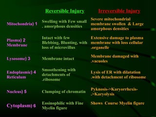
  • 1. Reversible Injury Irreversible Injury Severe mitochondrial Swelling with Few small Mitochondria( 1 membrane swollen & Large . amorphous densities amorphous densities Intact with few Extensive damage to plasma Plasma( 2 Blebbing, Blunting, with membrane with loss cellular Membrane loss of microvillus .organelle Membrane damaged with Lysosome( 3 Membrane intact .vacuoles Smoothening with Endoplasmic( 4 Lysis of ER with dilatation detachments of Reticulum .with detachment of ribosome .ribosome Pyknosis->Karyorrhexis- Nucleus( 5 Clumping of chromatin .>Karyolysis Eosinophilic with Fine Shows Course Myelin figure Cytoplasm( 6 Myelin figure
  • 2. Cell swelling - Light Microscopy Cellular Swelling = hydropic change Normal epithelium
  • 3. Nuclear Pyknosis Karyorrhexis Karyolysis
  • 4. Necrosis Necrosis refers to a spectrum of morphologic changes that follow cell death in living tissue, largely resulting from the progressive degradative action of enzymes on the lethally injured cell  occurs in the irreversible exogenous injury  Causes inflammation in the surrounding tissue as leakage of cellular organelle from damaged cell membrane occurs. occurs
  • 5. Morphology of Necrosis Cytoplasm:  increased eosinophilia because of the normal basophilia imparted by the RNA in the cytoplasm because of increased binding of eosin to denatured Intracytoplasmic proteins  the cytoplasm becomes vacuolated and appears moth-eaten.  Calcification of the dead cells. Dead cells ultimately replaced by large, whorled phospholipids masses called myelin figures Nuclear:  Pyknosis characterized by nuclear shrinkage and increased basophilia. the DNA apparently condenses into a solid, shrunken basophilic mass.  Karyolysis basophilia of the chromatin may fade.  Karyorrhexis pyknotic nucleus undergoes fragmentation. With the passage of time in a day or two, the nucleus in the necrotic cell totally disappears.
  • 6. By electron microscopy Necrotic cells are characterized by 1 , Damaged plasma and organelle membranes Marked dilation of mitochondria with the appearance of 2 , large amorphous densities 3 ,Intracytoplasmic myelin figures 4 ,Amorphous osmiophilic debris Aggregates of fluffy material probably representing 5 denatured protein
  • 7. Types of Necrosis :  Coagulative Necrosis ,  Liquifactive Necrosis,  Gangrenous Necrosis,  Caseous Necrosis,  Fat Necrosis.  Fibrinoid Necrosis.
  • 8. 1) Coagulative Necrosis : • Preservation of the basic outline of the coagulated cell for a span of at least some days. • Affected tissues exhibit a firm texture, • Preservation of the general tissue architecture, is characteristic of hypoxic death of cells in all tissues except the brain. A wedge-shaped kidney infarct (yellow). B, Microscopic view of the edge of the infarct, with normal kidney (N) and necrotic cells in the infarct (I) showing preserved cellular outlines with loss of nuclei and an inflammatory infiltrate
  • 9. 2) Liquefactive Necrosis : • Charestic of focal bacterial or fungal infection. • Often seen in hypoxic death of CNS. • liquefaction completely digests the dead cells. • The end result is transformation of the tissue into a liquid viscous mass. • If the process was initiated by acute inflammation, the material is frequently creamy yellow because of the presence of dead white cells and is called pus.
  • 10. 3) Gangrenous Necrosis (Surgical term): • Generally the lower leg, that has lost its blood supply and has undergone coagulation necrosis. When bacterial infection is superimposed, Coagulative necrosis is modified by the liquefactive action of the bacteria and the attracted leukocytes (so-called wet gangrene). • Dry gangrene: Is a form of infarction that results from ischemia and when there is secondary no infection because it gets dry. Characterized primarily by Coagulative necrosis without liquefaction. Dead tissue has mummified appearance (e.g. diabetic foot). Wet gangrene Dry gangrene
  • 11. 4) Caseous Necrosis : • A distinctive form of Coagulative necrosis, seen in tuberculous infection .The term caseous is derived from the cheesy white gross appearance of the area of necrosis. •On M/E: the necrotic focus appears as amorphous granular debris seemingly composed of fragmented, coagulated cells and amorphous granular debris enclosed within a distinctive inflammatory border known as a granulomatous reaction Epitheli oid cell Langha n’s giant A tuberculous lung with a large cell area of caseous necrosis. The caseous debris is yellow-white and cheesy.
  • 12. 5) Fat Necrosis : •This occurs in acute pancreatitis , activated pancreatic enzymes escape from acinar cells and ducts, the activated enzymes liquefy fat cell membranes. • Activated lipases split the triglyceride esters contained within fat cells. The released fatty acids combine with calcium to produce grossly visible chalky white areas as fat saponification. saponification • On M/E, the necrosis takes the form of foci of shadowy outlines of necrotic fat cells, with basophilic calcium deposits, surrounded by an inflammatory reaction. fat cells have lost their peripheral nuclei and their cytoplasm has converted to a mass of basophilic amorphous necrotic material.
  • 13. 6) Fibrinoid Necrosis : • Usually seen in immune reactions involving blood vessels. • This pattern of necrosis typically occurs when complexes of antigens and antibodies are deposited in the walls of arteries. • Deposits of these “immune complexes,” together with fibrin that has leaked out of vessels, result in a bright pink and amorphous appearance in H&E stains, called “fibrinoid” seen in vasculitis syndromes
  • 14. Apoptosis • From Greek meaning falling off. • Programmed and genetically controlled, enzyme dependent, specialized form of death of individual cells. • An active process and involves RNA and protein synthesis. • Plays role in – Physiologic cell death – Pathologic cell death. • Different from necrosis.
  • 15. Apoptosis  Apoptosis is a pathway of cell death that is induced by a tightly regulated suicide program in which cells die by activating enzymes that degrade the cells' own nuclear DNA and nuclear and cytoplasmic proteins.  Apoptotic cells break up into fragments, called apoptotic bodies, which contain portions of the cytoplasm and nucleus. The plasma membrane of the apoptotic cell and bodies remains intact.  Death by apoptosis is a normal phenomenon that serves to eliminate cells that are no longer needed, and to maintain a steady number of various cell populations in tissues.
  • 16. Remember ! IN APOPTOSIS: •Cells actually expend energy in order to die. •The cell membrane does not rupture. •The cell contents are not released into the extracellular space, and •Inflammation does not occur.
  • 17. Physiologic examples of apoptosis 1. Embryogenesis • Disappearance of Mullerian and Wolffian duct structures. • Development of lumen within hollow organs (e.g bowel and heart). 1. Hormone-dependent involution in adults – Endometrial breakdown in menstruation. – Post-lactational atrophy of breast. – Prostate atrophy following castration. 1. Involution of Thymus in the adult. 2. Cells that are programmed to die; for example, 1. The cells of the outer layers of epidermis, 2. Cells in the gut epithelium.
  • 18. Apoptosis in Physiologic Situations  Embryogenesis: programmed destruction of cells Hormone-dependent involution in the adult, such as endometrial cell breakdown during the menstrual cycle, the regression of the lactating breast after weaning, and prostatic atrophy after castration.  Cell deletion in proliferating cell populations ( intestinal epithelia).  Death of host cells that have served their useful purpose, such as neutrophils in an acute inflammatory response, and lymphocytes at the end of an immune response.  Elimination of potentially harmful self-reactive lymphocytes, either before or after they have completed their maturation  Cell death induced by cytotoxic T cells, a defense mechanism against viruses and tumors that serves to eliminate virus-infected and neoplastic cells.
  • 19. Apoptosis in Pathological conditions Apoptosis eliminates cells that are injured beyond repair without eliciting a host reaction. DNA damage. Radiation, cytotoxic anticancer drugs, and hypoxia can damage DNA, either directly or via production of free radicals. These injurious stimuli can cause apoptosis if the insult is mild. The damaged DNA, which may result in malignant transformation. Accumulation of misfolded proteins mutations in the genes encoding these proteins or because of extrinsic factors, such as damage caused by free radicals. Excessive accumulation of these proteins in the ER leads to a condition called ER stress, which culminates in apoptotic cell death. Cell death in certain in viral infections, (in Adenovirus and HIV infections) infections or by the host immune response (as in viral hepatitis). An important host response to viruses consists of cytotoxic T lymphocytes specific for viral proteins is responsible for cell death in viral, tumors and cellular rejection of transplants. Pathologic atrophy in parenchymal organs after duct obstruction , such as occurs in the pancreas, parotid gland, and kidney.
  • 20. Morphologic appearance 1. Apoptotic cells: 1. Have deeply pink staining cytoplasm. 2. Have pyknotic nucleus which fragment. 3. Are smaller in size. 4. Breakdown into fragments (apoptotic bodies). 5. Phagocytosis of apoptotic bodies by adjacent cells or macrophages. 6. A lack of inflammatory response.
  • 21. Removal of dead cells • Formation of cytoplasmic buds on the cell membrane containing – Nuclear fragments, mitochondria and protein fragments. • Breaking off of cytoplasmic buds  apoptotic bodies. • Phagocytosis of apoptotic bodies by neighboring cells or macrophages.
  • 23. Apoptosis of Apoptotic cell in epidermal liver cells
  • 24. Studies that the most effective way of prolonging life span is • Calorie restriction. effect of calorie restriction on longevity appears to be mediated by a family of proteins called sirtuins. •Sirtuins have histone deacetylase activity, reduce apoptosis, stimulate protein folding, and inhibit the harmful effects of oxygen free radicals. Sirtuins also increase insulin sensitivity and glucose metabolism. Food product like Vit-C, Vit-A, Vit-D, Green Tea, B-Carotiens and Red wine activate sirtuins and thus increase life span Or decreases Cellular Aging! • Growth factors, such as insulin-like growth factor, and intracellular signaling pathways triggered by these hormones also influence life span. • Transcription factors activated by insulin receptor signaling may induce genes that reduce longevity, and insulin receptor mutations are associated with increased lifespan.