Malignant tumors of skin
•Download as PPTX, PDF•
73 likes•16,407 views

ppt on basics of skin tumors, squamous cell carcinoma , bcc, melanoma for medical sudents.

Recommended
More Related Content
What's hot
What's hot (20)
Similar to Malignant tumors of skin
Similar to Malignant tumors of skin (20)
More from Mukhilesh Ramesh
More from Mukhilesh Ramesh (6)
Recently uploaded
Recently uploaded (20)
Malignant tumors of skin
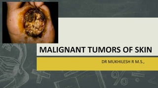
- 1. MALIGNANT TUMORS OF SKIN DR MUKHILESH R M.S.,
- 2. Salient Features Of Skin Malignancies Most commonly epidermal origin Basal cell carcinoma Squamous cell carcinoma Malignant melanoma Skin adnexal tumors are rare. Chemical carcinogens play a major role.
- 3. Basal Cell Carcinoma Most common skin tumor, originates from basal layer of epidermis Slowly growing , locally invasive – RODENT ULCER. 26 histological variants. Most common are Nodular Superficial speading Infiltrative Pigmented & Morpheaform
- 4. Predisposing factors UV rays Arsenics, coal , tar BCC White skin, genetics Middle aged, men
- 5. Pathogenesis No apparent precursor lesion Locally infiltrative. Rarely metastasise. Never lympatic spread Ovoid cells in nests with outer pallisading layer.
- 6. Contd… Nodulocystic Waxy , cream coloured with rolled, pearly borders surrounding central ulcer. Morpheaform Type IV collagenase and spread rapidly Flat, plaque like lesion Basosquamous variant Highly aggressive Metastasize similar to SCC and aggressive treatment required.
- 7. Prognosis High risk BCC >2cm Specific location – nose , ear, eyes Ill-defined margins Recurrent tumors immunosuppression
- 8. Management of BCC Surgical VS Non Surgical Non surgical Curettage Electrodessication Laser vapourisation Destroy any potential tissue sample for pathological confirmation and margin analysis
- 9. Surgical Management Complete tumor removal , with pathological confirmation and margin analysis. Large tumors invading adjacent structure with aggressive histology – WIDE LOCAL EXCISION 0.5-1cm margin Reconstructive procedures
- 10. MOHS Micrographic Surgery Excision of skin cancer under microscopic control. Minimise recurrent rates with maximum conservation. Indicated in Poorly demarcated, Recurrent / incompletely excised Near vital structures Can also be used for SCC, lentigo maligna,DFS
- 11. Contd… Under local anesthesia Saucerising excision of primary tumor Sample and defect are marked and oriented Stained with H&E. Examination of slide for residual tumor Excise more tissue from mapped area.
- 12. Contd…
- 13. Other Modalaties Radiotherapy Topical treatments 5-fluorouracil Imiquimod Cryotherapy
- 14. Cutaneous Squamous Cell Carcinoma Malignant tumor of keratinising epithelium of epidermis 2nd most common tumor Cumulative sun exposure and damage Associated with pre-existing scars, osetomyelitis, burn. Marjolin’s ulcer
- 15. Pathogenesis Sun exposure Scars and sinuses Chemical carcinogens SCC Tobacco use HPV 5 & HPV 16
- 16. Pathology Smooth nodular to verrucous , papillamatous and ulcerating lesions. Everted edges and surrounded by inflamed, indurated skin. Distant metastasis. Secondary lymph nodes involvement.
- 17. Differential Diagnosis Of SCC Actinic keratosis BCC Keratoacanthoma Pyoderma gangrenosum Warts
- 18. Microscopic Appearance Irregular masses of squamous epithelium proliferate and invade dermis. KERATIN PEARLS Perineural / vascular invasion Positive for cytokeratin 1 and 10 Border’s histological grading Ratio of pleomorphic and anaplastic to normal cells
- 19. Prognosis Invasion Depth – deeper lesion , worse the prognosis Surface size - >2 cm Histological grade Site Lips and ears – increase recurrent rate Immunosuppression Perineural and vascular involvement Aetiology
- 20. TNM Classification Size • T1 - <2cm • T2 - 2-5 cm • T3 - >5cm • T4 - muscle or bone involvement Nodes • N0 - no regional nodes • N1 - regional nodes Metastasis • M0 - no metastasis • M1- distant metastasis Grade • G1- low grade • G2moderately differentiated • G3- high grade
- 21. Management Surgical excision – accurate histology Margins to be assessed 4mm clearance for <2cm 1 cm clearance for >2cm Radiotherapy resistant – Veruccus carcinoma
- 22. Malignant Melanoma Cancer of melanocytes Wherever melanocytes exist Bowel mucosa Retina Leptomeninges
- 24. Macroscopic Features In Nevi Suggesting Malignant Melanoma
- 25. Contd… Tingling Itching Serosanguinous discharge Blood supply Melanomas >1mm have blood supply – doppler positive pigmented lesion
- 26. Types Of Malignant Melanoma Superficial spreading Nodular melanoma Lentigo maligna melanoma Acral lentiginous melanoma Amelanotic melanoma Desmoplastic melanoma
- 27. Superficial Spreading Melanoma Commonest type – 70% Arise from pre – existing nevus Rapid growth of darker pigmented are in a junctional nevus. Predominantly radial growth phase. Nodularity can occur – vertical growth phase.
- 28. Nodular Melanoma More aggressive Increased vertical growth than radial phase Middle age men. Usually trunk. Sharply demarcated, blue-black papules 1-2cm. Lack horizontal growth phase.
- 29. Lentigo Maligna Melanoma Hutchinson’s melanotic freckle Slow growing, variegated, brown macule Intense sun exposure. Women > men Less metastaic potential Better prognosis
- 30. Acral Lentiginous Melanoma Soles of feet and palms of hand Rare in white skinned people Flat, irregular macule. Can mimic a fungal infection Biopsy of the nail matrix rather than just the pigment. Hutchinson’s sign nail-fold pigmentation then widens progressively to produce a triangular pigmented macule with nail dystrophy.
- 31. Miscellaneous Amelanotic melanoma Not pigmented Poor prognosis Desmoplastic melanoma Head and neck Perineural invasion High recurrent rate
- 32. Histology Malignant changes of melanocytes in basal epidermis Horizontal growth phase – cells spread along the dermo-epidermal junction Vertical growth phase – dermis may be invaded and increased metastatic potential.
- 33. Satellite nodules Lesions situated with in 2-5cm of the primary Intransit lesions Situated >5cm , proximal to lymphnode basin
- 34. Management History and clinical examination Excision biopsy with 2mm margin of skin and subdermal fat. Incisional biopsy – large lesion / facial lesions where excision results in scarring. Staging of melanoma Clarkes’ staging Breslows’ classification
- 37. Management Of Malignant Melanoma Pigmented lesion Biopsy Diagnosis of melanoma <1mm depth Excision - 1cm margin 2-4mm depth Excision -2cm margin >4mm depth Excision – 3cm margin
- 38. Management of lymphnodes Based on breslow thickness. <1mm least beneficial with prophylactic dissection. >4mm increased chance of both lymphatic and distant metastasis. Intermediate thickness Elective prophylactic lymph node dissection Sentinel lymphnode biopsy
- 41. Other Modalities Chemotherapy Melphalan Vemurafenib Isolated limb perfusion therapy Immunotherapy IFN / TNF ALPHA Radiotherapy