SlideShare a Scribd company logo
1 of 104
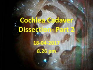
Cochlea Cadaver
Dissection- Part 2
18-04-2017
8.26 pm
Part-1 of this PPT
present at weblink
www.skullbase360.in
Middle cranial fossa
approach for
Cochlear implant
APICAL TURN / SUPERIOR TURN
cochleostomy in middle cranial
fossa approach
So the indication of middle cranial fossa superior
cochleostomy insertion is in infected cases after CWD
+ SP ( subtotal petrosectomy ) We can do redo by fat
risnced in rifampacin . But if you want to go by sterile
area middle cranial fossa superior cochleostomy &
reverse insertion & reverse programming
Superior cochleostomy in middle cranial fossa is just below origin
of GSPN Don't fear about carotid ( metal probe ) . Petrous carotid
is 1 cm anterior to origin of GSPN
Superior cochleostomy in middle cranial fossa is just below origin
of GSPN Don't fear about carotid ( metal probe ) . Petrous carotid
is 1 cm anterior to origin of GSPN
Superior cochleostomy in middle cranial fossa is just below origin
of GSPN Don't fear about carotid ( metal probe ) . Petrous carotid
is 1 cm anterior to origin of GSPN
Probe in Superior cochleostomy in
middle cranial fossa is just below origin
of GSPN
Superior cochleostomy in middle
cranial fossa is just below origin of
GSPN
Superior cochleostomy in middle
cranial fossa is just below origin of
GSPN
See the probe inserted through superior cochleostomy from
middle cranial fossa exactly corresponds to superior
cochleostomy just below tensor tympani from middle ear
See the probe inserted through superior cochleostomy from
middle cranial fossa exactly corresponds to superior
cochleostomy just below tensor tympani from middle ear
See the probe inserted through superior cochleostomy from middle cranial
fossa exactly corresponds to superior cochleostomy just below tensor
tympani from middle ear
See the probe inserted through superior cochleostomy
from middle cranial fossa exactly corresponds to
superior cochleostomy just below tensor tympani from
middle ear
Labyrinthine part of facial nerve
in middle cranial fossa
Just now i fractured
tegmen of middle ear with
my finger nail … it is so thin
…………..So identify ossicles
of middle ear through very
thin middle ear tegmen &
then identify horizontal
facial nerve & then 1st
genu & then labyrinthine
facial nerve ...... simplest
way to decompress
labyrinthine Or else if you
come from medically you
may injure cochlea or SSC
Note horizontal part of facial nerve
through middle cranial fossa as
continuation of GSPN
tegmen of middle ear is so thin …………..So identify ossicles of
middle ear through very thin middle ear tegmen & then identify
horizontal facial nerve & then 1st genu & then labyrinthine facial
nerve ...... simplest way to decompress labyrinthine Or else if you
come from medically you may injure cochlea or SSC
Note horizontal part of facial nerve
through middle cranial fossa as
continuation of GSPN
facial nerve in lateral part of IAC decompression is difficult even in middle
cranial fossa. It is between two solid bones of cochlea & SSC
facial nerve in lateral part of IAC decompression is difficult even
in middle cranial fossa. It is between two solid bones of cochlea
& SSC
IAC [ Internal Auditory Canal ]
Drilling
IAC conical tube present in angle of
SSC crest & GSPN ( more than 50 %
dehiscent )
IAC conical tube present in angle of
SSC crest & GSPN ( more than 50 %
dehiscent )
IAC has to be drilled from medial to lateral IAC first must be opened medially & then
only tracked along the direction of IAC ( postero-laterally ) Unless you injure cochlea
basal & medial turns
IAC has to be drilled from medial to lateral IAC first must be
opened medially & then only tracked along the direction of IAC (
postero-laterally ) Unless you injure cochlea basal & medial turns
KAWASE APPROACH
The pit infront of cochlea & IAC is
kawase approach
The pit infront of cochlea & IAC is
kawase approach
Here I am expanding kawase approach . In few minutes I show
you COA ( cochlear aperture)
Here I am expanding kawase approach . In few minutes
I show you COA ( cochlear aperture)
Probing in middle turn
Observe metal probe in perisiers (
dangerous) triangle
Observe metal probe in perisiers (
dangerous) triangle
Observe metal probe in perisiers (
dangerous) triangle
Perisiers triangle corresponds to
labyrinthine part of facial nerve
So the metal probe in perisiers triangle goes to the middle turn of cochlea & exactly
corresponds to labyrinthine part of facial nerve So middle turn array of CI stimulates
labyrinthine part of facial nerve causing twitchings in post-op . Then we have to switch
off those electrodes in software programming .
So the metal probe in perisiers triangle goes to the middle turn of cochlea &
exactly corresponds to labyrinthine part of facial nerve So middle turn array
of CI stimulates labyrinthine part of facial nerve causing twitchings in post-op
. Then we have to switch off those electrodes in software programming .
So the metal probe in perisiers triangle goes to the middle turn of cochlea & exactly corresponds
to labyrinthine part of facial nerve So middle turn array of CI stimulates labyrinthine part of facial
nerve causing twitchings in post-op . Then we have to switch off those electrodes in software
programming .
So the metal probe in perisiers triangle goes to the middle turn of cochlea &
exactly corresponds to labyrinthine part of facial nerve So middle turn array
of CI stimulates labyrinthine part of facial nerve causing twitchings in post-op
. Then we have to switch off those electrodes in software programming .
Probing in basal turn
Probe in basal turn opens into basal
turn cochleostomy in middle cranial
fossa
Probe in basal turn opens into basal
turn cochleostomy in middle cranial
fossa
Probe in basal turn opens into basal
turn cochleostomy in middle cranial
fossa
Probe in basal turn opens into basal
turn cochleostomy in middle cranial
fossa
Probe in basal turn opens into basal
turn cochleostomy in middle cranial
fossa
Probe in basal turn opens into basal
turn cochleostomy in middle cranial
fossa
Probe in basal turn opens into basal
turn cochleostomy in middle cranial
fossa
Probe in basal turn opens into basal
turn cochleostomy in middle cranial
fossa
See all the turns of cochlea from
middle fossa
SVN & FN converge
Superior Vestibular nerve ( SVN ) & facial nerve separatedby bills bar , that I
drilled . Observe here SVN & FN converge . Where as IVN & cochlear nerve
diverge ……….. This convergence of SVN & FN very useful in MRI reading
See horizontal Septum in IAC below SVN & FN ;
I cut superior Vestibular nerve ( SVN ) & FN
IVN & CN diverge
Observe here the IVN & cochlear nerve diverge ( not so clear in
cadaver )
Observe here the IVN & cochlear nerve diverge ( not so clear in
cadaver )
Observe here the IVN & cochlear nerve diverge ( not so clear in
cadaver )
Observe here the IVN & cochlear nerve diverge ( not so clear in
cadaver )
COA [ Cochlear aperture ]
Observe here cochlear nerve fibres going through the cibriform
area ( entry point of modiolus ) In COA ( cochlear aperture )
Observe in this one COA is 2.5 to 3 mm roughtly.
If COA less than 1.5 mm it is cochlear nerve aplasia
Cochlear implant
after
Translabyrinthine
approach
PSC is deeper than LSC & SSC is deeper
than PSC
Intact facial canal technique of Skull base .
If you transpose grade 3 facial palsy comes .
Ampulla of PSC bisects vertical part of
facial nerve exactly at midpoint
See probe coming to Sinus tympani So while clearing Sinus tympani PSC
exposed ... becareful
CI after LABYRINTHECTOMY
Only two is enough for CI – one is
cochlea & another cochlear nerve –
so even in vestibular schwannoma
excision by translabyrinthine
exposure we can do CI & patient
hears
Bills bar between FN & SVN
Labyrinthectomy done to enter
Posterior cranial fossa
VA [ Vestibular Aqueduct ]
https://drive.google.com/file/d/0B7F_FcpOJCfpS2lJa3NBNkVDeV
E/view?usp=sharing
IAC & VA are two eyes of baby in
temporal boone
IAC & VA are two eyes of baby in
temporal boone
Endolymphatic duct & Vestibular aqueduct both are same or not ........ I have to refer .
....... but clearly there is duct from vestibule to endolymphatic sac area . If it is more
than 1.5 mm it is " dilated Vestibular aqueduct " Another 1.5mm is ........, if COA (
cochlear aperture ) less than 1.5mm it is cochlear nerve aplasia.
Mario sanna book mention >1.5 mm VA dilated . For mnemonic sake 1.5 mm
is there at both VA & COA . One is more & one is less respectively
Radiologically if the width of the Vestibular aqueduct is more than the width
of the PSC, then it is dilated. -----Satish jain sir says >2mm VA dilated in any
section .
In HRCT Temporal bone Vestibular aqueduct ( VA )is seen parallel
to PSC ( Posterior semi circular canal ) Here also after drilling PSC
we are seeing VA
anatomically also after drilling PSC we are seeing VA .... so radiologically also
both sizes same [ my mnemonic & philosophy ] ..... if VA more than PSC it is
dilated
Abnormal cochleas dissection photos
added later in few days
Essence of abnormal cochleas
1. IP 2 is exactly like normal cochlea
2. IP 3 - wide cochleostomy & precurved electrode
3. cochlear hypoplasia -- outcomes depends on how many number
of electrodes inserted . Minimum 10 electrodes insertion should
be there to get better outcome
4. IP 1 - lateral wall electrode
5. common cavity - lateral wall electrode
6. CHARGE - still try CI , not working then ABI.
7. michel - ABI directly
In all abnormalities see cochlear nerve aplasia .... even absent in MRI ,
do EABR & keep CI
Part-1 of this PPT
present at weblink
www.skullbase360.in

More Related Content

What's hot

Endoscopic tranasglabellar & supraorbital approach
Endoscopic tranasglabellar & supraorbital approachEndoscopic tranasglabellar & supraorbital approach
Endoscopic tranasglabellar & supraorbital approachMurali Chand Nallamothu
 
Transoral & Transorbital approaches of skull base
Transoral & Transorbital approaches of skull baseTransoral & Transorbital approaches of skull base
Transoral & Transorbital approaches of skull baseMurali Chand Nallamothu
 
Dr neeraj suri ,AssoProf,Gmers medical college ,Gandhinagar.
Dr neeraj suri ,AssoProf,Gmers medical college ,Gandhinagar.Dr neeraj suri ,AssoProf,Gmers medical college ,Gandhinagar.
Dr neeraj suri ,AssoProf,Gmers medical college ,Gandhinagar.neerajmsuri
 
Anatomy of Skullbase
Anatomy of SkullbaseAnatomy of Skullbase
Anatomy of SkullbaseJinu Iype
 
Scalp incision and blood supply
Scalp incision and blood supplyScalp incision and blood supply
Scalp incision and blood supplydrajay02
 
Anatomy and Xray of PNS and orbit
Anatomy and Xray  of PNS and  orbitAnatomy and Xray  of PNS and  orbit
Anatomy and Xray of PNS and orbitSandeepAwal2
 

What's hot (20)

Endoscopic tranasglabellar & supraorbital approach
Endoscopic tranasglabellar & supraorbital approachEndoscopic tranasglabellar & supraorbital approach
Endoscopic tranasglabellar & supraorbital approach
 
Transoral & Transorbital approaches of skull base
Transoral & Transorbital approaches of skull baseTransoral & Transorbital approaches of skull base
Transoral & Transorbital approaches of skull base
 
Combined approaches of skull base 360°
Combined approaches of skull base 360°Combined approaches of skull base 360°
Combined approaches of skull base 360°
 
IAC 360°
IAC 360°IAC 360°
IAC 360°
 
Endoscopic lateral skull base
Endoscopic lateral skull baseEndoscopic lateral skull base
Endoscopic lateral skull base
 
Skull base 360°- part 2
Skull base 360°- part 2Skull base 360°- part 2
Skull base 360°- part 2
 
Skull base recontruction 360°
Skull base recontruction 360°Skull base recontruction 360°
Skull base recontruction 360°
 
Dr neeraj suri ,AssoProf,Gmers medical college ,Gandhinagar.
Dr neeraj suri ,AssoProf,Gmers medical college ,Gandhinagar.Dr neeraj suri ,AssoProf,Gmers medical college ,Gandhinagar.
Dr neeraj suri ,AssoProf,Gmers medical college ,Gandhinagar.
 
Skull base imaging
Skull base imagingSkull base imaging
Skull base imaging
 
REZ 360°
REZ 360°REZ 360°
REZ 360°
 
10 triangles 360°
10 triangles 360°10 triangles 360°
10 triangles 360°
 
Inferior petrosal sinus 360°
Inferior petrosal sinus 360°Inferior petrosal sinus 360°
Inferior petrosal sinus 360°
 
Anatomy of Skullbase
Anatomy of SkullbaseAnatomy of Skullbase
Anatomy of Skullbase
 
Pituitary 360°
Pituitary 360°Pituitary 360°
Pituitary 360°
 
Imaging requirements for cochlear implantation
Imaging requirements for cochlear implantationImaging requirements for cochlear implantation
Imaging requirements for cochlear implantation
 
Endoscopic orbital surgery
Endoscopic orbital surgeryEndoscopic orbital surgery
Endoscopic orbital surgery
 
Radiology in ENT
Radiology in ENTRadiology in ENT
Radiology in ENT
 
Anterior cranial fossa 360°
Anterior cranial fossa 360°Anterior cranial fossa 360°
Anterior cranial fossa 360°
 
Scalp incision and blood supply
Scalp incision and blood supplyScalp incision and blood supply
Scalp incision and blood supply
 
Anatomy and Xray of PNS and orbit
Anatomy and Xray  of PNS and  orbitAnatomy and Xray  of PNS and  orbit
Anatomy and Xray of PNS and orbit
 

Similar to Cochlea cadaver dissection - part 2

Hrct temporal bone pk1 ppt
Hrct temporal bone pk1 pptHrct temporal bone pk1 ppt
Hrct temporal bone pk1 pptDr pradeep Kumar
 
Anatomy of neuroaxial system final
Anatomy of neuroaxial system finalAnatomy of neuroaxial system final
Anatomy of neuroaxial system finaldr anurag giri
 
IMAGING OF TEMPORAL BONE
IMAGING OF TEMPORAL BONEIMAGING OF TEMPORAL BONE
IMAGING OF TEMPORAL BONESameer Peer
 
Radiology in ENT
Radiology in ENTRadiology in ENT
Radiology in ENTAnwaaar
 
Temporal bone Radiologic anatomy.. In depth
Temporal bone Radiologic anatomy.. In depthTemporal bone Radiologic anatomy.. In depth
Temporal bone Radiologic anatomy.. In depthAhmed Hamdy Hamdy
 
Posterio anterior cephalometrics / dental implant courses by Indian dental ac...
Posterio anterior cephalometrics / dental implant courses by Indian dental ac...Posterio anterior cephalometrics / dental implant courses by Indian dental ac...
Posterio anterior cephalometrics / dental implant courses by Indian dental ac...Indian dental academy
 
ppMRIARTIFACTS.pptx
ppMRIARTIFACTS.pptxppMRIARTIFACTS.pptx
ppMRIARTIFACTS.pptxKeerthan39
 
Postero anterior cephalometrics /certified fixed orthodontic courses by Indi...
Postero anterior  cephalometrics /certified fixed orthodontic courses by Indi...Postero anterior  cephalometrics /certified fixed orthodontic courses by Indi...
Postero anterior cephalometrics /certified fixed orthodontic courses by Indi...Indian dental academy
 
Postero - Anterior cephalometry basics/cosmetic dentistry courses
Postero - Anterior cephalometry basics/cosmetic dentistry coursesPostero - Anterior cephalometry basics/cosmetic dentistry courses
Postero - Anterior cephalometry basics/cosmetic dentistry coursesIndian dental academy
 
Revision of the Anatomy of the Posterior Aspect of the Eyeball - An Essentia...
Revision of the Anatomy of the Posterior Aspect of the Eyeball -  An Essentia...Revision of the Anatomy of the Posterior Aspect of the Eyeball -  An Essentia...
Revision of the Anatomy of the Posterior Aspect of the Eyeball - An Essentia...DrAbdelLatifsiam
 
Postero - Anterior Chphalometry /cosmetic dentistry courses
Postero - Anterior Chphalometry /cosmetic dentistry coursesPostero - Anterior Chphalometry /cosmetic dentistry courses
Postero - Anterior Chphalometry /cosmetic dentistry coursesIndian dental academy
 

Similar to Cochlea cadaver dissection - part 2 (20)

Orbit 360°
Orbit 360° Orbit 360°
Orbit 360°
 
Infratemporal fossa 360°
Infratemporal fossa 360°Infratemporal fossa 360°
Infratemporal fossa 360°
 
Hrct temporal bone pk1 ppt
Hrct temporal bone pk1 pptHrct temporal bone pk1 ppt
Hrct temporal bone pk1 ppt
 
Cephalometrics (2)
Cephalometrics (2)Cephalometrics (2)
Cephalometrics (2)
 
Anatomy of neuroaxial system final
Anatomy of neuroaxial system finalAnatomy of neuroaxial system final
Anatomy of neuroaxial system final
 
IMAGING OF TEMPORAL BONE
IMAGING OF TEMPORAL BONEIMAGING OF TEMPORAL BONE
IMAGING OF TEMPORAL BONE
 
L-OCR & M-OCR 360°
L-OCR & M-OCR 360°L-OCR & M-OCR 360°
L-OCR & M-OCR 360°
 
Radiology in ENT
Radiology in ENTRadiology in ENT
Radiology in ENT
 
Thyroid Gland
Thyroid GlandThyroid Gland
Thyroid Gland
 
Temporal bone Radiologic anatomy.. In depth
Temporal bone Radiologic anatomy.. In depthTemporal bone Radiologic anatomy.. In depth
Temporal bone Radiologic anatomy.. In depth
 
Imaging in ent
Imaging in entImaging in ent
Imaging in ent
 
Posterio anterior cephalometrics / dental implant courses by Indian dental ac...
Posterio anterior cephalometrics / dental implant courses by Indian dental ac...Posterio anterior cephalometrics / dental implant courses by Indian dental ac...
Posterio anterior cephalometrics / dental implant courses by Indian dental ac...
 
ppMRIARTIFACTS.pptx
ppMRIARTIFACTS.pptxppMRIARTIFACTS.pptx
ppMRIARTIFACTS.pptx
 
Postero anterior cephalometrics /certified fixed orthodontic courses by Indi...
Postero anterior  cephalometrics /certified fixed orthodontic courses by Indi...Postero anterior  cephalometrics /certified fixed orthodontic courses by Indi...
Postero anterior cephalometrics /certified fixed orthodontic courses by Indi...
 
Postero - Anterior cephalometry basics/cosmetic dentistry courses
Postero - Anterior cephalometry basics/cosmetic dentistry coursesPostero - Anterior cephalometry basics/cosmetic dentistry courses
Postero - Anterior cephalometry basics/cosmetic dentistry courses
 
Revision of the Anatomy of the Posterior Aspect of the Eyeball - An Essentia...
Revision of the Anatomy of the Posterior Aspect of the Eyeball -  An Essentia...Revision of the Anatomy of the Posterior Aspect of the Eyeball -  An Essentia...
Revision of the Anatomy of the Posterior Aspect of the Eyeball - An Essentia...
 
Postero - Anterior Chphalometry /cosmetic dentistry courses
Postero - Anterior Chphalometry /cosmetic dentistry coursesPostero - Anterior Chphalometry /cosmetic dentistry courses
Postero - Anterior Chphalometry /cosmetic dentistry courses
 
Cranial nerves 360°
Cranial nerves 360°Cranial nerves 360°
Cranial nerves 360°
 
Carotid 360°
Carotid 360°Carotid 360°
Carotid 360°
 
Clivus 360°
Clivus 360°Clivus 360°
Clivus 360°
 

More from Murali Chand Nallamothu

More from Murali Chand Nallamothu (12)

Cochlear implant systems
Cochlear implant systemsCochlear implant systems
Cochlear implant systems
 
Electro Acoustic Stimulation ( EAS )
Electro Acoustic Stimulation ( EAS ) Electro Acoustic Stimulation ( EAS )
Electro Acoustic Stimulation ( EAS )
 
Cochlear implant imaging
Cochlear implant imagingCochlear implant imaging
Cochlear implant imaging
 
Cochlear implant in a 2nd & 3 tier city
Cochlear implant in a 2nd & 3 tier cityCochlear implant in a 2nd & 3 tier city
Cochlear implant in a 2nd & 3 tier city
 
Line diagrams - skull base 360 - part 1
Line diagrams - skull base 360 - part 1Line diagrams - skull base 360 - part 1
Line diagrams - skull base 360 - part 1
 
Craniopharyngioma - What is the best approach
Craniopharyngioma - What is the best approach Craniopharyngioma - What is the best approach
Craniopharyngioma - What is the best approach
 
Decision making between anterior skull base & lateral skull base
Decision making between anterior skull base & lateral skull baseDecision making between anterior skull base & lateral skull base
Decision making between anterior skull base & lateral skull base
 
Skull base 360°- videos
Skull base 360°- videosSkull base 360°- videos
Skull base 360°- videos
 
Cranio vertebral junction / Foramen magnum 360°
Cranio vertebral junction / Foramen magnum 360°Cranio vertebral junction / Foramen magnum 360°
Cranio vertebral junction / Foramen magnum 360°
 
Cisterns 360°
Cisterns 360°Cisterns 360°
Cisterns 360°
 
Open skull base approaches
Open skull base approachesOpen skull base approaches
Open skull base approaches
 
Vertebral artery 360°
Vertebral artery 360°Vertebral artery 360°
Vertebral artery 360°
 

Recently uploaded

Grant Readiness 101 TechSoup and Remy Consulting
Grant Readiness 101 TechSoup and Remy ConsultingGrant Readiness 101 TechSoup and Remy Consulting
Grant Readiness 101 TechSoup and Remy ConsultingTechSoup
 
Kodo Millet PPT made by Ghanshyam bairwa college of Agriculture kumher bhara...
Kodo Millet  PPT made by Ghanshyam bairwa college of Agriculture kumher bhara...Kodo Millet  PPT made by Ghanshyam bairwa college of Agriculture kumher bhara...
Kodo Millet PPT made by Ghanshyam bairwa college of Agriculture kumher bhara...pradhanghanshyam7136
 
2024-NATIONAL-LEARNING-CAMP-AND-OTHER.pptx
2024-NATIONAL-LEARNING-CAMP-AND-OTHER.pptx2024-NATIONAL-LEARNING-CAMP-AND-OTHER.pptx
2024-NATIONAL-LEARNING-CAMP-AND-OTHER.pptxMaritesTamaniVerdade
 
On National Teacher Day, meet the 2024-25 Kenan Fellows
On National Teacher Day, meet the 2024-25 Kenan FellowsOn National Teacher Day, meet the 2024-25 Kenan Fellows
On National Teacher Day, meet the 2024-25 Kenan FellowsMebane Rash
 
Mixin Classes in Odoo 17 How to Extend Models Using Mixin Classes
Mixin Classes in Odoo 17  How to Extend Models Using Mixin ClassesMixin Classes in Odoo 17  How to Extend Models Using Mixin Classes
Mixin Classes in Odoo 17 How to Extend Models Using Mixin ClassesCeline George
 
ComPTIA Overview | Comptia Security+ Book SY0-701
ComPTIA Overview | Comptia Security+ Book SY0-701ComPTIA Overview | Comptia Security+ Book SY0-701
ComPTIA Overview | Comptia Security+ Book SY0-701bronxfugly43
 
Accessible Digital Futures project (20/03/2024)
Accessible Digital Futures project (20/03/2024)Accessible Digital Futures project (20/03/2024)
Accessible Digital Futures project (20/03/2024)Jisc
 
Python Notes for mca i year students osmania university.docx
Python Notes for mca i year students osmania university.docxPython Notes for mca i year students osmania university.docx
Python Notes for mca i year students osmania university.docxRamakrishna Reddy Bijjam
 
psychiatric nursing HISTORY COLLECTION .docx
psychiatric  nursing HISTORY  COLLECTION  .docxpsychiatric  nursing HISTORY  COLLECTION  .docx
psychiatric nursing HISTORY COLLECTION .docxPoojaSen20
 
Seal of Good Local Governance (SGLG) 2024Final.pptx
Seal of Good Local Governance (SGLG) 2024Final.pptxSeal of Good Local Governance (SGLG) 2024Final.pptx
Seal of Good Local Governance (SGLG) 2024Final.pptxnegromaestrong
 
Unit-V; Pricing (Pharma Marketing Management).pptx
Unit-V; Pricing (Pharma Marketing Management).pptxUnit-V; Pricing (Pharma Marketing Management).pptx
Unit-V; Pricing (Pharma Marketing Management).pptxVishalSingh1417
 
Explore beautiful and ugly buildings. Mathematics helps us create beautiful d...
Explore beautiful and ugly buildings. Mathematics helps us create beautiful d...Explore beautiful and ugly buildings. Mathematics helps us create beautiful d...
Explore beautiful and ugly buildings. Mathematics helps us create beautiful d...christianmathematics
 
1029 - Danh muc Sach Giao Khoa 10 . pdf
1029 -  Danh muc Sach Giao Khoa 10 . pdf1029 -  Danh muc Sach Giao Khoa 10 . pdf
1029 - Danh muc Sach Giao Khoa 10 . pdfQucHHunhnh
 
The basics of sentences session 3pptx.pptx
The basics of sentences session 3pptx.pptxThe basics of sentences session 3pptx.pptx
The basics of sentences session 3pptx.pptxheathfieldcps1
 
General Principles of Intellectual Property: Concepts of Intellectual Proper...
General Principles of Intellectual Property: Concepts of Intellectual  Proper...General Principles of Intellectual Property: Concepts of Intellectual  Proper...
General Principles of Intellectual Property: Concepts of Intellectual Proper...Poonam Aher Patil
 
Introduction to Nonprofit Accounting: The Basics
Introduction to Nonprofit Accounting: The BasicsIntroduction to Nonprofit Accounting: The Basics
Introduction to Nonprofit Accounting: The BasicsTechSoup
 
Magic bus Group work1and 2 (Team 3).pptx
Magic bus Group work1and 2 (Team 3).pptxMagic bus Group work1and 2 (Team 3).pptx
Magic bus Group work1and 2 (Team 3).pptxdhanalakshmis0310
 
Food safety_Challenges food safety laboratories_.pdf
Food safety_Challenges food safety laboratories_.pdfFood safety_Challenges food safety laboratories_.pdf
Food safety_Challenges food safety laboratories_.pdfSherif Taha
 
Spellings Wk 3 English CAPS CARES Please Practise
Spellings Wk 3 English CAPS CARES Please PractiseSpellings Wk 3 English CAPS CARES Please Practise
Spellings Wk 3 English CAPS CARES Please PractiseAnaAcapella
 

Recently uploaded (20)

Grant Readiness 101 TechSoup and Remy Consulting
Grant Readiness 101 TechSoup and Remy ConsultingGrant Readiness 101 TechSoup and Remy Consulting
Grant Readiness 101 TechSoup and Remy Consulting
 
Kodo Millet PPT made by Ghanshyam bairwa college of Agriculture kumher bhara...
Kodo Millet  PPT made by Ghanshyam bairwa college of Agriculture kumher bhara...Kodo Millet  PPT made by Ghanshyam bairwa college of Agriculture kumher bhara...
Kodo Millet PPT made by Ghanshyam bairwa college of Agriculture kumher bhara...
 
2024-NATIONAL-LEARNING-CAMP-AND-OTHER.pptx
2024-NATIONAL-LEARNING-CAMP-AND-OTHER.pptx2024-NATIONAL-LEARNING-CAMP-AND-OTHER.pptx
2024-NATIONAL-LEARNING-CAMP-AND-OTHER.pptx
 
On National Teacher Day, meet the 2024-25 Kenan Fellows
On National Teacher Day, meet the 2024-25 Kenan FellowsOn National Teacher Day, meet the 2024-25 Kenan Fellows
On National Teacher Day, meet the 2024-25 Kenan Fellows
 
Asian American Pacific Islander Month DDSD 2024.pptx
Asian American Pacific Islander Month DDSD 2024.pptxAsian American Pacific Islander Month DDSD 2024.pptx
Asian American Pacific Islander Month DDSD 2024.pptx
 
Mixin Classes in Odoo 17 How to Extend Models Using Mixin Classes
Mixin Classes in Odoo 17  How to Extend Models Using Mixin ClassesMixin Classes in Odoo 17  How to Extend Models Using Mixin Classes
Mixin Classes in Odoo 17 How to Extend Models Using Mixin Classes
 
ComPTIA Overview | Comptia Security+ Book SY0-701
ComPTIA Overview | Comptia Security+ Book SY0-701ComPTIA Overview | Comptia Security+ Book SY0-701
ComPTIA Overview | Comptia Security+ Book SY0-701
 
Accessible Digital Futures project (20/03/2024)
Accessible Digital Futures project (20/03/2024)Accessible Digital Futures project (20/03/2024)
Accessible Digital Futures project (20/03/2024)
 
Python Notes for mca i year students osmania university.docx
Python Notes for mca i year students osmania university.docxPython Notes for mca i year students osmania university.docx
Python Notes for mca i year students osmania university.docx
 
psychiatric nursing HISTORY COLLECTION .docx
psychiatric  nursing HISTORY  COLLECTION  .docxpsychiatric  nursing HISTORY  COLLECTION  .docx
psychiatric nursing HISTORY COLLECTION .docx
 
Seal of Good Local Governance (SGLG) 2024Final.pptx
Seal of Good Local Governance (SGLG) 2024Final.pptxSeal of Good Local Governance (SGLG) 2024Final.pptx
Seal of Good Local Governance (SGLG) 2024Final.pptx
 
Unit-V; Pricing (Pharma Marketing Management).pptx
Unit-V; Pricing (Pharma Marketing Management).pptxUnit-V; Pricing (Pharma Marketing Management).pptx
Unit-V; Pricing (Pharma Marketing Management).pptx
 
Explore beautiful and ugly buildings. Mathematics helps us create beautiful d...
Explore beautiful and ugly buildings. Mathematics helps us create beautiful d...Explore beautiful and ugly buildings. Mathematics helps us create beautiful d...
Explore beautiful and ugly buildings. Mathematics helps us create beautiful d...
 
1029 - Danh muc Sach Giao Khoa 10 . pdf
1029 -  Danh muc Sach Giao Khoa 10 . pdf1029 -  Danh muc Sach Giao Khoa 10 . pdf
1029 - Danh muc Sach Giao Khoa 10 . pdf
 
The basics of sentences session 3pptx.pptx
The basics of sentences session 3pptx.pptxThe basics of sentences session 3pptx.pptx
The basics of sentences session 3pptx.pptx
 
General Principles of Intellectual Property: Concepts of Intellectual Proper...
General Principles of Intellectual Property: Concepts of Intellectual  Proper...General Principles of Intellectual Property: Concepts of Intellectual  Proper...
General Principles of Intellectual Property: Concepts of Intellectual Proper...
 
Introduction to Nonprofit Accounting: The Basics
Introduction to Nonprofit Accounting: The BasicsIntroduction to Nonprofit Accounting: The Basics
Introduction to Nonprofit Accounting: The Basics
 
Magic bus Group work1and 2 (Team 3).pptx
Magic bus Group work1and 2 (Team 3).pptxMagic bus Group work1and 2 (Team 3).pptx
Magic bus Group work1and 2 (Team 3).pptx
 
Food safety_Challenges food safety laboratories_.pdf
Food safety_Challenges food safety laboratories_.pdfFood safety_Challenges food safety laboratories_.pdf
Food safety_Challenges food safety laboratories_.pdf
 
Spellings Wk 3 English CAPS CARES Please Practise
Spellings Wk 3 English CAPS CARES Please PractiseSpellings Wk 3 English CAPS CARES Please Practise
Spellings Wk 3 English CAPS CARES Please Practise
 

Cochlea cadaver dissection - part 2

  • 1. Cochlea Cadaver Dissection- Part 2 18-04-2017 8.26 pm
  • 2. Part-1 of this PPT present at weblink www.skullbase360.in
  • 3. Middle cranial fossa approach for Cochlear implant
  • 4. APICAL TURN / SUPERIOR TURN cochleostomy in middle cranial fossa approach So the indication of middle cranial fossa superior cochleostomy insertion is in infected cases after CWD + SP ( subtotal petrosectomy ) We can do redo by fat risnced in rifampacin . But if you want to go by sterile area middle cranial fossa superior cochleostomy & reverse insertion & reverse programming
  • 5. Superior cochleostomy in middle cranial fossa is just below origin of GSPN Don't fear about carotid ( metal probe ) . Petrous carotid is 1 cm anterior to origin of GSPN
  • 6. Superior cochleostomy in middle cranial fossa is just below origin of GSPN Don't fear about carotid ( metal probe ) . Petrous carotid is 1 cm anterior to origin of GSPN
  • 7. Superior cochleostomy in middle cranial fossa is just below origin of GSPN Don't fear about carotid ( metal probe ) . Petrous carotid is 1 cm anterior to origin of GSPN
  • 8. Probe in Superior cochleostomy in middle cranial fossa is just below origin of GSPN
  • 9. Superior cochleostomy in middle cranial fossa is just below origin of GSPN
  • 10. Superior cochleostomy in middle cranial fossa is just below origin of GSPN
  • 11. See the probe inserted through superior cochleostomy from middle cranial fossa exactly corresponds to superior cochleostomy just below tensor tympani from middle ear
  • 12. See the probe inserted through superior cochleostomy from middle cranial fossa exactly corresponds to superior cochleostomy just below tensor tympani from middle ear
  • 13. See the probe inserted through superior cochleostomy from middle cranial fossa exactly corresponds to superior cochleostomy just below tensor tympani from middle ear
  • 14. See the probe inserted through superior cochleostomy from middle cranial fossa exactly corresponds to superior cochleostomy just below tensor tympani from middle ear
  • 15. Labyrinthine part of facial nerve in middle cranial fossa
  • 16. Just now i fractured tegmen of middle ear with my finger nail … it is so thin …………..So identify ossicles of middle ear through very thin middle ear tegmen & then identify horizontal facial nerve & then 1st genu & then labyrinthine facial nerve ...... simplest way to decompress labyrinthine Or else if you come from medically you may injure cochlea or SSC
  • 17. Note horizontal part of facial nerve through middle cranial fossa as continuation of GSPN
  • 18. tegmen of middle ear is so thin …………..So identify ossicles of middle ear through very thin middle ear tegmen & then identify horizontal facial nerve & then 1st genu & then labyrinthine facial nerve ...... simplest way to decompress labyrinthine Or else if you come from medically you may injure cochlea or SSC
  • 19. Note horizontal part of facial nerve through middle cranial fossa as continuation of GSPN
  • 20.
  • 21.
  • 22.
  • 23.
  • 24. facial nerve in lateral part of IAC decompression is difficult even in middle cranial fossa. It is between two solid bones of cochlea & SSC
  • 25. facial nerve in lateral part of IAC decompression is difficult even in middle cranial fossa. It is between two solid bones of cochlea & SSC
  • 26. IAC [ Internal Auditory Canal ] Drilling
  • 27. IAC conical tube present in angle of SSC crest & GSPN ( more than 50 % dehiscent )
  • 28. IAC conical tube present in angle of SSC crest & GSPN ( more than 50 % dehiscent )
  • 29. IAC has to be drilled from medial to lateral IAC first must be opened medially & then only tracked along the direction of IAC ( postero-laterally ) Unless you injure cochlea basal & medial turns
  • 30. IAC has to be drilled from medial to lateral IAC first must be opened medially & then only tracked along the direction of IAC ( postero-laterally ) Unless you injure cochlea basal & medial turns
  • 32. The pit infront of cochlea & IAC is kawase approach
  • 33. The pit infront of cochlea & IAC is kawase approach
  • 34. Here I am expanding kawase approach . In few minutes I show you COA ( cochlear aperture)
  • 35. Here I am expanding kawase approach . In few minutes I show you COA ( cochlear aperture)
  • 37. Observe metal probe in perisiers ( dangerous) triangle
  • 38. Observe metal probe in perisiers ( dangerous) triangle
  • 39. Observe metal probe in perisiers ( dangerous) triangle
  • 40. Perisiers triangle corresponds to labyrinthine part of facial nerve
  • 41. So the metal probe in perisiers triangle goes to the middle turn of cochlea & exactly corresponds to labyrinthine part of facial nerve So middle turn array of CI stimulates labyrinthine part of facial nerve causing twitchings in post-op . Then we have to switch off those electrodes in software programming .
  • 42. So the metal probe in perisiers triangle goes to the middle turn of cochlea & exactly corresponds to labyrinthine part of facial nerve So middle turn array of CI stimulates labyrinthine part of facial nerve causing twitchings in post-op . Then we have to switch off those electrodes in software programming .
  • 43. So the metal probe in perisiers triangle goes to the middle turn of cochlea & exactly corresponds to labyrinthine part of facial nerve So middle turn array of CI stimulates labyrinthine part of facial nerve causing twitchings in post-op . Then we have to switch off those electrodes in software programming .
  • 44. So the metal probe in perisiers triangle goes to the middle turn of cochlea & exactly corresponds to labyrinthine part of facial nerve So middle turn array of CI stimulates labyrinthine part of facial nerve causing twitchings in post-op . Then we have to switch off those electrodes in software programming .
  • 45. So the metal probe in perisiers triangle goes to the middle turn of cochlea & exactly corresponds to labyrinthine part of facial nerve So middle turn array of CI stimulates labyrinthine part of facial nerve causing twitchings in post-op . Then we have to switch off those electrodes in software programming .
  • 46. So the metal probe in perisiers triangle goes to the middle turn of cochlea & exactly corresponds to labyrinthine part of facial nerve So middle turn array of CI stimulates labyrinthine part of facial nerve causing twitchings in post-op . Then we have to switch off those electrodes in software programming .
  • 47. So the metal probe in perisiers triangle goes to the middle turn of cochlea & exactly corresponds to labyrinthine part of facial nerve So middle turn array of CI stimulates labyrinthine part of facial nerve causing twitchings in post-op . Then we have to switch off those electrodes in software programming .
  • 48. So the metal probe in perisiers triangle goes to the middle turn of cochlea & exactly corresponds to labyrinthine part of facial nerve So middle turn array of CI stimulates labyrinthine part of facial nerve causing twitchings in post-op . Then we have to switch off those electrodes in software programming .
  • 49. So the metal probe in perisiers triangle goes to the middle turn of cochlea & exactly corresponds to labyrinthine part of facial nerve So middle turn array of CI stimulates labyrinthine part of facial nerve causing twitchings in post-op . Then we have to switch off those electrodes in software programming .
  • 51. Probe in basal turn opens into basal turn cochleostomy in middle cranial fossa
  • 52. Probe in basal turn opens into basal turn cochleostomy in middle cranial fossa
  • 53. Probe in basal turn opens into basal turn cochleostomy in middle cranial fossa
  • 54. Probe in basal turn opens into basal turn cochleostomy in middle cranial fossa
  • 55. Probe in basal turn opens into basal turn cochleostomy in middle cranial fossa
  • 56. Probe in basal turn opens into basal turn cochleostomy in middle cranial fossa
  • 57. Probe in basal turn opens into basal turn cochleostomy in middle cranial fossa
  • 58. Probe in basal turn opens into basal turn cochleostomy in middle cranial fossa
  • 59. See all the turns of cochlea from middle fossa
  • 60. SVN & FN converge
  • 61. Superior Vestibular nerve ( SVN ) & facial nerve separatedby bills bar , that I drilled . Observe here SVN & FN converge . Where as IVN & cochlear nerve diverge ……….. This convergence of SVN & FN very useful in MRI reading
  • 62. See horizontal Septum in IAC below SVN & FN ; I cut superior Vestibular nerve ( SVN ) & FN
  • 63. IVN & CN diverge
  • 64. Observe here the IVN & cochlear nerve diverge ( not so clear in cadaver )
  • 65. Observe here the IVN & cochlear nerve diverge ( not so clear in cadaver )
  • 66. Observe here the IVN & cochlear nerve diverge ( not so clear in cadaver )
  • 67. Observe here the IVN & cochlear nerve diverge ( not so clear in cadaver )
  • 68. COA [ Cochlear aperture ]
  • 69.
  • 70.
  • 71.
  • 72.
  • 73.
  • 74. Observe here cochlear nerve fibres going through the cibriform area ( entry point of modiolus ) In COA ( cochlear aperture )
  • 75.
  • 76.
  • 77. Observe in this one COA is 2.5 to 3 mm roughtly. If COA less than 1.5 mm it is cochlear nerve aplasia
  • 79.
  • 80.
  • 81.
  • 82. PSC is deeper than LSC & SSC is deeper than PSC
  • 83. Intact facial canal technique of Skull base . If you transpose grade 3 facial palsy comes .
  • 84. Ampulla of PSC bisects vertical part of facial nerve exactly at midpoint
  • 85. See probe coming to Sinus tympani So while clearing Sinus tympani PSC exposed ... becareful
  • 86. CI after LABYRINTHECTOMY Only two is enough for CI – one is cochlea & another cochlear nerve – so even in vestibular schwannoma excision by translabyrinthine exposure we can do CI & patient hears
  • 87.
  • 88. Bills bar between FN & SVN
  • 89.
  • 90.
  • 91. Labyrinthectomy done to enter Posterior cranial fossa
  • 92. VA [ Vestibular Aqueduct ]
  • 94. IAC & VA are two eyes of baby in temporal boone
  • 95. IAC & VA are two eyes of baby in temporal boone
  • 96.
  • 97. Endolymphatic duct & Vestibular aqueduct both are same or not ........ I have to refer . ....... but clearly there is duct from vestibule to endolymphatic sac area . If it is more than 1.5 mm it is " dilated Vestibular aqueduct " Another 1.5mm is ........, if COA ( cochlear aperture ) less than 1.5mm it is cochlear nerve aplasia.
  • 98. Mario sanna book mention >1.5 mm VA dilated . For mnemonic sake 1.5 mm is there at both VA & COA . One is more & one is less respectively
  • 99. Radiologically if the width of the Vestibular aqueduct is more than the width of the PSC, then it is dilated. -----Satish jain sir says >2mm VA dilated in any section .
  • 100. In HRCT Temporal bone Vestibular aqueduct ( VA )is seen parallel to PSC ( Posterior semi circular canal ) Here also after drilling PSC we are seeing VA
  • 101. anatomically also after drilling PSC we are seeing VA .... so radiologically also both sizes same [ my mnemonic & philosophy ] ..... if VA more than PSC it is dilated
  • 102.
  • 103. Abnormal cochleas dissection photos added later in few days Essence of abnormal cochleas 1. IP 2 is exactly like normal cochlea 2. IP 3 - wide cochleostomy & precurved electrode 3. cochlear hypoplasia -- outcomes depends on how many number of electrodes inserted . Minimum 10 electrodes insertion should be there to get better outcome 4. IP 1 - lateral wall electrode 5. common cavity - lateral wall electrode 6. CHARGE - still try CI , not working then ABI. 7. michel - ABI directly In all abnormalities see cochlear nerve aplasia .... even absent in MRI , do EABR & keep CI
  • 104. Part-1 of this PPT present at weblink www.skullbase360.in