Lipoproteins- structure, classification, metabolism and clinical significance
•Download as PPT, PDF•
346 likes•172,156 views

Lipoproteins transport lipids between cells and tissues. They consist of a nonpolar lipid core surrounded by a surface layer of phospholipids and proteins. Lipoproteins are classified based on density into chylomicrons, VLDL, IDL, LDL, and HDL. Chylomicrons transport dietary lipids from the intestine. The liver secretes VLDL, which circulates and is converted to IDL and LDL through lipolysis. HDL transports cholesterol from tissues to the liver. Apolipoproteins associated with each lipoprotein determine its function and metabolism.

Recommended
More Related Content
What's hot
What's hot (20)
Viewers also liked
Viewers also liked (20)
Similar to Lipoproteins- structure, classification, metabolism and clinical significance
Similar to Lipoproteins- structure, classification, metabolism and clinical significance (20)
More from Namrata Chhabra
More from Namrata Chhabra (20)
Recently uploaded
Recently uploaded (20)
Lipoproteins- structure, classification, metabolism and clinical significance
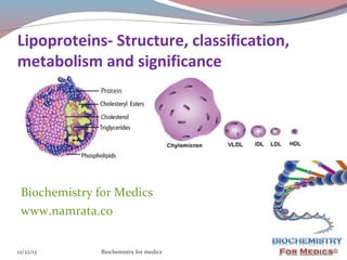
- 1. Lipoproteins- Structure, classification, metabolism and significance Biochemistry for Medics www.namrata.co 12/22/13 Biochemistry for medics 1
- 2. Lipids absorbed from the diet and synthesized by the liver and adipose tissue must be transported between various cells and organs for utilization and storage. Lipids are insoluble in water, the problem of transportation in the aqueous plasma is solved by associating nonpolar lipids (triacylglycerols and cholesteryl esters) with amphipathic lipids (phospholipids and cholesterol) and proteins to make water-miscible lipoproteins.
- 3. General Structure of Lipo proteins Lipoproteins consist of a nonpolar core and a single surface layer of amphipathic lipids The nonpolar lipid core consists of mainly triacylglycerol and cholesteryl ester and is surrounded by a single surface layer of amphipathic phospholipid and cholesterol molecules These are oriented so that their polar groups face outward to the aqueous medium. The protein moiety of a lipoprotein is known as an apolipoprotein or apoprotein. 12/22/13 Biochemistry for medics 3
- 4. General Structure of Lipo proteins Some apolipoproteins are integral and cannot be removed, whereas others can be freely transferred to other lipoproteins. 12/22/13 Biochemistry for medics 4
- 5. Classification of Lipoproteins Lipoproteins can be classified in three ways1) Based on density- They are separated by Ultracentrifugation. Depending upon the floatation constant (Sf), Five major groups of lipoproteins have been identified that are important physiologically and in clinical diagnosis. (i) Chylomicons, derived from intestinal absorption of triacylglycerol and other lipids; Density is generally less than 0.95 while the mean diameter lies between 100- 500 nm 12/22/13 Biochemistry for medics 5
- 6. Classification of Lipoproteins 1) Based on density (contd.) (ii) Very low density lipoproteins (VLDL), derived from the liver for the export of triacylglycerol; density lies between 0.95- 1.006 and the mean diameter lies between 30-80 nm. (iii) Intermediate density lipoproteins (IDL) are derived from the catabolism of VLDL,with a density ranging intermediate between Very low density and Low density lipoproteins i.e. ranging between 1.0061.019 and the mean diameter ranges between 2550nm. 12/22/13 Biochemistry for medics 6
- 7. Classification of Lipoproteins Based on density (contd.) iv) Low-density lipoproteins (LDL), representing a final stage in the catabolism of VLDL; density lies between 1.019-1.063 and mean diameter lies between 18-28 nm (iv) High-density lipoproteins (HDL), involved in cholesterol transport and also in VLDL and chylomicron metabolism. Density ranges between 1.063-1.121 and the mean diameter varies between 5-15 nm. 12/22/13 Biochemistry for medics 7
- 8. Classification of Lipoproteins Lipoproteins with high lipid content will have low density, larger size and so float on centrifugation. Those with high protein content sediment easily, have compact size and have a high density. 12/22/13 Biochemistry for medics 8
- 9. Classification of Lipoproteins 2) Based on electrophoretic mobilities Lipoproteins may be separated according to their electrophoretic properties into - α, pre β, β, and broad beta lipoproteins. The mobility of a lipoprotein is mainly dependent upon protein content. Those with higher protein content will move faster towards the anode and those with minimum protein content will have minimum mobility. 12/22/13 Biochemistry for medics 9
- 10. Classification of Lipoproteins Lipoproteins may be separated according to their electrophoretic properties into - α, pre β, β, and broad beta lipoproteins. 12/22/13 Biochemistry for medics 10
- 11. Classification of Lipoproteins 2) Based on electrophoretic mobilities (contd.) HDL are -α , VLDL pre- β, LDL-β , and IDL are broad beta lipoproteins. Free fatty acid and albumin complex although not a lipoprotein is an important lipid fraction in serum and is the fastest moving fraction. Chylomicrons remain at the origin since they have more lipid content. VLDLs with less protein content than LDL move faster than LDL, this is due to nature of apoprotein present. 12/22/13 Biochemistry for medics 11
- 12. Classification of Lipoproteins As the lipid content increases, density decreases and size increases, that is why Chylomicrons are least dense but biggest in size, while HDL are rich in proteins , hence most dense but smallest in size. 12/22/13 Biochemistry for medics 12
- 13. Classification of Lipoproteins 3) Based on nature of Apo- protein content One or more apolipoproteins (proteins or polypeptides) are present in each lipoprotein. The major apolipoproteins of HDL (α-lipoprotein) are designated A. The main apolipoprotein of LDL (β -lipoprotein) is apolipoprotein B (B-100), which is found also in VLDL. Chylomicons contain a truncated form of apo B (B-48) that is synthesized in the intestine, while B-100 is synthesized in the liver. Apo E is found in VLDL, HDL, Chylomicons, and chylomicron remnants. 12/22/13 Biochemistry for medics 13
- 14. Functions of Apo proteins (1) They can form part of the structure of the lipoprotein, e.g. apo B, structural component of VLDL and Chylomicons (2) They are enzyme cofactors, e.g. C-II for lipoprotein lipase, A-I for lecithin: cholesterol acyl transferase (LCAT), or enzyme inhibitors, eg, apo A-II and apo CIII for lipoprotein lipase, apo C-I for cholesteryl ester transfer protein (3) They act as ligands for interaction with lipoprotein receptors in tissues, e.g. apo B-100 and apo E for the LDL receptor, apo A-I for the HDL receptor. 12/22/13 Biochemistry for medics 14
- 15. Metabolism of chylomicrons Chylomicrons are found in chyle formed only by the lymphatic system draining the intestine. They are responsible for the transport of all dietary lipids into the circulation. Synthesis of Chylomicrons 1) Synthesis of Apo B48 Chylomicrons contain Apo B48, synthesized in the rough endoplasmic reticulum (RER). The synthesis of apo B48 is the result of RNA editing process. Coding information can be changed at the mRNA level by RNA editing. 12/22/13 Biochemistry for medics 15
- 16. Synthesis of Chylomicrons The synthesis of apo B48 is the result of RNA editing process. 12/22/13 Biochemistry for medics 16
- 17. Metabolism of chylomicrons Synthesis of Chylomicrons In liver, the single apo B gene is transcribed into an mRNA that directs the synthesis of a 100-kDa protein, apoB100. In the intestine, the same gene directs the synthesis of the primary transcript; however, a Cytidine deaminase converts a CAA codon in the mRNA to UAA at a single specific site. Rather than encoding glutamine, this codon becomes a termination signal, and a 48-kDa protein (apoB48) is the result. ApoB100 and apoB48 have different functions in the two organs 12/22/13 Biochemistry for medics 17
- 18. Metabolism of chylomicrons Apolipoprotein B, synthesized in the RER, is incorporated into lipoproteins in the SER, the main site of synthesis of triacylglycerol. After addition of carbohydrate residues in G, they are released from the cell by reverse pinocytosis. Chylomicrons pass into the lymphatic system. Clinical Significance- In Abetalipoproteinemia (a rare disease), lipoproteins containing apo B are not formed and lipid droplets accumulate in the intestine and liver(Due to non formation of VLDL) 12/22/13 Biochemistry for medics 18
- 19. Synthesis of Chylomicrons (contd.) 2) Synthesis of lipids and formation of lipoprotein Long-chain fatty acids are esterified to yield to triacylglycerol in the mucosal cells and together with the other products of lipid digestion, are incorporated into lipoproteins in the SER, the main site of synthesis of triacylglycerol. 3) Addition of Carbohydrate-Carbohydrate residues are added in the golgi apparatus. 4) Release of Chylomicrons- After addition of carbohydrate residues in golgi apparatus, they are released from the cell by reverse pinocytosis. 5) Transportation of Chylomicrons- Chylomicrons pass into the lymphatic system and eventually enter the systemic circulation. 12/22/13 Biochemistry for medics 19
- 20. Catabolism of Chylomicrons Chylomicrons are acted upon by the enzyme lipoprotein lipase . Reaction with lipoprotein lipase results in the loss of approximately 90% of the triacylglycerol of chylomicrons and in the loss of apo C (which returns to HDL) but not apo E, which is retained. The resulting chylomicron remnant is about half the diameter of the parent chylomicron and is relatively enriched in cholesterol and cholesteryl esters because of the loss of triacylglycerol. 12/22/13 Biochemistry for medics 20
- 21. Catabolism of Chylomicrons Chylomicron remnants are taken up by the liver by receptor-mediated endocytosis, and the cholesteryl esters and triacylglycerols are hydrolyzed and metabolized. Uptake is mediated by apo E . Hepatic lipase has a dual role: (1) it acts as a ligand to facilitate remnant uptake and (2) it hydrolyzes remnant triacylglycerol and phospholipid. The products released are re utilized for the synthesis of VLDL. 12/22/13 Biochemistry for medics 21
- 22. Catabolism of Chylomicrons 12/22/13 Biochemistry for medics 22
- 23. Metabolism of VLDL There are striking similarities in the mechanisms of formation of chylomicrons by intestinal cells and of VLDL by hepatic parenchymal cells Apart from the mammary gland, the intestine and liver are the only tissues from which particulate lipid is secreted. Newly secreted or "nascent" VLDL contain only a small amount of apolipoproteins C and E, and the full complement is acquired from HDL in the circulation Apo B 100 is essential for VLDL formation. 12/22/13 Biochemistry for medics 23
- 24. Metabolism of VLDL VLDL are secreted into the space of Disse and then into the hepatic sinusoids through fenestrae in the endothelial lining. for medics 12/22/13 24 Biochemistry
- 25. Catabolism of VLDL Catabolism of VLDL is similar to chylomicrons Both phospholipids and apo C-II are required as cofactors for lipoprotein lipase activity, while apo A-II and apo C-III act as inhibitors. Hydrolysis takes place while the VLDLs are attached to the enzyme on the endothelium. Triacylglycerol is hydrolyzed progressively through a diacyl glycerol to a monoacylglycerol and finally to free fatty acids plus glycerol. Some of the released free fatty acids return to the circulation, attached to albumin, but the bulk is transported into the tissue . 12/22/13 Biochemistry for medics 25
- 26. Catabolism of VLDL Reaction with lipoprotein lipase results in the loss of approximately 90% of the triacylglycerol of VLDLs and in the loss of apo C (which returns to HDL) but not apo E, which is retained. These changes occurring to VLDL, lead to the formation of VLDL remnants or IDL (intermediate-density lipoprotein) After metabolism to IDL, VLDL may be taken up by the liver directly via the LDL (apo B-100, E) receptor, or it may be converted to LDL. Only one molecule of apo B-100 is present in each of these lipoprotein particles, and this is conserved during the transformations. Thus, each LDL particle is derived from a single precursor VLDL particle. In humans, a relatively large proportion of IDL forms LDL, accounting for the increased concentrations of LDL in humans compared with many other mammals. 12/22/13 Biochemistry for medics 26
- 27. Metabolism of LDL The liver and many extra hepatic tissues express the LDL (apo B-100, E) receptor. It is so designated because it is specific for apo B-100 but not B-48, which lacks the carboxyl terminal domain of B100 containing the LDL receptor ligand, and it also takes up lipoproteins rich in apo E. This receptor is defective in familial hypercholesterolemia. Approximately 30% of LDL is degraded in extra-hepatic tissues and 70% in the liver. A positive correlation exists between the incidence of coronary atherosclerosis and the plasma concentration of LDL cholesterol. 12/22/13 Biochemistry for medics 27
- 28. Metabolism of VLDL The liver and many extrahepatic tissues express the LDL (apo B100, E) receptor. It is so designated because it is specific for apo B-100 but not B-48, which lacks the carboxyl terminal domain of B-100 containing the LDL receptor ligand, and it also takes up lipoproteins rich in apo E. 12/22/13 Biochemistry for medics 28
- 29. Metabolism of HDL Synthesis of HDL HDL is synthesized and secreted from both liver and intestine . However, apo C and apo E are synthesized in the liver and transferred from liver HDL to intestinal HDL when the latter enters the plasma. A major function of HDL is to act as a repository for the apo C and apo E required in the metabolism of chylomicrons and VLDL. Nascent HDL consists of discoid phospholipid bilayer containing apo A and free cholesterol. 12/22/13 Biochemistry for medics 29
- 30. Metabolism of HDL LCAT and the LCAT activator apo A-I—bind to the discoidal particles, and the surface phospholipid and free cholesterol are converted into cholesteryl esters and lysolecithin . The nonpolar cholesteryl esters move into the hydrophobic interior of the bilayer, whereas lysolecithin is transferred to plasma albumin. Thus, a nonpolar core is generated, forming a spherical, pseudomicellar HDL covered by a surface film of polar lipids and apolipoproteins. This aids the removal of excess unesterified cholesterol from lipoproteins and tissues . 12/22/13 Biochemistry for medics 30
- 31. Metabolism of HDL Role of LCAT LCAT( Lecithin Cholesterol Acyl Transferase) enzyme catalyzes the esterification of cholesterol to form Cholesteryl ester. The reaction can be represented as followsLecithin + Cholesterol Lysolecithin + Cholesteryl Ester 12/22/13 Biochemistry for medics 31
- 32. Reverse cholesterol transport The cholesterol efflux is brought about by esterification of cholesterol under the effect of LCAT. The cholesteryl ester rich HDL (HDL2) gains entry through Scavenger receptor (SR-B1) 12/22/13 Biochemistry for medics 32
- 33. Metabolism of HDL The class B scavenger receptor B1 (SR-B1) has been identified as an HDL receptor with a dual role in HDL metabolism. In the liver and in steroidogenic tissues, it binds HDL via apo A-I, and cholesteryl ester is selectively delivered to the cells, although the particle itself, including apo A-I, is not taken up. In the tissues, on the other hand, SR-B1 mediates the acceptance of cholesterol from the cells by HDL, which then transports it to the liver for excretion via the bile (either as cholesterol or after conversion to bile acids) in the process known as reverse 12/22/13 33 Biochemistry for medics cholesterol transport
- 34. HDL- cycle HDL3, generated from discoidal HDL by the action of LCAT, accepts cholesterol from the tissues via the SRB1 and the cholesterol is then esterified by LCAT, increasing the size of the particles to form the less dense HDL2. HDL3 is then reformed, either after selective delivery of cholesteryl ester to the liver via the SR-B1 or by hydrolysis of HDL2 phospholipid and triacylglycerol by hepatic lipase. This interchange of HDL2 and HDL3 is called the HDL cycle. Free apo A-I is released by these processes and forms pre -HDL after associating with a minimum amount of 12/22/13 Biochemistry for medics 34
- 35. Metabolism of HDL A second important mechanism for reverse cholesterol transport involves the ATP-binding cassette transporter A1 (ABCA1). ABCA1 is a member of a family of transporter proteins that couple the hydrolysis of ATP to the binding of a substrate, enabling it to be transported across the membrane. ABCA1 preferentially transfer cholesterol from cells to poorly lipidated particles such as pre -HDL or apo A-1, which are then converted to HDL3 via discoidal HDL Pre -HDL is the most potent form of HDL inducing cholesterol efflux from the tissues. 12/22/13 Biochemistry for medics 35
- 36. Functions of HDL Scavenging action- HDL scavenges extra cholesterol from peripheral tissues by reverse cholesterol transport HDL, with the help of apo E competes with LDL for binding sites on the membranes and prevents internalization of LDL cholesterol in the smooth cells of the arterial walls HDL contributes its apo C and E to nascent VLDL and chylomicrons for receptor mediated endocytosis HDL stimulated prostacyclin synthesis by the endothelial cells, which prevent thrombus formation HDL also helps in the removal of macrophages from 12/22/13 36 Biochemistry the arterial walls . for medics
- 37. Summary of formation and fate of lipoproteins Chylomicrons is a 12/22/13 Biochemistry for medics transporter of dietary lipids whereas VLDL is a transporter of endogenous lipids(mainly TGs). LDL transports cholesterol to peripheral cells while HDL transports cholesterol from peripheral cells back 37 to liver.
- 38. Role of HDL in receptor mediated endocytosis HDL contributes its apo C and E to nascent VLDL and chylomicrons for receptor mediated endocytosis 12/22/13 Biochemistry for medics 38
- 39. Clinical Significance of lipoprotein metabolism Fatty Liver is an abnormal accumulation of certain fats (triglycerides) inside liver cells. Hepatic triacylglycerol synthesis provides the immediate stimulus for the formation and secretion of VLDL. Impaired VLDL formation or secretion leads to nonmobilization of lipid components from the liver, results in fatty liver. 12/22/13 Biochemistry for medics 39
- 40. Fatty Liver (contd.) Fatty livers fall into two main categoriesA)More synthesis of Triglycerides High carbohydrate diet High fat feeding Starvation Diabetes mellitus High carbohydrate diet stimulates de novo fatty acid synthesis by providing excess of Acetyl CoA and high fat feeding provides more flux of fatty acids from the diet that can be esterifies to provide excess 12/22/13 triglycerides.Biochemistry for medics 40
- 41. Fatty Liver (contd.) B) Defective VLDL synthesis -The second type of fatty liver is usually due to a metabolic block in the production of plasma lipoproteins, thus allowing triacylglycerol to accumulate. The lesion may be due to – (1)A block in apolipoproteins synthesis a) Protein energy Malnutrition b) Impaired absorption c) Presence of inhibitors of endogenous protein synthesis e.g.Carbon tetra chloride, Puromycin, Ethionine , Heavy metals etc. d) Hypobetalipoproteinemia- Defective apo B gene can cause impaired synthesis of apo B protein. 12/22/13 Biochemistry for medics 41
- 42. Fatty Liver (contd.) (2) A failure in provision of phospholipids that are found in lipoproteins a)A deficiency of choline, a lipotropic factor can cause impaired formation of phosphatidyl choline (Lecithin),a glycerophospholipid. b)Methionine deficiency can also cause impaired choline synthesis c) Inositol deficiency d)Deficiency of essential fatty acids can also cause impaired PL synthesis 12/22/13 Biochemistry for medics 42
- 43. Fatty Liver (contd.) (3) Impaired Glycosylation- Orotic acid also causes fatty liver; it interferes with glycosylation of the lipoprotein, thus inhibiting release, and may also impair the recruitment of triacylglycerol to the particles. In conditions of orotic aciduria(disorder of pyrimidine nucleotide biosynthesis), fatty liver can be observed. 4) Impaired secretion of VLDL- oxidative stress is a common cause for membrane disruption of lipoproteins. 12/22/13 Biochemistry for medics 43
- 44. Fatty Liver (contd.) 2) Alcoholic fatty liver Alcoholism leads to fat accumulation in the liver, hyperlipidemia, and ultimately cirrhosis. The fatty liver is caused by a combination of impaired fatty acid oxidation and increased lipogenesis, which is thought to be due to changes in the [NADH]/ [NAD+] redox potential in the liver, and also to interference with the action of transcription factors regulating the expression of the enzymes involved in the pathways. 12/22/13 Biochemistry for medics 44
- 46. Fatty Liver and Lipotropic agents Lipotropic agents- Agents such as Choline Inositol Methionine and other essential amino acids, Essential fatty acids, Anti oxidant vitamins, Vitamin B12, folic acid and Synthetic antioxidants which have the apparent effect of removal of fats from the liver cells, and thus prevent the formation of fatty liver are called lipotropic agents. for medics 12/22/13 Biochemistry 46
- 47. Primary Disorders of Plasma Lipoproteins (Dyslipoproteinemias) Inherited defects in lipoprotein metabolism lead to the primary condition of either hypo- or hyperlipoproteinemia . In addition, diseases such as diabetes mellitus, hypothyroidism, nephrotic syndrome, and atherosclerosis are associated with secondary abnormal lipoprotein patterns that are very similar to one or another of the primary inherited conditions. All of the primary conditions are due to a defect at a stage in lipoprotein formation, transport, or degradation. 12/22/13 Biochemistry for medics 47
- 48. Primary Disorders of Plasma Lipoproteins (Dyslipoproteinemias) Name Defect Characteristics Abetalipoproteinemia No chylomicrons, VLDL, or LDL are formed because of defect in the loading of apo B with lipid. Rare; blood acylglycerols low; intestine and liver accumulate acylglycerols. Intestinal malabsorption. Familial alphalipoprotein deficiency All have low or near absence of HDL. Hypertriacylglycerolemia due to absence of apo CII, Low LDL levels. Atherosclerosis in the elderly. Hypolipoproteinemias Tangier disease Fish-eye disease Apo-A-I deficiencies 12/22/13 Biochemistry for medics 48
- 49. Primary Disorders of Plasma Lipoproteins (Dyslipoproteinemias) Name Defect Characteristics Familial lipoprotein lipase deficiency (type I) Hypertriacylglycerolemia due to deficiency of LPL, abnormal LPL, or apo CII deficiency causing inactive LPL. Slow clearance of chylomicrons and VLDL. Low levels of LDL and HDL. No increased risk of coronary disease. Familial hypercholesterolemia (type II a) Defective LDL receptors or mutation in ligand region of apo B-100. Elevated LDL levels and hypercholesterolemia, resulting in atherosclerosis and coronary disease. Hyperlipoproteinemia 12/22/13 Biochemistry for medics 49
- 50. Primary Disorders of Plasma Lipoproteins (Dyslipoproteinemias)- contd. Name Defect Characteristics Familial type III hyperlipoproteinemia (broad beta disease, remnant removal disease, familial dysbetalipoproteinemia) Deficiency in remnant clearance by the liver is due to abnormality in apo E. Increase in chylomicron and VLDL remnants , Causes hypercholesterolemia, xanthomas, and atherosclerosis. Familial Hypertriacylglycerolemia (type IV) Overproduction of VLDL often associated with glucose intolerance and hyperinsulinemia. High cholesterol, VLDL, Subnormal LDL and HDL. Associated with Alcoholism, diabetes mellitus and obesity. Hepatic lipase deficiency 12/22/13 Deficiency of the enzyme Patients have xanthomas leads to accumulation of and coronary heart large triacylglycerol-rich disease. HDL and VLDL remnants 50 Biochemistry for medics
- 51. Atherosclerosis Read the details at http://www.namrata.co/atherosclerosis-a-review-power-poin 12/22/13 Biochemistry for medics 51
- 52. For further details Refer A Case Oriented Approach Towards Biochemistry by: Namrata Chhabra, Sahil Chhabra http://www.jaypeedigital.com/BookDetails.aspx?id=9789350 And visit our web sitehttp://www.namrata.co/category/metabolism-lipids/subject 12/22/13 Biochemistry for medics 52