SlideShare a Scribd company logo
1 of 69
CAP PROTOCOL
CAP PROTOCOL - URINARY
BLADDER
PROCEDURES-NOTE A
 Bladder biopsy
 Transurethral resection of bladder tumour specimen
 Cystectomy (Partial, Total)
- Radical Cystoprostatectomy
-Pelvic exenteration
SUMMARY OF CHANGES
Bladder biopsy and TURBT
 The word checklist was changed to case summary
 Histological grade-squamous cell carcinoma and
adenocarcinoma as changed to squamous cell
carcinoma or adenocarcinoma
 Microscopic extension-“Urothelial carcinoma in situ”
was changed to “Urothelial carcinoma” as follows:
1. Urothelial carcinoma involving prostatic urethra in
prostatic chips sampled by TURBT
2. Urothelial carcinoma involving prostatic ducts and
acini in prostatic chips sampled by TURBT
 “Cystitis cystic glandularis” was changed to “Cystitis
cystica et glandularis.”
SUMMARY OF CHANGES-
CYSTECTOMY AND ANTERIOR RESECTION
Microscopic Tumor Extension
Reporting on this element was changed to the following:
 Cannot be assessed
 No evidence of primary tumor
 Noninvasive papillary carcinoma
 Carcinoma in situ: “flat tumor”
 Tumor invades lamina propria
 Tumor invades muscularis propria
1. Tumor invades superficial muscularis propria (inner
half)
2. Tumor invades deep muscularis propria (outer half)
 Tumor invades perivesical tissue
Microscopically
Macroscopically (extravesical mass)
SUMMARY OF CHANGES-CYSTECTOMY AND
ANTERIOR RESECTION
 Prostatic stroma
 Seminal vesicles
 Uterus
 Tumor invades adjacent structures
 Vagina
 Adnexae
 Pelvis wall
 Abdominal wall
 Rectum
 Other (specify)
MARGINS
Reporting on margins was changed to include noninvasive
high-grade urothelial carcinoma and other significant changes
at the margin and a note was added, as follows:
Margins (select all that apply)
1. Cannot be assessed
2. Margin(s) involved by invasive carcinoma
 Ureteral margin
 Distal urethral margin
 Deep soft tissue margin
 Other margin(s) (specify)
Margins(s) involved by carcinoma in situ/noninvasive high-
grade urothelial carcinoma
 Ureteral margin
 Distal urethral margin
 Other margin(s) (specify)
MARGINS
Margins uninvolved by invasive
carcinoma/carcinoma in situ/noninvasive high-
grade urothelial carcinoma
 Distance of carcinoma from closest margin: ___
mm
 Specify margin:
____________________________
 Other significant changes at margin (specify
margin): ________________________
 Low-grade dysplasia
 Non-invasive low-grade urothelial carcinoma
NOTE A -PROCEDURE
Procedure (required only for TURBT)
 Biopsy
 TURBT
 Other (specify)
 Not specified
NOTE A-PROCEDURE
 Partial cystectomy
First time tumour recurrence with a solitary tumour
Tumour located at the dome
Carcinoma of urachus or diverticulum
 Total cystectomy
Non muscle invasive carcinoma with non functional
bladder
High grade Pt1 non responsive to therapy
 Radical cystectomy
 Radical cystoprostatectomy
 Anterior exenteration
 Other (specify)
 Not specified
TUMOUR SITE-NOTE A
 Trigone
 Right lateral wall
 Left lateral wall
 Anterior wall
 Posterior wall
 Dome
 Other (specify)
 Not specified
TUMOUR SIZE- NOTE A
 Greatest dimension: ___ cm
 Additional dimensions: __x___ cm
 Cannot be determined
NOTE A - HISTORY
 Symptoms with duration
 History of renal stones
 History or recent urinary tract procedures
 Cystoscopic findings
NOTE B- HISTOLOGICAL TYPE
 Urothelial (transitional cell) carcinoma
 Urothelial (transitional cell) carcinoma with
squamous differentiation
 Urothelial (transitional cell) carcinoma with
glandular differentiation
 Urothelial (transitional cell) carcinoma with variant
histology (specify)
 Squamous cell carcinoma, typical
 Squamous cell carcinoma, variant histology
HISTOLOGICAL TYPE
 Adenocarcinoma, typical
 Adenocarcinoma, variant histology (specify):
 Small cell carcinoma
 Undifferentiated carcinoma (specify)
 Mixed cell type (specify)
 Other (specify)
 Carcinoma, type cannot be determined
CLASSIFICATION OF NEOPLASMS OF THE
URINARY BLADDER
Urothelial (Transitional Cell) Neoplasia
Benign
 Urothelial papilloma (World Health Organization
[WHO] 2004/ International Society of Urologic
Pathology [ISUP]), WHO, 1973, grade 0)
 Inverted papilloma
 Papillary urothelial neoplasm of low malignant
potential (WHO 2004/ISUP); WHO, 1973, grade I)
CLASSIFICATION OF NEOPLASMS OF THE
URINARY BLADDER
Malignant
Papillary
 Typical, noninvasive
 Typical, with invasion
Variant
 With squamous or glandular differentiation
 Micropapillary
CLASSIFICATION OF NEOPLASMS OF THE
URINARY BLADDER
Non papillary
1. Carcinoma in situ
2. Invasive carcinoma
Variants containing or exhibiting Deceptively benign
features
 Nested pattern (resembling von Brunn’s nests)
 Small tubular pattern
 Microcystic pattern
 Inverted pattern
 Squamous differentiation
 Glandular differentiation
 Micropapillary histology
 Sarcomatoid foci (“sarcomatoid carcinoma”)
CLASSIFICATION OF NEOPLASMS OF THE
URINARY BLADDER
Urothelial carcinoma with unusual cytoplasmic
features
 Clear cell(glycogen rich)
 Plasmacytoid
 Rhabdoid
 Lipoid rich
Urothelial carcinoma with syncytiotrophoblasts
Unusual stromal reactions
 Pseudosarcomatous stroma
 Stromal osseous or cartilaginous metaplasia
 Osteoclast-type giant cells
 With prominent lymphoid infiltrate
CLASSIFICATION OF NEOPLASMS OF THE
URINARY BLADDER
Squamous Cell Carcinoma
1. Typical
2. Variant
 Verrucous carcinoma
 Basaloid squamous cell carcinoma
 Sarcomatoid carcinoma
Adenocarcinoma
Anatomic variants
 Bladder mucosa
 Urachal
 With exstrophy
 From endometriosis
CLASSIFICATION OF NEOPLASMS OF THE
URINARY BLADDER
Histologic variants of adenocarcinoma
 Typical intestinal type
 Mucinous (including colloid)
 Signet-ring cell
 Clear cell
 Hepatoid
 Mixture of above patterns – adenocarcinoma not
otherwise specified (NOS)
Tumors of Mixed Cell Types
CLASSIFICATION OF NEOPLASMS OF THE
URINARY BLADDER
Undifferentiated Carcinoma
 Small cell carcinoma
 Large cell neuroendocrine carcinoma
 Lymphoepithelioma-like carcinoma
 Osteoclast-rich carcinoma
 Giant cell carcinoma
 Not otherwise specified
Metastatic Carcinoma
GRADING
 Histologic Grade (select all that apply) (Note C)
 Not applicable
 Cannot be determined
 Urothelial carcinoma
 Low-grade
 High-grade
 Other (specify):
HISTOLOGICAL GRADE- NOTE C
Squamous cell carcinoma or adenocarcinoma
 GX: Cannot be assessed
 G1: Well differentiated
 G2: Moderately differentiated
 G3: Poorly differentiated
Other (specify)
 Other carcinoma
 Low-grade
 High-grade
 Other (specify)
WORLD HEALTH ORGANIZATION (WHO) 2004/ INTERNATIONAL
SOCIETY OF UROLOGIC PATHOLOGY (ISUP) CLASSIFICATION
FOR UROTHELIAL (TRANSITIONAL CELL) LESIONS
 Hyperplasia
Flat hyperplasia
Papillary hyperplasia
 Flat Lesions with Atypia
Reactive (inflammatory) atypia
Atypia of unknown significance
Dysplasia (low-grade intraurothelial neoplasia)
Carcinoma in situ (high-grade intraurothelial
neoplasia)
PAPILLARY NEOPLASMS
 Papilloma
 Inverted papilloma
 Papillary neoplasm of low malignant potential
 Papillary carcinoma, low-grade
 Papillary carcinoma, high-grade
INVASIVE NEOPLASMS
 Lamina propria invasion
 Muscularis propria (detrusor muscle) invasion
NOTE C- ASSOCIATED EPITHELIAL
LESIONS
 None identified
 Urothelial (transitional cell) papilloma (World Health
Organization [WHO] 2004/ International Society of
Urologic Pathology [ISUP])
 Urothelial (transitional cell) papilloma, inverted type
 Papillary urothelial (transitional cell) neoplasm, low
malignant potential (WHO 2004/ISUP)
 Cannot be determined
TUMOUR CONFIGURATION-NOTE C
 Papillary
 Solid/nodule
 Flat
 Ulcerated
 Indeterminate
 Other (specify)
ADEQUACY OF MATERIAL FOR DETERMINING
MUSCULARIS PROPRIA INVASION- NOTE D IN
BIOPSY AND TURBT
 Muscularis propria (detrusor muscle) not identified
 Muscularis propria (detrusor muscle) present
 Presence of muscularis propria indeterminate
MICROSCOPIC TUMOR EXTENSION (SELECT
ALL THAT APPLY) (NOTE D)
 Cannot be assessed
 No evidence of primary tumor
 Noninvasive papillary carcinoma
 Carcinoma in situ: “flat tumor”
 Tumor invades lamina propria
 Tumor invades muscularis propria
Tumor invades superficial muscularis propria
(inner half)
Tumor invades deep muscularis propria (outer
half)
MICROSCOPIC TUMOUR EXTENSION
 Tumor invades perivesical tissue
Microscopically
Macroscopically (extravesical mass)
 Tumor invades adjacent structures
Prostatic stroma
Seminal vesicles
Uterus
Vagina
Adnexae
Pelvis wall
Abdominal wall
Rectum
Other (specify):
MUSCLE INVASION
 Muscularis mucosa or muscularis propria
 Muscle invasion indeterminate for type of muscle
invasion
 Tissue distortion, cautery artefact, poor orientation,
fibrosis, inflammation.
 Sub staging of muscle invasion.
INVOLVEMENT OF PROSTATE
 Prostatic urethra(flat carcinoma in situ, papillary or
invasive carcinoma)
 Prostatic gland involvement
Involvement of prostatic ducts and acini
without stromal invasion (carcinoma in situ
involving prostate glands).
Urothelial carcinoma involving prostatic
stroma (either from prostatic urethral carcinoma,
carcinoma extending directly through the bladder
wall, or carcinoma involving prostatic ducts and
acini additionally with stromal invasion
ADDITIONAL PATHOLOGICAL FINDINGS
 Urothelial dysplasia (low-grade intraurothelial
neoplasia)
 Inflammation/regenerative changes
 Therapy-related changes
 Cautery artifact
 Cystitis cystica et glandularis
 Keratinizing squamous metaplasia
 Intestinal metaplasia
 Other (specify)
NOTE E- LYMHOVASCULAR INVASION
 Not identified
 Present
 Indeterminate
STAGING- NOTE F
 Pathologic staging is usually performed after surgical
resection of the primary tumor. Pathologic staging
depends on pathologic documentation of the anatomic
extent of disease, whether or not the primary
tumor has been completely removed.
 If a biopsied tumor is not resected for any reason (eg,
when technically unfeasible) and if the highest T and N
categories or the M1 category of the tumor can be
confirmed microscopically, the criteria for pathologic
classification and staging have been satisfied
without total removal of the primary cancer.
NOTE F- PATHOLOGICAL STAGING
 Primary Tumor (pT)
 pTX: Primary tumor cannot be assessed
 pT0: No evidence of primary tumor
 pTa: Noninvasive papillary carcinoma
 pTis: Carcinoma in situ: “flat tumor”
 pT1: Tumor invades subepithelial connective tissue
(lamina propria)
 pT2: Tumor invades muscularis propria (detrusor
muscle)
pT2a: Tumor invades superficial muscularis
propria (inner half)
TNM STAGING
pT2b: Tumor invades deep muscularis propria (outer
half)
 pT3: Tumor invades perivesical tissue
pT3a: Microscopically
pT3b: Macroscopically (extravesicular mass)
 pT4: Tumor invades any of the following: prostatic
stroma, seminal vesicles, uterus, vagina, pelvic wall,
abdominal wall
pT4a: Tumor invades prostatic stroma or uterus or
vagina
pT4b: Tumor invades pelvic wall or abdominal wall
Residual Tumor (R)
Tumor remaining in a patient after therapy with
curative intent (eg, surgical resection for cure) is
categorized by a system known as R classification,
shown below.
 RX Presence of residual tumor cannot be assessed
 R0 No residual tumor
 R1 Microscopic residual tumor
 R2 Macroscopic residual tumor
STAGING
 The “m” suffix indicates the presence of multiple
primary tumors in a single site and is recorded in
parentheses: pT(m)NM.
 The “y” prefix indicates those cases in which
classification is performed during or following initial
multimodality therapy (ie, neoadjuvant
chemotherapy, radiation therapy, or both
chemotherapy and radiation therapy.
 The “r” prefix indicates a recurrent tumor when
staged after a documented disease-free interval,
and is identified by the “r” prefix: rTNM.
NOTE G- MARGINS
 Cannot be assessed
 Margin(s) involved by invasive carcinoma
Ureteral margin
Distal urethral margin
Deep soft tissue margin
Other margin(s) (specify)
 Margins(s) involved by carcinoma in situ/noninvasive
high-grade urothelial carcinoma
Ureteral margin
Distal urethral margin
Other margin(s) (specify)
 Margins uninvolved by invasive carcinoma/carcinoma in
situ/noninvasive high-grade urothelial carcinoma
STAGING
 The “m” suffix indicates the presence of multiple primary
tumors in a single site and is recorded in parentheses:
pT(m)NM.
 The “y” prefix indicates those cases in which
classification is performed during or following initial
multimodality therapy (ie, neoadjuvant chemotherapy,
radiation therapy, or both chemotherapy and radiation
therapy). The cTNM or pTNM category is identified by a
“y” prefix. The ycTNM or ypTNM categorizes the extent
of tumor actually present at the time of that
examination.).
 The “r” prefix indicates a recurrent tumor when staged
after a documented disease-free interval, and is
identified by the “r” prefix: rTNM.
REGIONAL NODES (PN) LYMPH
 pNX: Lymph nodes cannot be assessed
 pN0: No lymph node metastasis
 pN1: Single regional lymph node metastasis in the
true pelvis (hypogastric, obturator, external iliac or
presacral lymph node)
 pN2: Multiple regional lymph node metastasis in
the true pelvis (hypogastric, obrutrator, external iliac
or presacral lymph node metastasis)
 pN3: Lymph node metastasis to the common iliac
lymph nodes
 No nodes submitted or found
LYMPH NODES
 Number of Lymph Nodes Examined
 Specify
 Number cannot be determined (explain)
 Number of Lymph Nodes Involved (any size)
 Specify
 Number cannot be determined (explain)
METASTASIS
 Distant Metastasis (pM)
 Not applicable
 pM1: Distant metastasis
 Specify site(s), if known
G -SECTIONS FOR MICROSCOPIC
EVALUATION
Bladder
Sections of bladder for microscopic evaluation are as
follows.
 In TURBT specimens, submit 1 section per
centimeter of tumor diameter (up to 10 cassettes).
 If the tumor is noninvasive by the initial sampling,
additional submission of tissue (including possibly
submitting all tissue) is necessary to diagnose or rule
out the presence of invasion.
 If tumor is invasive into lamina propria in the initial
sampling, additional sections (including possibly
submitting the entire specimen) may be necessary to
diagnose or rule out the possibility of muscularis propria
invasion.
G -SECTIONS FOR MICROSCOPIC
EVALUATIONS
 In cystectomy specimens, several representative
sections of the tumor, including the macroscopically
deepest penetration, should be sampled.
 Submit several sections of the mucosa remote
from the carcinoma, especially if abnormal,
including the lateral wall(s), dome, and trigone.
 Submit 1 section of ureteral margin, unless
submitted separately as frozen section specimens,
and 1 section of urethral margin
 If a long segment of the ureter(s) is present, then
additional sections from the mid-portion may be
necessary, as urothelial cancer often is multifocal
PROSTATE AND PROSTATIC URETHRA
 Prostatic urethral involvement should be carefully
investigated in cystectomy specimens.
 Sections should include the prostatic urethra,
including at the margin and with the surrounding
prostatic parenchyma.
 Representative sections of the peripheral zone,
central zone, and seminal vesicles should
be included.
LYMPH NODES
 Submit 1 section from each grossly positive lymph
node.
 All other lymph nodes should be entirely
submitted, as presence of nodal disease may be
used as an indication for adjuvant therapy.
 Lymph nodes may be grossly or microscopically
detected in the perivesical fat.
OTHER TISSUES
 Submit 1 or more sections of uterus (as indicated)
and 1 or more sections of vagina, seminal vesicles,
and other organs (as indicated).
 If the tumor grossly appears to invade the prostate,
uterus, or vagina, sections should be targeted, such
that the relationship of the infiltrating tumor in the
bladder wall and
the adjacent viscus is clearly demonstrable
MARGINS (SELECT ALL THAT APPLY) (NOTE H)
 Cannot be assessed
 Margin(s) involved by invasive carcinoma
Ureteral margin
Distal urethral margin
Deep soft tissue margin
Other margin(s) (specify)
 Margins(s) involved by carcinoma in
situ/noninvasive high-grade urothelial carcinoma
Ureteral margin
Distal urethral margin
Other margin(s) (specify).
MARGINS
Margins uninvolved by invasive carcinoma/carcinoma
in situ/noninvasive high-grade urothelial carcinoma
 Distance of carcinoma from closest margin: ___
mm
 Specify margin#:
____________________________
 Other significant changes at margin (specify
margin)#: ________________________
 Low-grade dysplasia
 Non-invasive low-grade urothelial carcinoma
For partial cystectomies, if the specimen is received
unoriented precluding identification of specific
margins, it should be denoted here.
THANK YOU

More Related Content

What's hot

Bethesda system for cervix cytology
Bethesda system for cervix cytologyBethesda system for cervix cytology
Bethesda system for cervix cytologyRavi Kumar Meena
 
Spindle cell lesions of the breast diagnostic issues 2019 (1)
Spindle cell lesions of the breast  diagnostic issues 2019 (1)Spindle cell lesions of the breast  diagnostic issues 2019 (1)
Spindle cell lesions of the breast diagnostic issues 2019 (1)Alejandro Palacio
 
Interpretation of endoscopic gastrointestinal biopsy
Interpretation of endoscopic gastrointestinal biopsyInterpretation of endoscopic gastrointestinal biopsy
Interpretation of endoscopic gastrointestinal biopsyAppy Akshay Agarwal
 
Immunohistochemistry in Gynaecological Cancers
Immunohistochemistry in Gynaecological CancersImmunohistochemistry in Gynaecological Cancers
Immunohistochemistry in Gynaecological CancersSujoy Dasgupta
 
Atlas on bethesda system for reporting cervical cytology
Atlas on bethesda system for reporting cervical cytologyAtlas on bethesda system for reporting cervical cytology
Atlas on bethesda system for reporting cervical cytologyAshish Jawarkar
 
Liquid based cytology ( l b c)
Liquid  based  cytology ( l b c)Liquid  based  cytology ( l b c)
Liquid based cytology ( l b c)vikramsaraswat
 
Approach to undifferentiated tumors
Approach to undifferentiated tumorsApproach to undifferentiated tumors
Approach to undifferentiated tumorsDr. Varughese George
 
Giant cell lesions of bone
Giant cell lesions of boneGiant cell lesions of bone
Giant cell lesions of boneShreya D Prabhu
 
papillary lesions of the breast.pptx
papillary lesions of the breast.pptxpapillary lesions of the breast.pptx
papillary lesions of the breast.pptxSirnaEmana1
 
Breast pathology 1
Breast pathology 1Breast pathology 1
Breast pathology 1Prasad CSBR
 

What's hot (20)

Bethesda system for cervix cytology
Bethesda system for cervix cytologyBethesda system for cervix cytology
Bethesda system for cervix cytology
 
Cytology of bone lesions
Cytology of bone lesionsCytology of bone lesions
Cytology of bone lesions
 
Spindle cell lesions of the breast diagnostic issues 2019 (1)
Spindle cell lesions of the breast  diagnostic issues 2019 (1)Spindle cell lesions of the breast  diagnostic issues 2019 (1)
Spindle cell lesions of the breast diagnostic issues 2019 (1)
 
Interpretation of endoscopic gastrointestinal biopsy
Interpretation of endoscopic gastrointestinal biopsyInterpretation of endoscopic gastrointestinal biopsy
Interpretation of endoscopic gastrointestinal biopsy
 
Immunohistochemistry in Gynaecological Cancers
Immunohistochemistry in Gynaecological CancersImmunohistochemistry in Gynaecological Cancers
Immunohistochemistry in Gynaecological Cancers
 
Testicular tumors
Testicular tumorsTesticular tumors
Testicular tumors
 
Atlas on bethesda system for reporting cervical cytology
Atlas on bethesda system for reporting cervical cytologyAtlas on bethesda system for reporting cervical cytology
Atlas on bethesda system for reporting cervical cytology
 
Renal pediatric tumors
Renal pediatric tumorsRenal pediatric tumors
Renal pediatric tumors
 
Liquid based cytology ( l b c)
Liquid  based  cytology ( l b c)Liquid  based  cytology ( l b c)
Liquid based cytology ( l b c)
 
Liver cytology
Liver cytologyLiver cytology
Liver cytology
 
Approach to undifferentiated tumors
Approach to undifferentiated tumorsApproach to undifferentiated tumors
Approach to undifferentiated tumors
 
Bethesda system for reporting
Bethesda system for reportingBethesda system for reporting
Bethesda system for reporting
 
Giant cell lesions of bone
Giant cell lesions of boneGiant cell lesions of bone
Giant cell lesions of bone
 
Grossing colon.pptx
Grossing colon.pptxGrossing colon.pptx
Grossing colon.pptx
 
Approach to testicular tumors
Approach to testicular tumorsApproach to testicular tumors
Approach to testicular tumors
 
Pancreas cytology
Pancreas cytologyPancreas cytology
Pancreas cytology
 
papillary lesions of the breast.pptx
papillary lesions of the breast.pptxpapillary lesions of the breast.pptx
papillary lesions of the breast.pptx
 
Prostate biopsy
Prostate biopsyProstate biopsy
Prostate biopsy
 
Liquid based cytology
Liquid based cytologyLiquid based cytology
Liquid based cytology
 
Breast pathology 1
Breast pathology 1Breast pathology 1
Breast pathology 1
 

Viewers also liked

Rare Solid Cancers: An Introduction - Slide 10 - V. Kataja - Rare urological ...
Rare Solid Cancers: An Introduction - Slide 10 - V. Kataja - Rare urological ...Rare Solid Cancers: An Introduction - Slide 10 - V. Kataja - Rare urological ...
Rare Solid Cancers: An Introduction - Slide 10 - V. Kataja - Rare urological ...European School of Oncology
 
Transitional cell carcinoma of urinary bladeder
Transitional cell carcinoma of urinary bladederTransitional cell carcinoma of urinary bladeder
Transitional cell carcinoma of urinary bladederrezauro
 
Case Report – Small Cell Carcinoma Bladder
Case Report – Small Cell Carcinoma BladderCase Report – Small Cell Carcinoma Bladder
Case Report – Small Cell Carcinoma BladderKholiwe Skosana
 
Problems In Urothelial Pathology
Problems In Urothelial PathologyProblems In Urothelial Pathology
Problems In Urothelial Pathologygvanleenders
 
Bladder Cancer NMIBC [Dr.Edmond Wong]
Bladder Cancer NMIBC [Dr.Edmond Wong]Bladder Cancer NMIBC [Dr.Edmond Wong]
Bladder Cancer NMIBC [Dr.Edmond Wong]Edmond Wong
 
The international federation for cervical pathology and colposcopy course
The international federation for cervical pathology and colposcopy courseThe international federation for cervical pathology and colposcopy course
The international federation for cervical pathology and colposcopy courseTariq Mohammed
 
Lower urinarytract csbrp
Lower urinarytract csbrpLower urinarytract csbrp
Lower urinarytract csbrpPrasad CSBR
 
Cevical intraepithelial neoplasia
Cevical intraepithelial neoplasiaCevical intraepithelial neoplasia
Cevical intraepithelial neoplasiaWhitney Joseph
 
Pre-invasive and Invasive Lesions of the Cervix
Pre-invasive and Invasive Lesions of the CervixPre-invasive and Invasive Lesions of the Cervix
Pre-invasive and Invasive Lesions of the CervixDJ CrissCross
 
Pathology of cervix &uterus
Pathology of cervix &uterusPathology of cervix &uterus
Pathology of cervix &uterusayeayetun08
 
Pathology of cervix
Pathology of cervixPathology of cervix
Pathology of cervixPrasad CSBR
 
Renal pathology..at glance
Renal pathology..at glanceRenal pathology..at glance
Renal pathology..at glanceraj kumar
 
Introduction to Renal Pathology
Introduction to Renal PathologyIntroduction to Renal Pathology
Introduction to Renal Pathologyedwinchowyw
 

Viewers also liked (20)

Bladder cancer
Bladder cancerBladder cancer
Bladder cancer
 
Rare Solid Cancers: An Introduction - Slide 10 - V. Kataja - Rare urological ...
Rare Solid Cancers: An Introduction - Slide 10 - V. Kataja - Rare urological ...Rare Solid Cancers: An Introduction - Slide 10 - V. Kataja - Rare urological ...
Rare Solid Cancers: An Introduction - Slide 10 - V. Kataja - Rare urological ...
 
Transitional cell carcinoma of urinary bladeder
Transitional cell carcinoma of urinary bladederTransitional cell carcinoma of urinary bladeder
Transitional cell carcinoma of urinary bladeder
 
Case Report – Small Cell Carcinoma Bladder
Case Report – Small Cell Carcinoma BladderCase Report – Small Cell Carcinoma Bladder
Case Report – Small Cell Carcinoma Bladder
 
Bladder tumor
Bladder tumorBladder tumor
Bladder tumor
 
Problems In Urothelial Pathology
Problems In Urothelial PathologyProblems In Urothelial Pathology
Problems In Urothelial Pathology
 
Bladder Cancer NMIBC [Dr.Edmond Wong]
Bladder Cancer NMIBC [Dr.Edmond Wong]Bladder Cancer NMIBC [Dr.Edmond Wong]
Bladder Cancer NMIBC [Dr.Edmond Wong]
 
The international federation for cervical pathology and colposcopy course
The international federation for cervical pathology and colposcopy courseThe international federation for cervical pathology and colposcopy course
The international federation for cervical pathology and colposcopy course
 
Lower urinarytract csbrp
Lower urinarytract csbrpLower urinarytract csbrp
Lower urinarytract csbrp
 
Bladder cancer
Bladder cancerBladder cancer
Bladder cancer
 
Bladder cancer
Bladder cancerBladder cancer
Bladder cancer
 
Cevical intraepithelial neoplasia
Cevical intraepithelial neoplasiaCevical intraepithelial neoplasia
Cevical intraepithelial neoplasia
 
Placental pathology
Placental pathologyPlacental pathology
Placental pathology
 
Pre-invasive and Invasive Lesions of the Cervix
Pre-invasive and Invasive Lesions of the CervixPre-invasive and Invasive Lesions of the Cervix
Pre-invasive and Invasive Lesions of the Cervix
 
Pathology of cervix &uterus
Pathology of cervix &uterusPathology of cervix &uterus
Pathology of cervix &uterus
 
Pathology of cervix
Pathology of cervixPathology of cervix
Pathology of cervix
 
Pathology Of Kidney
Pathology Of KidneyPathology Of Kidney
Pathology Of Kidney
 
Renal pathology..at glance
Renal pathology..at glanceRenal pathology..at glance
Renal pathology..at glance
 
Introduction to Renal Pathology
Introduction to Renal PathologyIntroduction to Renal Pathology
Introduction to Renal Pathology
 
Glomerular diseases
Glomerular diseases Glomerular diseases
Glomerular diseases
 

Similar to Cap protocol bladder

COLORECTAL CANCER.pdf
COLORECTAL CANCER.pdfCOLORECTAL CANCER.pdf
COLORECTAL CANCER.pdfShapi. MD
 
Carcinoma stomach seminar
Carcinoma stomach seminarCarcinoma stomach seminar
Carcinoma stomach seminarRushabh Shah
 
Urinary bladder pathology radiology
Urinary bladder pathology radiologyUrinary bladder pathology radiology
Urinary bladder pathology radiologyDr pradeep Kumar
 
Gall Bladder Carcinoma
Gall Bladder CarcinomaGall Bladder Carcinoma
Gall Bladder CarcinomaDr.Mohsin Khan
 
Imaging of urinary bladder carcinoma
Imaging of urinary bladder carcinomaImaging of urinary bladder carcinoma
Imaging of urinary bladder carcinomaMilan Silwal
 
GASTRIC CARCINOMA- EPIGASTRIC LUMPS- Abdominal Lumps.pptx
GASTRIC CARCINOMA- EPIGASTRIC LUMPS- Abdominal Lumps.pptxGASTRIC CARCINOMA- EPIGASTRIC LUMPS- Abdominal Lumps.pptx
GASTRIC CARCINOMA- EPIGASTRIC LUMPS- Abdominal Lumps.pptxSelvaraj Balasubramani
 
CARCINOMA URINARY BLADDER
CARCINOMA URINARY BLADDERCARCINOMA URINARY BLADDER
CARCINOMA URINARY BLADDERVikas Kumar
 
Pancreatic neoplasms
Pancreatic neoplasmsPancreatic neoplasms
Pancreatic neoplasmsAjai Sasidhar
 
BREAST CANCER DIAGNOSIS AND MANAGEMENT.pdf
BREAST CANCER DIAGNOSIS AND MANAGEMENT.pdfBREAST CANCER DIAGNOSIS AND MANAGEMENT.pdf
BREAST CANCER DIAGNOSIS AND MANAGEMENT.pdfShapi. MD
 
Bladder Cancer2.ppt
Bladder Cancer2.pptBladder Cancer2.ppt
Bladder Cancer2.pptOlfatHammam1
 
Testicular tumors-Cassification, Biomarkers and Staging by Dr Rajesh
Testicular tumors-Cassification, Biomarkers and Staging by Dr RajeshTesticular tumors-Cassification, Biomarkers and Staging by Dr Rajesh
Testicular tumors-Cassification, Biomarkers and Staging by Dr RajeshRajesh Sinwer
 
Carcinoma urinary bladder
Carcinoma urinary bladderCarcinoma urinary bladder
Carcinoma urinary bladderIsha Shah
 
Carcinomarectum 111113085726-phpapp01
Carcinomarectum 111113085726-phpapp01Carcinomarectum 111113085726-phpapp01
Carcinomarectum 111113085726-phpapp01Drrajan Paliwal
 

Similar to Cap protocol bladder (20)

COLORECTAL CANCER.pdf
COLORECTAL CANCER.pdfCOLORECTAL CANCER.pdf
COLORECTAL CANCER.pdf
 
Carcinoma stomach seminar
Carcinoma stomach seminarCarcinoma stomach seminar
Carcinoma stomach seminar
 
4 renal tumors written
4  renal tumors written4  renal tumors written
4 renal tumors written
 
Rectal Cancer
Rectal CancerRectal Cancer
Rectal Cancer
 
Urinary bladder pathology radiology
Urinary bladder pathology radiologyUrinary bladder pathology radiology
Urinary bladder pathology radiology
 
Gall Bladder Carcinoma
Gall Bladder CarcinomaGall Bladder Carcinoma
Gall Bladder Carcinoma
 
Imaging of urinary bladder carcinoma
Imaging of urinary bladder carcinomaImaging of urinary bladder carcinoma
Imaging of urinary bladder carcinoma
 
GASTRIC CARCINOMA- EPIGASTRIC LUMPS- Abdominal Lumps.pptx
GASTRIC CARCINOMA- EPIGASTRIC LUMPS- Abdominal Lumps.pptxGASTRIC CARCINOMA- EPIGASTRIC LUMPS- Abdominal Lumps.pptx
GASTRIC CARCINOMA- EPIGASTRIC LUMPS- Abdominal Lumps.pptx
 
Gastric cancer
Gastric cancerGastric cancer
Gastric cancer
 
CARCINOMA URINARY BLADDER
CARCINOMA URINARY BLADDERCARCINOMA URINARY BLADDER
CARCINOMA URINARY BLADDER
 
Pancreatic neoplasms
Pancreatic neoplasmsPancreatic neoplasms
Pancreatic neoplasms
 
8 bladder tumor
8 bladder tumor8 bladder tumor
8 bladder tumor
 
BREAST CANCER DIAGNOSIS AND MANAGEMENT.pdf
BREAST CANCER DIAGNOSIS AND MANAGEMENT.pdfBREAST CANCER DIAGNOSIS AND MANAGEMENT.pdf
BREAST CANCER DIAGNOSIS AND MANAGEMENT.pdf
 
Bladder Cancer2.ppt
Bladder Cancer2.pptBladder Cancer2.ppt
Bladder Cancer2.ppt
 
Gallblader carcinoma
Gallblader carcinomaGallblader carcinoma
Gallblader carcinoma
 
Testicular tumors-Cassification, Biomarkers and Staging by Dr Rajesh
Testicular tumors-Cassification, Biomarkers and Staging by Dr RajeshTesticular tumors-Cassification, Biomarkers and Staging by Dr Rajesh
Testicular tumors-Cassification, Biomarkers and Staging by Dr Rajesh
 
Carcinoma stomach
Carcinoma stomachCarcinoma stomach
Carcinoma stomach
 
Carcinoma urinary bladder
Carcinoma urinary bladderCarcinoma urinary bladder
Carcinoma urinary bladder
 
Carcinomarectum 111113085726-phpapp01
Carcinomarectum 111113085726-phpapp01Carcinomarectum 111113085726-phpapp01
Carcinomarectum 111113085726-phpapp01
 
Ca rectum
Ca rectumCa rectum
Ca rectum
 

More from namrathrs87

Special stains in Bone marrow examination
Special stains in Bone marrow examinationSpecial stains in Bone marrow examination
Special stains in Bone marrow examinationnamrathrs87
 
Adnexal tumours of the skin and familial syndromes.
Adnexal tumours of the skin and familial syndromes.Adnexal tumours of the skin and familial syndromes.
Adnexal tumours of the skin and familial syndromes.namrathrs87
 
Effusion cytology - Diagnosis.
Effusion cytology - Diagnosis.Effusion cytology - Diagnosis.
Effusion cytology - Diagnosis.namrathrs87
 
Acute myeloid leukemia
Acute myeloid leukemiaAcute myeloid leukemia
Acute myeloid leukemianamrathrs87
 
IHC in breast pathology
IHC in breast pathologyIHC in breast pathology
IHC in breast pathologynamrathrs87
 
Comparitive genomic hybridisation
Comparitive genomic hybridisationComparitive genomic hybridisation
Comparitive genomic hybridisationnamrathrs87
 

More from namrathrs87 (8)

Special stains in Bone marrow examination
Special stains in Bone marrow examinationSpecial stains in Bone marrow examination
Special stains in Bone marrow examination
 
Adnexal tumours of the skin and familial syndromes.
Adnexal tumours of the skin and familial syndromes.Adnexal tumours of the skin and familial syndromes.
Adnexal tumours of the skin and familial syndromes.
 
Effusion cytology - Diagnosis.
Effusion cytology - Diagnosis.Effusion cytology - Diagnosis.
Effusion cytology - Diagnosis.
 
Acute myeloid leukemia
Acute myeloid leukemiaAcute myeloid leukemia
Acute myeloid leukemia
 
Artefacts
ArtefactsArtefacts
Artefacts
 
Ppt cyst lung
Ppt cyst lungPpt cyst lung
Ppt cyst lung
 
IHC in breast pathology
IHC in breast pathologyIHC in breast pathology
IHC in breast pathology
 
Comparitive genomic hybridisation
Comparitive genomic hybridisationComparitive genomic hybridisation
Comparitive genomic hybridisation
 

Recently uploaded

independent Call Girls Sarjapur Road - 7001305949 with real photos and phone ...
independent Call Girls Sarjapur Road - 7001305949 with real photos and phone ...independent Call Girls Sarjapur Road - 7001305949 with real photos and phone ...
independent Call Girls Sarjapur Road - 7001305949 with real photos and phone ...narwatsonia7
 
Gurgaon Sushant Lok Phase 2 Call Girls Service ( 9873940964 ) unlimited hard...
Gurgaon Sushant Lok Phase 2 Call Girls Service ( 9873940964 )  unlimited hard...Gurgaon Sushant Lok Phase 2 Call Girls Service ( 9873940964 )  unlimited hard...
Gurgaon Sushant Lok Phase 2 Call Girls Service ( 9873940964 ) unlimited hard...ggsonu500
 
Hi,Fi Call Girl In Whitefield - [ Cash on Delivery ] Contact 7001305949 Escor...
Hi,Fi Call Girl In Whitefield - [ Cash on Delivery ] Contact 7001305949 Escor...Hi,Fi Call Girl In Whitefield - [ Cash on Delivery ] Contact 7001305949 Escor...
Hi,Fi Call Girl In Whitefield - [ Cash on Delivery ] Contact 7001305949 Escor...narwatsonia7
 
EMS and Extrication: Coordinating Critical Care
EMS and Extrication: Coordinating Critical CareEMS and Extrication: Coordinating Critical Care
EMS and Extrication: Coordinating Critical CareRommie Duckworth
 
Russian Escorts Service Delhi 9711199171 SONI VIP & HOT BOOK NOW
Russian Escorts Service Delhi 9711199171 SONI VIP & HOT BOOK NOWRussian Escorts Service Delhi 9711199171 SONI VIP & HOT BOOK NOW
Russian Escorts Service Delhi 9711199171 SONI VIP & HOT BOOK NOWsangeevkumar5478
 
Russian Escorts Delhi | 9711199171 | all area service available
Russian Escorts Delhi | 9711199171 | all area service availableRussian Escorts Delhi | 9711199171 | all area service available
Russian Escorts Delhi | 9711199171 | all area service availablesandeepkumar69420
 
Call Girls Dwarka 9999965857 Cheap & Best with original Photos
Call Girls Dwarka 9999965857 Cheap & Best with original PhotosCall Girls Dwarka 9999965857 Cheap & Best with original Photos
Call Girls Dwarka 9999965857 Cheap & Best with original Photosparshadkalavatidevi7
 
MVP Health Care City of Schenectady Presentation
MVP Health Care City of Schenectady PresentationMVP Health Care City of Schenectady Presentation
MVP Health Care City of Schenectady PresentationMVP Health Care
 
Pregnancy and Breastfeeding Dental Considerations.pptx
Pregnancy and Breastfeeding Dental Considerations.pptxPregnancy and Breastfeeding Dental Considerations.pptx
Pregnancy and Breastfeeding Dental Considerations.pptxcrosalofton
 
Call Girls Ghaziabad 9999965857 Cheap and Best with original Photos
Call Girls Ghaziabad 9999965857 Cheap and Best with original PhotosCall Girls Ghaziabad 9999965857 Cheap and Best with original Photos
Call Girls Ghaziabad 9999965857 Cheap and Best with original Photosparshadkalavatidevi7
 
Rohini Sector 30 Call Girls ( 9873940964 ) Book Hot And Sexy Girls In A Few C...
Rohini Sector 30 Call Girls ( 9873940964 ) Book Hot And Sexy Girls In A Few C...Rohini Sector 30 Call Girls ( 9873940964 ) Book Hot And Sexy Girls In A Few C...
Rohini Sector 30 Call Girls ( 9873940964 ) Book Hot And Sexy Girls In A Few C...ddev2574
 
Russian Call Girls Sadashivanagar | 7001305949 At Low Cost Cash Payment Booking
Russian Call Girls Sadashivanagar | 7001305949 At Low Cost Cash Payment BookingRussian Call Girls Sadashivanagar | 7001305949 At Low Cost Cash Payment Booking
Russian Call Girls Sadashivanagar | 7001305949 At Low Cost Cash Payment Bookingnarwatsonia7
 
Models Call Girls Electronic City | 7001305949 At Low Cost Cash Payment Booking
Models Call Girls Electronic City | 7001305949 At Low Cost Cash Payment BookingModels Call Girls Electronic City | 7001305949 At Low Cost Cash Payment Booking
Models Call Girls Electronic City | 7001305949 At Low Cost Cash Payment Bookingnarwatsonia7
 
2025 Inpatient Prospective Payment System (IPPS) Proposed Rule
2025 Inpatient Prospective Payment System (IPPS) Proposed Rule2025 Inpatient Prospective Payment System (IPPS) Proposed Rule
2025 Inpatient Prospective Payment System (IPPS) Proposed RuleShelby Lewis
 
Globalny raport: „Prawdziwe piękno 2024" od Dove
Globalny raport: „Prawdziwe piękno 2024" od DoveGlobalny raport: „Prawdziwe piękno 2024" od Dove
Globalny raport: „Prawdziwe piękno 2024" od Doveagatadrynko
 
Disaster Management Cycle (DMC)| Ms. Pooja Sharma , Department of Hospital A...
Disaster Management Cycle (DMC)| Ms. Pooja Sharma , Department of  Hospital A...Disaster Management Cycle (DMC)| Ms. Pooja Sharma , Department of  Hospital A...
Disaster Management Cycle (DMC)| Ms. Pooja Sharma , Department of Hospital A...Era University , Lucknow
 
Gurgaon DLF Phase 5 Call Girls ( 9873940964 ) Book Hot And Sexy Girls In A Fe...
Gurgaon DLF Phase 5 Call Girls ( 9873940964 ) Book Hot And Sexy Girls In A Fe...Gurgaon DLF Phase 5 Call Girls ( 9873940964 ) Book Hot And Sexy Girls In A Fe...
Gurgaon DLF Phase 5 Call Girls ( 9873940964 ) Book Hot And Sexy Girls In A Fe...ggsonu500
 
Biology class 12 assignment neet level practise chapter wise
Biology class 12 assignment neet level practise chapter wiseBiology class 12 assignment neet level practise chapter wise
Biology class 12 assignment neet level practise chapter wiseNAGKINGRAPELLY
 
Call Girls Sawda 9999965857 Cheap and Best with original Photos
Call Girls Sawda 9999965857 Cheap and Best with original PhotosCall Girls Sawda 9999965857 Cheap and Best with original Photos
Call Girls Sawda 9999965857 Cheap and Best with original Photoskartikkumark7k7
 

Recently uploaded (20)

independent Call Girls Sarjapur Road - 7001305949 with real photos and phone ...
independent Call Girls Sarjapur Road - 7001305949 with real photos and phone ...independent Call Girls Sarjapur Road - 7001305949 with real photos and phone ...
independent Call Girls Sarjapur Road - 7001305949 with real photos and phone ...
 
Gurgaon Sushant Lok Phase 2 Call Girls Service ( 9873940964 ) unlimited hard...
Gurgaon Sushant Lok Phase 2 Call Girls Service ( 9873940964 )  unlimited hard...Gurgaon Sushant Lok Phase 2 Call Girls Service ( 9873940964 )  unlimited hard...
Gurgaon Sushant Lok Phase 2 Call Girls Service ( 9873940964 ) unlimited hard...
 
Hi,Fi Call Girl In Whitefield - [ Cash on Delivery ] Contact 7001305949 Escor...
Hi,Fi Call Girl In Whitefield - [ Cash on Delivery ] Contact 7001305949 Escor...Hi,Fi Call Girl In Whitefield - [ Cash on Delivery ] Contact 7001305949 Escor...
Hi,Fi Call Girl In Whitefield - [ Cash on Delivery ] Contact 7001305949 Escor...
 
EMS and Extrication: Coordinating Critical Care
EMS and Extrication: Coordinating Critical CareEMS and Extrication: Coordinating Critical Care
EMS and Extrication: Coordinating Critical Care
 
Russian Escorts Service Delhi 9711199171 SONI VIP & HOT BOOK NOW
Russian Escorts Service Delhi 9711199171 SONI VIP & HOT BOOK NOWRussian Escorts Service Delhi 9711199171 SONI VIP & HOT BOOK NOW
Russian Escorts Service Delhi 9711199171 SONI VIP & HOT BOOK NOW
 
Russian Escorts Delhi | 9711199171 | all area service available
Russian Escorts Delhi | 9711199171 | all area service availableRussian Escorts Delhi | 9711199171 | all area service available
Russian Escorts Delhi | 9711199171 | all area service available
 
Russian Call Girls South Delhi 9711199171 discount on your booking
Russian Call Girls South Delhi 9711199171 discount on your bookingRussian Call Girls South Delhi 9711199171 discount on your booking
Russian Call Girls South Delhi 9711199171 discount on your booking
 
Call Girls Dwarka 9999965857 Cheap & Best with original Photos
Call Girls Dwarka 9999965857 Cheap & Best with original PhotosCall Girls Dwarka 9999965857 Cheap & Best with original Photos
Call Girls Dwarka 9999965857 Cheap & Best with original Photos
 
MVP Health Care City of Schenectady Presentation
MVP Health Care City of Schenectady PresentationMVP Health Care City of Schenectady Presentation
MVP Health Care City of Schenectady Presentation
 
Pregnancy and Breastfeeding Dental Considerations.pptx
Pregnancy and Breastfeeding Dental Considerations.pptxPregnancy and Breastfeeding Dental Considerations.pptx
Pregnancy and Breastfeeding Dental Considerations.pptx
 
Call Girls Ghaziabad 9999965857 Cheap and Best with original Photos
Call Girls Ghaziabad 9999965857 Cheap and Best with original PhotosCall Girls Ghaziabad 9999965857 Cheap and Best with original Photos
Call Girls Ghaziabad 9999965857 Cheap and Best with original Photos
 
Rohini Sector 30 Call Girls ( 9873940964 ) Book Hot And Sexy Girls In A Few C...
Rohini Sector 30 Call Girls ( 9873940964 ) Book Hot And Sexy Girls In A Few C...Rohini Sector 30 Call Girls ( 9873940964 ) Book Hot And Sexy Girls In A Few C...
Rohini Sector 30 Call Girls ( 9873940964 ) Book Hot And Sexy Girls In A Few C...
 
Russian Call Girls Sadashivanagar | 7001305949 At Low Cost Cash Payment Booking
Russian Call Girls Sadashivanagar | 7001305949 At Low Cost Cash Payment BookingRussian Call Girls Sadashivanagar | 7001305949 At Low Cost Cash Payment Booking
Russian Call Girls Sadashivanagar | 7001305949 At Low Cost Cash Payment Booking
 
Models Call Girls Electronic City | 7001305949 At Low Cost Cash Payment Booking
Models Call Girls Electronic City | 7001305949 At Low Cost Cash Payment BookingModels Call Girls Electronic City | 7001305949 At Low Cost Cash Payment Booking
Models Call Girls Electronic City | 7001305949 At Low Cost Cash Payment Booking
 
2025 Inpatient Prospective Payment System (IPPS) Proposed Rule
2025 Inpatient Prospective Payment System (IPPS) Proposed Rule2025 Inpatient Prospective Payment System (IPPS) Proposed Rule
2025 Inpatient Prospective Payment System (IPPS) Proposed Rule
 
Globalny raport: „Prawdziwe piękno 2024" od Dove
Globalny raport: „Prawdziwe piękno 2024" od DoveGlobalny raport: „Prawdziwe piękno 2024" od Dove
Globalny raport: „Prawdziwe piękno 2024" od Dove
 
Disaster Management Cycle (DMC)| Ms. Pooja Sharma , Department of Hospital A...
Disaster Management Cycle (DMC)| Ms. Pooja Sharma , Department of  Hospital A...Disaster Management Cycle (DMC)| Ms. Pooja Sharma , Department of  Hospital A...
Disaster Management Cycle (DMC)| Ms. Pooja Sharma , Department of Hospital A...
 
Gurgaon DLF Phase 5 Call Girls ( 9873940964 ) Book Hot And Sexy Girls In A Fe...
Gurgaon DLF Phase 5 Call Girls ( 9873940964 ) Book Hot And Sexy Girls In A Fe...Gurgaon DLF Phase 5 Call Girls ( 9873940964 ) Book Hot And Sexy Girls In A Fe...
Gurgaon DLF Phase 5 Call Girls ( 9873940964 ) Book Hot And Sexy Girls In A Fe...
 
Biology class 12 assignment neet level practise chapter wise
Biology class 12 assignment neet level practise chapter wiseBiology class 12 assignment neet level practise chapter wise
Biology class 12 assignment neet level practise chapter wise
 
Call Girls Sawda 9999965857 Cheap and Best with original Photos
Call Girls Sawda 9999965857 Cheap and Best with original PhotosCall Girls Sawda 9999965857 Cheap and Best with original Photos
Call Girls Sawda 9999965857 Cheap and Best with original Photos
 

Cap protocol bladder

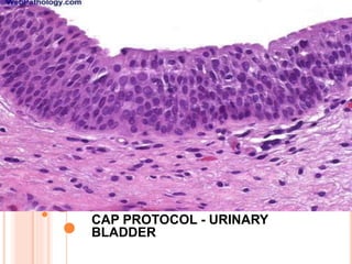
  • 1. CAP PROTOCOL CAP PROTOCOL - URINARY BLADDER
  • 2. PROCEDURES-NOTE A  Bladder biopsy  Transurethral resection of bladder tumour specimen  Cystectomy (Partial, Total) - Radical Cystoprostatectomy -Pelvic exenteration
  • 3. SUMMARY OF CHANGES Bladder biopsy and TURBT  The word checklist was changed to case summary  Histological grade-squamous cell carcinoma and adenocarcinoma as changed to squamous cell carcinoma or adenocarcinoma  Microscopic extension-“Urothelial carcinoma in situ” was changed to “Urothelial carcinoma” as follows: 1. Urothelial carcinoma involving prostatic urethra in prostatic chips sampled by TURBT 2. Urothelial carcinoma involving prostatic ducts and acini in prostatic chips sampled by TURBT  “Cystitis cystic glandularis” was changed to “Cystitis cystica et glandularis.”
  • 4. SUMMARY OF CHANGES- CYSTECTOMY AND ANTERIOR RESECTION Microscopic Tumor Extension Reporting on this element was changed to the following:  Cannot be assessed  No evidence of primary tumor  Noninvasive papillary carcinoma  Carcinoma in situ: “flat tumor”  Tumor invades lamina propria  Tumor invades muscularis propria 1. Tumor invades superficial muscularis propria (inner half) 2. Tumor invades deep muscularis propria (outer half)  Tumor invades perivesical tissue Microscopically Macroscopically (extravesical mass)
  • 5. SUMMARY OF CHANGES-CYSTECTOMY AND ANTERIOR RESECTION  Prostatic stroma  Seminal vesicles  Uterus  Tumor invades adjacent structures  Vagina  Adnexae  Pelvis wall  Abdominal wall  Rectum  Other (specify)
  • 6. MARGINS Reporting on margins was changed to include noninvasive high-grade urothelial carcinoma and other significant changes at the margin and a note was added, as follows: Margins (select all that apply) 1. Cannot be assessed 2. Margin(s) involved by invasive carcinoma  Ureteral margin  Distal urethral margin  Deep soft tissue margin  Other margin(s) (specify) Margins(s) involved by carcinoma in situ/noninvasive high- grade urothelial carcinoma  Ureteral margin  Distal urethral margin  Other margin(s) (specify)
  • 7. MARGINS Margins uninvolved by invasive carcinoma/carcinoma in situ/noninvasive high- grade urothelial carcinoma  Distance of carcinoma from closest margin: ___ mm  Specify margin: ____________________________  Other significant changes at margin (specify margin): ________________________  Low-grade dysplasia  Non-invasive low-grade urothelial carcinoma
  • 8. NOTE A -PROCEDURE Procedure (required only for TURBT)  Biopsy  TURBT  Other (specify)  Not specified
  • 9. NOTE A-PROCEDURE  Partial cystectomy First time tumour recurrence with a solitary tumour Tumour located at the dome Carcinoma of urachus or diverticulum  Total cystectomy Non muscle invasive carcinoma with non functional bladder High grade Pt1 non responsive to therapy  Radical cystectomy  Radical cystoprostatectomy  Anterior exenteration  Other (specify)  Not specified
  • 10. TUMOUR SITE-NOTE A  Trigone  Right lateral wall  Left lateral wall  Anterior wall  Posterior wall  Dome  Other (specify)  Not specified
  • 11. TUMOUR SIZE- NOTE A  Greatest dimension: ___ cm  Additional dimensions: __x___ cm  Cannot be determined
  • 12. NOTE A - HISTORY  Symptoms with duration  History of renal stones  History or recent urinary tract procedures  Cystoscopic findings
  • 13. NOTE B- HISTOLOGICAL TYPE  Urothelial (transitional cell) carcinoma  Urothelial (transitional cell) carcinoma with squamous differentiation  Urothelial (transitional cell) carcinoma with glandular differentiation  Urothelial (transitional cell) carcinoma with variant histology (specify)  Squamous cell carcinoma, typical  Squamous cell carcinoma, variant histology
  • 14. HISTOLOGICAL TYPE  Adenocarcinoma, typical  Adenocarcinoma, variant histology (specify):  Small cell carcinoma  Undifferentiated carcinoma (specify)  Mixed cell type (specify)  Other (specify)  Carcinoma, type cannot be determined
  • 15. CLASSIFICATION OF NEOPLASMS OF THE URINARY BLADDER Urothelial (Transitional Cell) Neoplasia Benign  Urothelial papilloma (World Health Organization [WHO] 2004/ International Society of Urologic Pathology [ISUP]), WHO, 1973, grade 0)  Inverted papilloma  Papillary urothelial neoplasm of low malignant potential (WHO 2004/ISUP); WHO, 1973, grade I)
  • 16.
  • 17.
  • 18.
  • 19. CLASSIFICATION OF NEOPLASMS OF THE URINARY BLADDER Malignant Papillary  Typical, noninvasive  Typical, with invasion Variant  With squamous or glandular differentiation  Micropapillary
  • 20.
  • 21.
  • 22.
  • 23. CLASSIFICATION OF NEOPLASMS OF THE URINARY BLADDER Non papillary 1. Carcinoma in situ 2. Invasive carcinoma Variants containing or exhibiting Deceptively benign features  Nested pattern (resembling von Brunn’s nests)  Small tubular pattern  Microcystic pattern  Inverted pattern  Squamous differentiation  Glandular differentiation  Micropapillary histology  Sarcomatoid foci (“sarcomatoid carcinoma”)
  • 24.
  • 25.
  • 26.
  • 27.
  • 28. CLASSIFICATION OF NEOPLASMS OF THE URINARY BLADDER Urothelial carcinoma with unusual cytoplasmic features  Clear cell(glycogen rich)  Plasmacytoid  Rhabdoid  Lipoid rich Urothelial carcinoma with syncytiotrophoblasts Unusual stromal reactions  Pseudosarcomatous stroma  Stromal osseous or cartilaginous metaplasia  Osteoclast-type giant cells  With prominent lymphoid infiltrate
  • 29.
  • 30. CLASSIFICATION OF NEOPLASMS OF THE URINARY BLADDER Squamous Cell Carcinoma 1. Typical 2. Variant  Verrucous carcinoma  Basaloid squamous cell carcinoma  Sarcomatoid carcinoma Adenocarcinoma Anatomic variants  Bladder mucosa  Urachal  With exstrophy  From endometriosis
  • 31.
  • 32.
  • 33.
  • 34.
  • 35. CLASSIFICATION OF NEOPLASMS OF THE URINARY BLADDER Histologic variants of adenocarcinoma  Typical intestinal type  Mucinous (including colloid)  Signet-ring cell  Clear cell  Hepatoid  Mixture of above patterns – adenocarcinoma not otherwise specified (NOS) Tumors of Mixed Cell Types
  • 36. CLASSIFICATION OF NEOPLASMS OF THE URINARY BLADDER Undifferentiated Carcinoma  Small cell carcinoma  Large cell neuroendocrine carcinoma  Lymphoepithelioma-like carcinoma  Osteoclast-rich carcinoma  Giant cell carcinoma  Not otherwise specified Metastatic Carcinoma
  • 37. GRADING  Histologic Grade (select all that apply) (Note C)  Not applicable  Cannot be determined  Urothelial carcinoma  Low-grade  High-grade  Other (specify):
  • 38. HISTOLOGICAL GRADE- NOTE C Squamous cell carcinoma or adenocarcinoma  GX: Cannot be assessed  G1: Well differentiated  G2: Moderately differentiated  G3: Poorly differentiated Other (specify)  Other carcinoma  Low-grade  High-grade  Other (specify)
  • 39. WORLD HEALTH ORGANIZATION (WHO) 2004/ INTERNATIONAL SOCIETY OF UROLOGIC PATHOLOGY (ISUP) CLASSIFICATION FOR UROTHELIAL (TRANSITIONAL CELL) LESIONS  Hyperplasia Flat hyperplasia Papillary hyperplasia  Flat Lesions with Atypia Reactive (inflammatory) atypia Atypia of unknown significance Dysplasia (low-grade intraurothelial neoplasia) Carcinoma in situ (high-grade intraurothelial neoplasia)
  • 40. PAPILLARY NEOPLASMS  Papilloma  Inverted papilloma  Papillary neoplasm of low malignant potential  Papillary carcinoma, low-grade  Papillary carcinoma, high-grade
  • 41. INVASIVE NEOPLASMS  Lamina propria invasion  Muscularis propria (detrusor muscle) invasion
  • 42. NOTE C- ASSOCIATED EPITHELIAL LESIONS  None identified  Urothelial (transitional cell) papilloma (World Health Organization [WHO] 2004/ International Society of Urologic Pathology [ISUP])  Urothelial (transitional cell) papilloma, inverted type  Papillary urothelial (transitional cell) neoplasm, low malignant potential (WHO 2004/ISUP)  Cannot be determined
  • 43. TUMOUR CONFIGURATION-NOTE C  Papillary  Solid/nodule  Flat  Ulcerated  Indeterminate  Other (specify)
  • 44. ADEQUACY OF MATERIAL FOR DETERMINING MUSCULARIS PROPRIA INVASION- NOTE D IN BIOPSY AND TURBT  Muscularis propria (detrusor muscle) not identified  Muscularis propria (detrusor muscle) present  Presence of muscularis propria indeterminate
  • 45. MICROSCOPIC TUMOR EXTENSION (SELECT ALL THAT APPLY) (NOTE D)  Cannot be assessed  No evidence of primary tumor  Noninvasive papillary carcinoma  Carcinoma in situ: “flat tumor”  Tumor invades lamina propria  Tumor invades muscularis propria Tumor invades superficial muscularis propria (inner half) Tumor invades deep muscularis propria (outer half)
  • 46. MICROSCOPIC TUMOUR EXTENSION  Tumor invades perivesical tissue Microscopically Macroscopically (extravesical mass)  Tumor invades adjacent structures Prostatic stroma Seminal vesicles Uterus Vagina Adnexae Pelvis wall Abdominal wall Rectum Other (specify):
  • 47. MUSCLE INVASION  Muscularis mucosa or muscularis propria  Muscle invasion indeterminate for type of muscle invasion  Tissue distortion, cautery artefact, poor orientation, fibrosis, inflammation.  Sub staging of muscle invasion.
  • 48. INVOLVEMENT OF PROSTATE  Prostatic urethra(flat carcinoma in situ, papillary or invasive carcinoma)  Prostatic gland involvement Involvement of prostatic ducts and acini without stromal invasion (carcinoma in situ involving prostate glands). Urothelial carcinoma involving prostatic stroma (either from prostatic urethral carcinoma, carcinoma extending directly through the bladder wall, or carcinoma involving prostatic ducts and acini additionally with stromal invasion
  • 49. ADDITIONAL PATHOLOGICAL FINDINGS  Urothelial dysplasia (low-grade intraurothelial neoplasia)  Inflammation/regenerative changes  Therapy-related changes  Cautery artifact  Cystitis cystica et glandularis  Keratinizing squamous metaplasia  Intestinal metaplasia  Other (specify)
  • 50. NOTE E- LYMHOVASCULAR INVASION  Not identified  Present  Indeterminate
  • 51. STAGING- NOTE F  Pathologic staging is usually performed after surgical resection of the primary tumor. Pathologic staging depends on pathologic documentation of the anatomic extent of disease, whether or not the primary tumor has been completely removed.  If a biopsied tumor is not resected for any reason (eg, when technically unfeasible) and if the highest T and N categories or the M1 category of the tumor can be confirmed microscopically, the criteria for pathologic classification and staging have been satisfied without total removal of the primary cancer.
  • 52.
  • 53. NOTE F- PATHOLOGICAL STAGING  Primary Tumor (pT)  pTX: Primary tumor cannot be assessed  pT0: No evidence of primary tumor  pTa: Noninvasive papillary carcinoma  pTis: Carcinoma in situ: “flat tumor”  pT1: Tumor invades subepithelial connective tissue (lamina propria)  pT2: Tumor invades muscularis propria (detrusor muscle) pT2a: Tumor invades superficial muscularis propria (inner half)
  • 54. TNM STAGING pT2b: Tumor invades deep muscularis propria (outer half)  pT3: Tumor invades perivesical tissue pT3a: Microscopically pT3b: Macroscopically (extravesicular mass)  pT4: Tumor invades any of the following: prostatic stroma, seminal vesicles, uterus, vagina, pelvic wall, abdominal wall pT4a: Tumor invades prostatic stroma or uterus or vagina pT4b: Tumor invades pelvic wall or abdominal wall
  • 55. Residual Tumor (R) Tumor remaining in a patient after therapy with curative intent (eg, surgical resection for cure) is categorized by a system known as R classification, shown below.  RX Presence of residual tumor cannot be assessed  R0 No residual tumor  R1 Microscopic residual tumor  R2 Macroscopic residual tumor
  • 56. STAGING  The “m” suffix indicates the presence of multiple primary tumors in a single site and is recorded in parentheses: pT(m)NM.  The “y” prefix indicates those cases in which classification is performed during or following initial multimodality therapy (ie, neoadjuvant chemotherapy, radiation therapy, or both chemotherapy and radiation therapy.  The “r” prefix indicates a recurrent tumor when staged after a documented disease-free interval, and is identified by the “r” prefix: rTNM.
  • 57. NOTE G- MARGINS  Cannot be assessed  Margin(s) involved by invasive carcinoma Ureteral margin Distal urethral margin Deep soft tissue margin Other margin(s) (specify)  Margins(s) involved by carcinoma in situ/noninvasive high-grade urothelial carcinoma Ureteral margin Distal urethral margin Other margin(s) (specify)  Margins uninvolved by invasive carcinoma/carcinoma in situ/noninvasive high-grade urothelial carcinoma
  • 58. STAGING  The “m” suffix indicates the presence of multiple primary tumors in a single site and is recorded in parentheses: pT(m)NM.  The “y” prefix indicates those cases in which classification is performed during or following initial multimodality therapy (ie, neoadjuvant chemotherapy, radiation therapy, or both chemotherapy and radiation therapy). The cTNM or pTNM category is identified by a “y” prefix. The ycTNM or ypTNM categorizes the extent of tumor actually present at the time of that examination.).  The “r” prefix indicates a recurrent tumor when staged after a documented disease-free interval, and is identified by the “r” prefix: rTNM.
  • 59. REGIONAL NODES (PN) LYMPH  pNX: Lymph nodes cannot be assessed  pN0: No lymph node metastasis  pN1: Single regional lymph node metastasis in the true pelvis (hypogastric, obturator, external iliac or presacral lymph node)  pN2: Multiple regional lymph node metastasis in the true pelvis (hypogastric, obrutrator, external iliac or presacral lymph node metastasis)  pN3: Lymph node metastasis to the common iliac lymph nodes  No nodes submitted or found
  • 60. LYMPH NODES  Number of Lymph Nodes Examined  Specify  Number cannot be determined (explain)  Number of Lymph Nodes Involved (any size)  Specify  Number cannot be determined (explain)
  • 61. METASTASIS  Distant Metastasis (pM)  Not applicable  pM1: Distant metastasis  Specify site(s), if known
  • 62. G -SECTIONS FOR MICROSCOPIC EVALUATION Bladder Sections of bladder for microscopic evaluation are as follows.  In TURBT specimens, submit 1 section per centimeter of tumor diameter (up to 10 cassettes).  If the tumor is noninvasive by the initial sampling, additional submission of tissue (including possibly submitting all tissue) is necessary to diagnose or rule out the presence of invasion.  If tumor is invasive into lamina propria in the initial sampling, additional sections (including possibly submitting the entire specimen) may be necessary to diagnose or rule out the possibility of muscularis propria invasion.
  • 63. G -SECTIONS FOR MICROSCOPIC EVALUATIONS  In cystectomy specimens, several representative sections of the tumor, including the macroscopically deepest penetration, should be sampled.  Submit several sections of the mucosa remote from the carcinoma, especially if abnormal, including the lateral wall(s), dome, and trigone.  Submit 1 section of ureteral margin, unless submitted separately as frozen section specimens, and 1 section of urethral margin  If a long segment of the ureter(s) is present, then additional sections from the mid-portion may be necessary, as urothelial cancer often is multifocal
  • 64. PROSTATE AND PROSTATIC URETHRA  Prostatic urethral involvement should be carefully investigated in cystectomy specimens.  Sections should include the prostatic urethra, including at the margin and with the surrounding prostatic parenchyma.  Representative sections of the peripheral zone, central zone, and seminal vesicles should be included.
  • 65. LYMPH NODES  Submit 1 section from each grossly positive lymph node.  All other lymph nodes should be entirely submitted, as presence of nodal disease may be used as an indication for adjuvant therapy.  Lymph nodes may be grossly or microscopically detected in the perivesical fat.
  • 66. OTHER TISSUES  Submit 1 or more sections of uterus (as indicated) and 1 or more sections of vagina, seminal vesicles, and other organs (as indicated).  If the tumor grossly appears to invade the prostate, uterus, or vagina, sections should be targeted, such that the relationship of the infiltrating tumor in the bladder wall and the adjacent viscus is clearly demonstrable
  • 67. MARGINS (SELECT ALL THAT APPLY) (NOTE H)  Cannot be assessed  Margin(s) involved by invasive carcinoma Ureteral margin Distal urethral margin Deep soft tissue margin Other margin(s) (specify)  Margins(s) involved by carcinoma in situ/noninvasive high-grade urothelial carcinoma Ureteral margin Distal urethral margin Other margin(s) (specify).
  • 68. MARGINS Margins uninvolved by invasive carcinoma/carcinoma in situ/noninvasive high-grade urothelial carcinoma  Distance of carcinoma from closest margin: ___ mm  Specify margin#: ____________________________  Other significant changes at margin (specify margin)#: ________________________  Low-grade dysplasia  Non-invasive low-grade urothelial carcinoma For partial cystectomies, if the specimen is received unoriented precluding identification of specific margins, it should be denoted here.