SlideShare a Scribd company logo
1 of 20
Classification, Nomenclature, Laboratory Identification
The Gram-Positive Cell
As previously mentioned, Gram-positive bacteria are characterized by their
blue-violet color reaction in the Gram-staining procedure . The blue-violet
color reaction is caused by crystal-violet, the primary Gram-stain dye,
complexing with the iodine mordant. When the decolorizer is applied, a slow
dehydration of the crystal-violet/iodine complex is observed due to the closing
of pores running through the cell wall. Because the crystal-violet is still present
in the cell, the counter stain is not incorporated, thus maintaining the cell's
blue-violet color. If you recall, most cell walls contain peptidoglycan, a
molecule made of amino acids and sugar. A distinguishing factor among Gram-
positive bacteria is that roughly 90% of their cell wall is comprised of
peptidoglycan and a Gram-positve bacteria can have more than 20 layers of
peptidoglycan stacked together to form the cell wall. That's pretty thick!
Examples of common Gram-positive cells are Staphylococcus aureus and
Streptococcus cremoris, a bacterium used in dairy production.
GRAM-POSITIVE COCCI
The Gram-positive cocci are grouped together based on their Gram-stain
reaction, thick cell wall composition, and spherical shape. Most of the
organisms in these groups are members of the Micrococcaceae family All of
the organisms in these groups are non-endospore forming chemosynthetic
heterotrophs. We will discuss only the clinically relevent bacteria from
Micrococcus and Staphylococcus of the Micrococcaceae family.
Streptococcus and Enterococcus(formerly a species of Streptococcus) are
discussed as well because of the many diseases they inflict on humans. The
chart below shows the paths to identification of the genera discussed.
GRAM-POSITIVE RODS
The Gram-positive rods in this section will be divided into three distinct
varieties based upon their ability to produce endospores and their
morphological appearance:
• ENDOSPORE-FORMING
o BACILLUS
• REGULAR, NON-ENDOSPORE-FORMING
o LACTOBACILLUS
o LISTERIA
o ERYSIPELOTHRIX
• IRREGULAR, NON-ENDOSPORE-FORMING
o CORYNEBACTERIUM
These bacteria are ubiquitous in nature and most are aerobic or facultatively
anaerobic.
The Gram-Negative Cell
Unlike Gram-positive bacteria, which asuume a violet color in Gram staining,
Gram negative bacteria incorporate the counterstain rather than the primary
stain. Because the cell wall of Gram(-) bacteria is high in lipid content and low
in peptidoglycan content, the primary crystal-violet escapes from the cell when
the decolorizer is added. This is because primary stains like to bind with
peptidoglycan- something the G(-) cell has very little of. Gram(-) bacteria cause
a lot of problems because many species are pathogenic. This pathogenic
capability is usually associated with certain components of their cell walls,
particularly the lipopolysaccharide (endotoxin) layer. Remember the Black
Plague which wiped out a a third of the population of Europe? It was caused by
the tiny G(-) rod, Yersinia pestis. Most enteric (bowel related) illnesses can
also be attributed to this group of bacteria. Some of my favorites are
Salmonella food poisoning. If you choose, you can directly access the diseases
section of this tutorial to learn more.
MISCELLANEOUS GRAM-NEGATIVE RODS
VIBRIO
CAMPYLOBACTER
HELICOBACTER
NEISSERIA
The Neisseria genus consists of aerobic, non-spore-forming Gram-negative
coccobacilli which inhabit the mucous membranes of many animals (and
humans). These non-motile microbes require a moist environment and warm
temperatures (human body temperature range) to achieve optimum growth. An
important means of identification of Neisseria species is the oxidase test, for
which all members test positive. Additionally, Neisseria grow well on
chocolate agar containing antibiotics that inhibit growth of Gram-negative
bacteria, Gram-positive bacteria, and molds. The two most clinically significant
members of the genus Neisseria are N. gonorrhoeae and N. meningitidis.
N. gonorrhoeae
Infection by the diplococcoid bacterium N. gonorrhoeae is referred to as a
gonococcal infection. Gonorrhea is transmitted between humans through
intimate contact of the mucous membrane. This sexually transmitted organism
can be carried by men and women for many years without any sign or
symptoms. In infected males, the disease is characterized by a urethral
discharge of pus and can eventually result in other complications such as
prostatitis and periurethral abscess. The incubation period of the bacterium can
last from a day to a week. Females with gonorrhea exhibit vaginal discharge,
abdominal pain, and abnormal non-menstrual bleeding. Ironically, the
widespread use of birth control devices such as the pill has actually increased
the number of gonococcal infections in the United States. Use of the pill can
lower the glycogen concentration of the vaginal membrane. This environmental
change inhibits the growth of acid-producing bacteria, such as Lactobacillus,
which are the natural flora of the vagina. The vaginal pH soon becomes less
acidic and a variety of organisms are able to grow there. As with most other
sexually transmitted diseases, gonorrhea is prevalent in young adult and
homosexual populations. This disease may sound really bad, but it is treatable.
N. gonorrheae is sensitive to ultraviolet radiation, drying, and antibiotics.
Because chlamydia infection is often associated with a gonococcal infection, a
regimen of ceftriaxone and doxycycline is used to kill both organisms.
LABORATORY INDICATIONS:
• Oxidase +
• Glucose fermentative
N. meningitidis
It doesn't take a genius to figure out that N. meningitidis causes meningitis,
inflammation of the membranes covering the central nervous system. The
different strains of N. meningitidis are classified by their capsular
polysaccharides. This bacterium is the second leading cause of meningitis in
the United States. Early symptoms may include headache, fever, and vomiting.
Death can quickly follow due to endotoxin shock or focal cerebral involvement.
Infection doesn't always lead to death, however. The organism can often
assume a carrier status with very few carriers actually developing the disease.
Infected patients can be treated with penicillin, while rifampin may be used
prophylactically as a means of preventing the disease state in carriers.
ENTEROBACTERIACEAE
Members of genera belonging to the Enterobacteriaceae family have earned a
reputation placing them among the most pathogenic and most often
encountered organisms in clinical microbiology. These large Gram- negative
rods are usually associated with intestinal infections, but can be found in almost
all natural habitats. They are the causative agents of such diseases as
meningitis, bacillary dysentery, typhoid, and food poisoning. As well as being
oxidase negative, all members of this family are glucose fermenters and nitrate
reducers. In most cases, the pathogenicity of a particular enteric bacterium can
be determined by its ability to metabolize lactose. Non-utilizers are usually
pathogenic while the lactose utilizers are not. Because many different species in
this family can cause similar symptoms, biochemical tests are crucial to the
identification, diagnosis, and treatment of infection. We will discuss the twelve
genera of the Enterobacteriaceae family which are most commonly encountered
in the clinical laboratory:
• ESCHERICHIA COLI
• SHIGELLA
• EDWARDSIELLA
• SALMONELLA
• CITROBACTER
• KLEBSIELLA
• ENTEROBACTER
• SERRATIA
• PROTEUS
• MORGANELLA
• PROVIDENCIA
• YERSINIA
Gram-staining Procedure
Gram-staining is a four part procedure which uses certain dyes to make a
bacterial cell stand out against against its background. The specimen should be
mounted and heat fixed on a slide before you procede to stain it. The reagents
you will need to successfully perform this operation are:
• Crystal Violet (the Primary Stain)
• Iodine Solution (the Mordant)
• Decolorizer (ethanol is a good choice)
• Safranin (the Counterstain)
• Water (preferably in a squirt bottle)
Before starting, make sure that all reagents, as well as the squirt-bottle of water,
are easily accessible because you won't have time to go get them during the
staining procedure. Also, make sure you are doing this near a sink because it
can get really messy. Wear the appropriate lab attire.
STEP 1: Place your slide on a slide holder or a rack. Flood (cover completely)
the entire slide with crystal violet. Let the crystal violet stand for about 60
seconds. When the time has elapsed, wash your slide for 5 seconds with the
water bottle. The specimen should appear blue-violet when observed with the
naked eye.
STEP 2: Now, flood your slide with the iodine solution. Let it stand about a
minute as well. When time has expired, rinse the slide with water for 5 seconds
and immediately procede to step three. At this point, the specimen should still
be blue-violet.
STEP 3: This step involves addition of the decolorizer, ethanol. Step 3 is
somewhat subjective because using too much decolorizer could result in a false
Gram (-) result. Likewise, not using enough decolorizer may yield a false Gram
(+) results. To be safe, add the ethanol dropwise until the blue-violet color is no
longer emitted from your specimen. As in the previous steps, rinse with the
water for 5 seconds.
STEP 4: The final step involves applying the counterstain, saffranin. Flood the
slide with the dye as you did in steps 1 and 2. Let this stand for about a minute
to allow the bacteria to incorporate the saffranin. Gram positive cells will
incorporate little or no counterstain and will remain blue-violet in appearance.
Gram negative bacteria, however, take on a pink color and are easily
distinguishable from the Gram positives. Again, rinse with water for 5 seconds
to remove any excess of dye.
After you have completed steps 1 through 4, you should dry the slide with
bibulous paper or allow it to air dry before viewing it under the microscope.
STAPHYLOCOCCUS
Clinically, the most important genus of the Micrococcaceae family is
Staphylococcus. The Staphylococcus genus is classified into two major groups:
aureus and non-aureus. S. aureus is a leading cause of soft tissue infections, as
well as toxic shock syndrome (TSS) and scalded skin syndrome. It can be
distinguished from other species of Staph by a positive result in a coagulase
test(all other species are negative).
The pathogenic effects of Staph are mainly asssociated with the toxins it
produces. Most of these toxins are produced in the stationary phase of the
bacterial growth curve. In fact, it is not uncommon for an infected site to
contain no viable Staph cells. The S. aureus enterotoxin causes quick onset
food poisoning which can lead to cramps and severe vomiting. Infection can be
traced to contaminated meats which have not been fully cooked. These
microbes also secrete leukocidin, a toxin which destroys white blood cells and
leads to the formation of pus and acne. Particularly, S. aureus has been found to
be the causative agent in such ailments as pneumonia, meningitis, boils,
arthritis, and osteomyelitis (chronic bone infection). Most S. aureus are
penicillin resistant, but vancomycin and nafcillin are known to be effective
against most strains.
Of the non-aureus species, S. epidermis is the most clinically significant. This
bacterium is an opportunistic pathogen which is a normal resident of human
skin. Those susceptible to infection by the bacterium are IV drug users,
newborns, elderly, and those using catheters or other artificial appliances.
Infection is easily treatable with vancomycin or rifampin.
LABORATORY INDICATIONS:
• Anaerobic glucose fermentation with acid production
• Catalase +
• Nitrate +
• Coagulase +
STREPTOCOCCUS
The Streptococcus genus consists of Gram-positive, aerobic bacteria which
appear as chains under microscopic observation. The organisms in this genus
are characterized by a coccus appearance, a thick cell wall, and aerobic action
on glucose. Four different classification systems exist for this important
microorganism:
CLINICAL
• Pyogenic Streptococci
• Oral Streptococci
• Enteric Streptococci,
HEMOLYSYS
• alpha-hemolysis
• beta-hemolysis
• gamma-hemolysis
SEROLOGICAL-Lancefield (A-H), (K-U)
BIOCHEMICAL(physiological)
GROUP A
The first group in the Lancefield classification system includes only one
species of Streptococcus, S. pyogenes. This particular opportunistic pathogen is
responsable for about 90% of all cases of pharyngitis. A common form of
pharyngitis is "Strep throat" which is characterized by inflamation and swelling
of the throat, as well as development of pus-filled regions on the tonsils.
Penicillin is usually administered to patients as soon as possible to quell the
possibility of the infection spreading from the upper respiratory system into the
lungs. Once in the lungs, the infection could give rise to pneumonia. Some
cases also develop into rheumatic fever if left untreated. Other diseases linked
to S. pyogenes are skin infections such as impetigo, cellulitis, and erysipelas.
LABORATORY INDICATIONS:
• Catalase -
• Beta-hemolysis
• Bacitracin sensitive
GROUP B
The B classification of Lansefield also includes only one bacterium, S.
agalactiae. For years this bacterium has been the causative agent in mastitis in
cows. Currently, it has been found to be a cause of sexually transmitted
urogenital infections in females. Although infection is easily treated with
penicillin, proper diagnosis is necessary for women nearing labor because the
infection can easily spread to the child via the birth canal.
LABORATORY INDICATIONS:
• CAMP +
• Beta-hemolysis
GROUP D
Type D Streptococcus is the next clinically important bacterium because of the
multitude of diseases it is known to cause. Although many are harmless, the
pathogenic strains cause complications of the human digestive tract. This group
has recently been reclassified into two divisions: Enterococcus and non-
Enterococcus. The Enterococci include E. faecalis, a cause of urinary tract
infections, and E. faecium, a bacterium resistant to many common antibiotics.
Diseases such as septicemia, endocarrditis, and appendicitis have also been
attributed to group D Strep. Fecal matter from infected individuals is a source
for isolation and identification techniques. Once identified, Group D Strep can
be treated with ampicillin alone or in combination with gentamicin.
LABORATORY INDICATIONS:
• Hydrolysis of bile esculin (dark brown medium)
-this indicates the ability of the bacteria to tolerate bile from the liver
• Growth in high salt conc.
OTHER IMPORTANT STREP
Streptococcus pneumoniae
Because its surface carbohydrate antigens do not correspond to a specific
Lancefield group, S. pneumonia is discussed separately. Although not given a
letter designation, S. pneumoniae can be considered a Pyogenic (pus-
producing) strain of Strep. It can be distinguished from other Pyogenic bacteria
by its high sensitivity to Optochin (no growth zone of inhibition). This
bacterium causes pneumonia (obviously!), meningitis, and otitis media. It also
demonstrates alpha-hemolytic growth on blood agar.
Viridans Group
The Viridans Streptococci, consisting of S. mutans and S. mitis, are alpha-
hemolytic bacteria. These bacteria inhabit the mouth. In fact, a large percentage
of tooth decay can be attributed to S. mutans.
BACILLUS
Bacillus represents a genus of Gram-positive bacteria which are ubiquitous in
nature (soil, water, and airborne dust). Some species are natural flora in the
human intestines. When grown on blood agar, Bacillus produces large,
spreading, gray-white colonies with irregular margins. A unique characteristic
of this bacterium is its ability to produce endospores when environmental
conditions are stressful. The only other known spore-producing bacterium is
Clostridium. Although most species of Bacillus are harmless saprophytes, two
species are considered medically significant: B.anthracis and B. cereus.
B. anthracis
B. anthracis is the bacterium which causes anthrax in cows, sheep, and
sometimes humans. Anthrax is transmitted to humans via direct contact with
animal products or inhalation of endospores. Under the microscope, B.
anthracis cells appear to have square ends and seem to be attached by a joint to
other cells. The spores are best observed when the bacterium is cultered on
artificial media. Sources of infection are usually industrial or agricultural and
the infection is classified as one of three types:
• CUTANEOUS INFECTION (95% of human cases)
• INHALATION ANTHRAX (rare)
• GASTROINTESTINAL ANTHRAX (very rare!)
LABORATORY INDICATIONS:
• Nonhemolytic (sheep blood agar)
• Non-motile
• Gel hydrolysis -
• Catalase +
B. cereus
Unlike B. anthracis, B.cereus is a motile bacterium which can cause toxin-
mediated food poisoning. It is known to inhabit many kinds of food including
stew, cereal, and milk. Most recently, however, it has been found in fried rice.
The two toxins released by the bacterium lead to vomiting and diarrhea,
symptoms similar to those of Staphylococcus food poisoning. Because toxin
production usually takes place after the infected foods are cooked, proper cold
storage of food is recommended immediately after preparation.
LABORATORY INDICATIONS:
• Hemolytic (sheep blood agar)
• Motile
• Gel hydrolysis +
• Glucose, maltose, & salicin fermentative
• Catalase +
Biochemical Tests, Media, Techniques
CATALASE TEST
Some bacteria and macrophages can reduce diatomic oxygen to hydrogen
peroxide or superoxide. Both of these molecules are toxic to bacteria. Some
bacteria, however, possess a defense mechanism which can minimize the harm
done by the two compounds. These resistant bacteria use two enzymes to
catalyze the conversion of hydrogen peroxide and superoxide back into
diatomic oxygen and water. One of these enzymes is catalase and its presence
can be detected by a simple test. The catalase test involves adding hydrogen
peroxide to a culture sample or agar slant. If the bacteria in question produce
catalase, they will convert the hydrogen peroxide and oxygen gas will be
evolved. The evolution of gas causes bubbles to form and is indicative of a
positive test.
CITRATE TEST
The citrate test is used to determine the ability of a bacterium to utilize citrate
as its only source of carbon. Bacteria can break the conjugate base salt of
citrate into organic acids and carbon dioxide. The carbon dioxide can combine
with the sodium from the conjugate base salt to form a basic compound,
sodium carbonate. A pH indicator in the medium detects the presence of this
compound by turning blue (a positive test).
COAGULASE TEST
Like the mannitol salts agar, the coagulase test is another
method for differienting between pathogenic and non-
pathogenic strains of Staphylococcus. Bacteria that produce coagulase use it as
a defense mechanism by clotting the areas of plasma around them, thereby
enabling themselves to resist phagocytosis by the host's immune system. The
sample in question is usually inoculated onto 0.5 ml of rabbit plasma and
incubated at 37 degrees celsius for one to four hours. A positive test is denoted
by a clot formation in the test tube after the allotted time.
OPTOCHIN TEST
The optochin test is a presumptive test that is used to identify strains of
Streptococcus pneumoniae. Optochin (ethyl hydrocupreine) disks are placed on
inoculated blood agar plates. Because S. pneumoniae is not optochin resistant, a
zone of inhibition will develop around the disk where the bacteria have been
lysed. This zone is typically 14mm from the disk or greater.
OXIDASE TEST
Cytochrome oxidase is an enzyme found in some bacteria that transfers
electrons to oxygen, the final electron acceptor in some electron transport
chains. Thus, the enzyme oxidizes reduced cytochrome c to make this transfer
of energy. Presence of cytochrome oxidase can be detected through the use of
an Oxidase Disk which acts as an electron donator to cytochrome oxidase. If
the bacteria oxidize the disk (remove electrons) the disk will turn purple,
indicating a positive test. No color change indicates a negative test.
UREASE TEST
Urease is an enzyme that breaks the carbon-nitrogen bond of amides to form
carbon dioxide, ammonia, and water. Members of genus Proteus are known to
produce urease. Urease can be detected by plating bacteria onto an amide
containing medium, specifically urea. When urea is broken down, ammonia is
released and the pH of the medium increases (becomes more basic). This pH
change is detected by a pH indicator that turns pink in a basic environment. A
pink medium indicates a positive test for urease.
MEDIA
CHOCOLATE AGAR
Chocolate agar is a nutrient medium which is used in culturing fastidious
organisms such as Haemophilus species and Neisseria. It is comprised of sheep
blood that provides the X and V factors necessary for Haemophilus growth.
Chocolate agar, however, does not reveal hemolysis data, so species
differentiation among the members of Haemophilus must be performed in
another manner.
EOSIN METHYLENE BLUE (EMB) AGAR
EMB agar is a differential medium used in identification and isolation of Gram-
negative enteric rods. EMB agar also inhibits the growth of Gram-positive
organisms. The differential basis of this medium involves two indicator dyes,
eosin and methylene blue, that distinguish between lactose fermenting and non-
lactose fermenting organisms. Lactose fermenters form colonies with dark
centers and clear borders while the non-lactose fermenters form completely
coloroless colonies.
MACCONKEY AGAR
MacConkey agar is probably the most popular solid differential medium in the
world. It is mainly used in identification of lactose fermenting, Gram-negative
enteric pathogens and for inhibiting growth of Gram-positive organisms.
Bacterial colonies that can ferment lactose turn the medium red. This red color
is due to the pH indicators response to the acidic environment created by
fermenting lactose. Organisms that do not ferment lactose do not cause a color
change.
MANNITOL SALTS AGAR
A common medium used for the isolation of pathogenic staphylococci is the
Mannitol Salts Agar. The high salt concentration of this medium inhibits the
growth of most other organisms. Pathogenic staphylococci not only grow on
the medium, but they also produce acid from it. This acid production turns the
pH indicator from red to yellow. Non-pathogenic staphylococci can grow on
the medium but produce no acid from it.
SMEAR PREPARATION
The preparation of a smear is required for many laboratory procedures,
including the Gram-stain. The purpose of making a smear is to fix the bacteria
onto the slide and to prevent the sample from being lost during a staining
procedure. A smear can be prepared from a solid or broth medium. Below are
some guidelines for preparing a smear for a Gram-stain.
1. Place one needle of solid bacterial growth or two loops
of liquid bacterial growth in the center of a clean slide.
2. If working from a solid medium, add one drop (and only one drop)
of water to your specimen with a water bottle. If using a broth
medium, do not add the water.
3. Now, with your inoculating loop, mix the specimen with the water
completely and spread the mixture out to cover about half of the
total slide area.
4. Place the slide on a slide warmer and wait for it to dry. The smear is now
ready for the staining procedure.

More Related Content

What's hot

Gram staining Principle, Procedure, Reagents required for Gram Staining and t...
Gram staining Principle, Procedure, Reagents required for Gram Staining and t...Gram staining Principle, Procedure, Reagents required for Gram Staining and t...
Gram staining Principle, Procedure, Reagents required for Gram Staining and t...Zunaira Gillani
 
Preparing smears for staining
Preparing smears for stainingPreparing smears for staining
Preparing smears for stainingMicrobiology
 
staining in medical laboratory
 staining in medical laboratory staining in medical laboratory
staining in medical laboratoryAhmedmenewer Abdu
 
Gram positive and Gram negetive Bacteria
Gram positive and Gram negetive BacteriaGram positive and Gram negetive Bacteria
Gram positive and Gram negetive BacteriaSilvia Shomba
 
STAINING TECHNIQUES IN MICROBIOLOGY & CELL BIOLOGY
STAINING TECHNIQUES IN MICROBIOLOGY & CELL BIOLOGYSTAINING TECHNIQUES IN MICROBIOLOGY & CELL BIOLOGY
STAINING TECHNIQUES IN MICROBIOLOGY & CELL BIOLOGYManu Bhardwaj
 
Gram stain demonstration
Gram stain demonstrationGram stain demonstration
Gram stain demonstrationsanjay singh
 
Gram positive and gram negitive bacteria
Gram positive and gram negitive bacteriaGram positive and gram negitive bacteria
Gram positive and gram negitive bacteriaCollege of Agriculture
 
403149684 stainingtechniquesgp-150203151421-conversion-gate02-pdf
403149684 stainingtechniquesgp-150203151421-conversion-gate02-pdf403149684 stainingtechniquesgp-150203151421-conversion-gate02-pdf
403149684 stainingtechniquesgp-150203151421-conversion-gate02-pdfssuserd72fc5
 
Identification of gram positive and gram negative bacteria
Identification of gram positive and gram negative bacteriaIdentification of gram positive and gram negative bacteria
Identification of gram positive and gram negative bacteriaMicrobiology
 
A presentaion on bacterial staining
A presentaion on bacterial  stainingA presentaion on bacterial  staining
A presentaion on bacterial stainingAyesha Choudhury
 
Ilovemicrobiology!!!!!! Ilana kovach
Ilovemicrobiology!!!!!! Ilana kovach Ilovemicrobiology!!!!!! Ilana kovach
Ilovemicrobiology!!!!!! Ilana kovach Ilana Kovach
 

What's hot (17)

Bacterial staining methods
Bacterial staining methodsBacterial staining methods
Bacterial staining methods
 
Gram staining Principle, Procedure, Reagents required for Gram Staining and t...
Gram staining Principle, Procedure, Reagents required for Gram Staining and t...Gram staining Principle, Procedure, Reagents required for Gram Staining and t...
Gram staining Principle, Procedure, Reagents required for Gram Staining and t...
 
Preparing smears for staining
Preparing smears for stainingPreparing smears for staining
Preparing smears for staining
 
staining in medical laboratory
 staining in medical laboratory staining in medical laboratory
staining in medical laboratory
 
Gram positive and Gram negetive Bacteria
Gram positive and Gram negetive BacteriaGram positive and Gram negetive Bacteria
Gram positive and Gram negetive Bacteria
 
Gram Stains
Gram StainsGram Stains
Gram Stains
 
STAINING TECHNIQUES IN MICROBIOLOGY & CELL BIOLOGY
STAINING TECHNIQUES IN MICROBIOLOGY & CELL BIOLOGYSTAINING TECHNIQUES IN MICROBIOLOGY & CELL BIOLOGY
STAINING TECHNIQUES IN MICROBIOLOGY & CELL BIOLOGY
 
Gram stain demonstration
Gram stain demonstrationGram stain demonstration
Gram stain demonstration
 
Gram's staining
Gram's stainingGram's staining
Gram's staining
 
Gram positive and gram negitive bacteria
Gram positive and gram negitive bacteriaGram positive and gram negitive bacteria
Gram positive and gram negitive bacteria
 
Gram stain
Gram stainGram stain
Gram stain
 
403149684 stainingtechniquesgp-150203151421-conversion-gate02-pdf
403149684 stainingtechniquesgp-150203151421-conversion-gate02-pdf403149684 stainingtechniquesgp-150203151421-conversion-gate02-pdf
403149684 stainingtechniquesgp-150203151421-conversion-gate02-pdf
 
Staining techniques
Staining techniquesStaining techniques
Staining techniques
 
Identification of gram positive and gram negative bacteria
Identification of gram positive and gram negative bacteriaIdentification of gram positive and gram negative bacteria
Identification of gram positive and gram negative bacteria
 
A presentaion on bacterial staining
A presentaion on bacterial  stainingA presentaion on bacterial  staining
A presentaion on bacterial staining
 
Ilovemicrobiology!!!!!! Ilana kovach
Ilovemicrobiology!!!!!! Ilana kovach Ilovemicrobiology!!!!!! Ilana kovach
Ilovemicrobiology!!!!!! Ilana kovach
 
Gram Staining
Gram StainingGram Staining
Gram Staining
 

Viewers also liked

Introduction to Selenium WebDriver
Introduction to Selenium WebDriverIntroduction to Selenium WebDriver
Introduction to Selenium WebDriverSpringPeople
 
Social Media for Data Sharing in Agricultural R&D
Social Media for Data Sharing in Agricultural R&DSocial Media for Data Sharing in Agricultural R&D
Social Media for Data Sharing in Agricultural R&DSudhir Kumar Soam
 
электроное портфолио
электроное портфолиоэлектроное портфолио
электроное портфолиоazzaq1
 
Psicologia social
Psicologia socialPsicologia social
Psicologia social26844369
 
عرض تنظيم العملية التعليمية
عرض تنظيم العملية التعليميةعرض تنظيم العملية التعليمية
عرض تنظيم العملية التعليميةwasmiah_almutairi
 
Climate Change Presentation Handout 2015
Climate Change Presentation Handout 2015Climate Change Presentation Handout 2015
Climate Change Presentation Handout 2015Cheyenne Tuller
 
Flipside Of Urban Structure And Behavior : A Case Of Gurgaon And Kapashera, D...
Flipside Of Urban Structure And Behavior : A Case Of Gurgaon And Kapashera, D...Flipside Of Urban Structure And Behavior : A Case Of Gurgaon And Kapashera, D...
Flipside Of Urban Structure And Behavior : A Case Of Gurgaon And Kapashera, D...Mostafizur Rahman
 
Data Integration and Data Warehousing for Cloud, Big Data and IoT: 
What’s Ne...
Data Integration and Data Warehousing for Cloud, Big Data and IoT: 
What’s Ne...Data Integration and Data Warehousing for Cloud, Big Data and IoT: 
What’s Ne...
Data Integration and Data Warehousing for Cloud, Big Data and IoT: 
What’s Ne...Rittman Analytics
 

Viewers also liked (14)

Introduction to Selenium WebDriver
Introduction to Selenium WebDriverIntroduction to Selenium WebDriver
Introduction to Selenium WebDriver
 
Social Media for Data Sharing in Agricultural R&D
Social Media for Data Sharing in Agricultural R&DSocial Media for Data Sharing in Agricultural R&D
Social Media for Data Sharing in Agricultural R&D
 
электроное портфолио
электроное портфолиоэлектроное портфолио
электроное портфолио
 
Psicologia social
Psicologia socialPsicologia social
Psicologia social
 
3d 프린터 부상과 창조경제 접목방안
3d 프린터 부상과 창조경제 접목방안3d 프린터 부상과 창조경제 접목방안
3d 프린터 부상과 창조경제 접목방안
 
3d printer 건설 생산방식의 혁신
3d printer   건설 생산방식의 혁신3d printer   건설 생산방식의 혁신
3d printer 건설 생산방식의 혁신
 
Reino animal completo
Reino  animal completoReino  animal completo
Reino animal completo
 
عرض تنظيم العملية التعليمية
عرض تنظيم العملية التعليميةعرض تنظيم العملية التعليمية
عرض تنظيم العملية التعليمية
 
3 d printer의 현재와 미래
3 d printer의 현재와 미래3 d printer의 현재와 미래
3 d printer의 현재와 미래
 
Climate Change Presentation Handout 2015
Climate Change Presentation Handout 2015Climate Change Presentation Handout 2015
Climate Change Presentation Handout 2015
 
Gestion de conflit
Gestion de conflitGestion de conflit
Gestion de conflit
 
Flipside Of Urban Structure And Behavior : A Case Of Gurgaon And Kapashera, D...
Flipside Of Urban Structure And Behavior : A Case Of Gurgaon And Kapashera, D...Flipside Of Urban Structure And Behavior : A Case Of Gurgaon And Kapashera, D...
Flipside Of Urban Structure And Behavior : A Case Of Gurgaon And Kapashera, D...
 
Data Integration and Data Warehousing for Cloud, Big Data and IoT: 
What’s Ne...
Data Integration and Data Warehousing for Cloud, Big Data and IoT: 
What’s Ne...Data Integration and Data Warehousing for Cloud, Big Data and IoT: 
What’s Ne...
Data Integration and Data Warehousing for Cloud, Big Data and IoT: 
What’s Ne...
 
Matrice leader
Matrice leaderMatrice leader
Matrice leader
 

Similar to Bacteria - FULL INFO-

Bacteria Classification By Gram Staining Essay
Bacteria Classification By Gram Staining EssayBacteria Classification By Gram Staining Essay
Bacteria Classification By Gram Staining EssayChristy Hunt
 
Gram Staining Paper
Gram Staining PaperGram Staining Paper
Gram Staining PaperJulie Brown
 
Assessing Gram-Stain Error Rates Within The Pharmaceutical Microbiology Labor...
Assessing Gram-Stain Error Rates Within The Pharmaceutical Microbiology Labor...Assessing Gram-Stain Error Rates Within The Pharmaceutical Microbiology Labor...
Assessing Gram-Stain Error Rates Within The Pharmaceutical Microbiology Labor...Joe Andelija
 
Presentation on Gram Staining.pptx
Presentation on Gram Staining.pptxPresentation on Gram Staining.pptx
Presentation on Gram Staining.pptxAdilAli756423
 
Bacteriology&immunology lab2
Bacteriology&immunology lab2Bacteriology&immunology lab2
Bacteriology&immunology lab2Brian Musalo
 
Bacterial staining
Bacterial stainingBacterial staining
Bacterial stainingdrsameh16
 
Gram’s Staining for medical students.pptx
Gram’s Staining for medical students.pptxGram’s Staining for medical students.pptx
Gram’s Staining for medical students.pptxNCRIMS, Meerut
 
Types of staining techniques
Types of staining techniquesTypes of staining techniques
Types of staining techniqueszankrutipatel2
 
Gram staining technique.pptx
Gram staining technique.pptxGram staining technique.pptx
Gram staining technique.pptxAteebUllahKhan
 
Gram stain for microbiology student
Gram stain for microbiology studentGram stain for microbiology student
Gram stain for microbiology studentRaNa MB
 
Gram positive and gram negative bacteria
Gram positive and gram negative bacteriaGram positive and gram negative bacteria
Gram positive and gram negative bacteriaMohit Hinsu
 
Identification of bacteria
Identification of bacteriaIdentification of bacteria
Identification of bacteriaDhanashri Mali
 

Similar to Bacteria - FULL INFO- (20)

Bacteraial staianing.pptx
Bacteraial staianing.pptxBacteraial staianing.pptx
Bacteraial staianing.pptx
 
Bacteria Classification By Gram Staining Essay
Bacteria Classification By Gram Staining EssayBacteria Classification By Gram Staining Essay
Bacteria Classification By Gram Staining Essay
 
Gram Staining Paper
Gram Staining PaperGram Staining Paper
Gram Staining Paper
 
Assessing Gram-Stain Error Rates Within The Pharmaceutical Microbiology Labor...
Assessing Gram-Stain Error Rates Within The Pharmaceutical Microbiology Labor...Assessing Gram-Stain Error Rates Within The Pharmaceutical Microbiology Labor...
Assessing Gram-Stain Error Rates Within The Pharmaceutical Microbiology Labor...
 
Presentation on Gram Staining.pptx
Presentation on Gram Staining.pptxPresentation on Gram Staining.pptx
Presentation on Gram Staining.pptx
 
Gram staining
Gram stainingGram staining
Gram staining
 
Bacteriology&immunology lab2
Bacteriology&immunology lab2Bacteriology&immunology lab2
Bacteriology&immunology lab2
 
Bacterial staining
Bacterial stainingBacterial staining
Bacterial staining
 
Gram’s Staining for medical students.pptx
Gram’s Staining for medical students.pptxGram’s Staining for medical students.pptx
Gram’s Staining for medical students.pptx
 
Types of staining techniques
Types of staining techniquesTypes of staining techniques
Types of staining techniques
 
Gram staining technique.pptx
Gram staining technique.pptxGram staining technique.pptx
Gram staining technique.pptx
 
Microbiology
MicrobiologyMicrobiology
Microbiology
 
GRAM STAINING- GLM.pptx
GRAM STAINING- GLM.pptxGRAM STAINING- GLM.pptx
GRAM STAINING- GLM.pptx
 
Strep. Agalactiae
Strep. AgalactiaeStrep. Agalactiae
Strep. Agalactiae
 
Gram stain for microbiology student
Gram stain for microbiology studentGram stain for microbiology student
Gram stain for microbiology student
 
Gram positive and gram negative bacteria
Gram positive and gram negative bacteriaGram positive and gram negative bacteria
Gram positive and gram negative bacteria
 
Gram staining
Gram staining Gram staining
Gram staining
 
Different stains
Different stains  Different stains
Different stains
 
Identification of bacteria
Identification of bacteriaIdentification of bacteria
Identification of bacteria
 
Neisseria
NeisseriaNeisseria
Neisseria
 

More from Muhammad Nasrullah (20)

UJIAN GARPU TALA
UJIAN GARPU TALAUJIAN GARPU TALA
UJIAN GARPU TALA
 
TONSILITIS
TONSILITISTONSILITIS
TONSILITIS
 
STRABISMUS
STRABISMUSSTRABISMUS
STRABISMUS
 
OFTALMOLOGI
OFTALMOLOGIOFTALMOLOGI
OFTALMOLOGI
 
KERATITIS - UVEITIS
KERATITIS - UVEITISKERATITIS - UVEITIS
KERATITIS - UVEITIS
 
CONJUNCTIVITIS
CONJUNCTIVITISCONJUNCTIVITIS
CONJUNCTIVITIS
 
BLEPHARITIS
BLEPHARITISBLEPHARITIS
BLEPHARITIS
 
BAKTERIOLOGI OFTALMOLOGI
BAKTERIOLOGI OFTALMOLOGIBAKTERIOLOGI OFTALMOLOGI
BAKTERIOLOGI OFTALMOLOGI
 
SIMTOMATOLOGI SISTEM SARAF
SIMTOMATOLOGI SISTEM SARAFSIMTOMATOLOGI SISTEM SARAF
SIMTOMATOLOGI SISTEM SARAF
 
RADIOLOGI CNS
RADIOLOGI CNSRADIOLOGI CNS
RADIOLOGI CNS
 
PARKINSONISME
PARKINSONISMEPARKINSONISME
PARKINSONISME
 
Migraine - SAKIT KEPALA
Migraine - SAKIT KEPALAMigraine - SAKIT KEPALA
Migraine - SAKIT KEPALA
 
EPILEPSI
EPILEPSIEPILEPSI
EPILEPSI
 
DEMENTIA
DEMENTIADEMENTIA
DEMENTIA
 
BELL'S PALSY
BELL'S PALSYBELL'S PALSY
BELL'S PALSY
 
UTERINE ABNORMALITIES
UTERINE ABNORMALITIESUTERINE ABNORMALITIES
UTERINE ABNORMALITIES
 
TUMOR
TUMORTUMOR
TUMOR
 
TOILET AND SUTURE
TOILET AND SUTURETOILET AND SUTURE
TOILET AND SUTURE
 
SISTA ( CYST )
SISTA ( CYST )SISTA ( CYST )
SISTA ( CYST )
 
PERDARAHAN MELALUI VAGINA
PERDARAHAN MELALUI VAGINAPERDARAHAN MELALUI VAGINA
PERDARAHAN MELALUI VAGINA
 

Recently uploaded

Call Girls Service Chennai Jiya 7001305949 Independent Escort Service Chennai
Call Girls Service Chennai Jiya 7001305949 Independent Escort Service ChennaiCall Girls Service Chennai Jiya 7001305949 Independent Escort Service Chennai
Call Girls Service Chennai Jiya 7001305949 Independent Escort Service ChennaiNehru place Escorts
 
Pharmaceutical Marketting: Unit-5, Pricing
Pharmaceutical Marketting: Unit-5, PricingPharmaceutical Marketting: Unit-5, Pricing
Pharmaceutical Marketting: Unit-5, PricingArunagarwal328757
 
Call Girls Thane Just Call 9910780858 Get High Class Call Girls Service
Call Girls Thane Just Call 9910780858 Get High Class Call Girls ServiceCall Girls Thane Just Call 9910780858 Get High Class Call Girls Service
Call Girls Thane Just Call 9910780858 Get High Class Call Girls Servicesonalikaur4
 
Call Girl Service Bidadi - For 7001305949 Cheap & Best with original Photos
Call Girl Service Bidadi - For 7001305949 Cheap & Best with original PhotosCall Girl Service Bidadi - For 7001305949 Cheap & Best with original Photos
Call Girl Service Bidadi - For 7001305949 Cheap & Best with original Photosnarwatsonia7
 
Russian Call Girls Gunjur Mugalur Road : 7001305949 High Profile Model Escort...
Russian Call Girls Gunjur Mugalur Road : 7001305949 High Profile Model Escort...Russian Call Girls Gunjur Mugalur Road : 7001305949 High Profile Model Escort...
Russian Call Girls Gunjur Mugalur Road : 7001305949 High Profile Model Escort...narwatsonia7
 
VIP Call Girls Lucknow Nandini 7001305949 Independent Escort Service Lucknow
VIP Call Girls Lucknow Nandini 7001305949 Independent Escort Service LucknowVIP Call Girls Lucknow Nandini 7001305949 Independent Escort Service Lucknow
VIP Call Girls Lucknow Nandini 7001305949 Independent Escort Service Lucknownarwatsonia7
 
Dwarka Sector 6 Call Girls ( 9873940964 ) Book Hot And Sexy Girls In A Few Cl...
Dwarka Sector 6 Call Girls ( 9873940964 ) Book Hot And Sexy Girls In A Few Cl...Dwarka Sector 6 Call Girls ( 9873940964 ) Book Hot And Sexy Girls In A Few Cl...
Dwarka Sector 6 Call Girls ( 9873940964 ) Book Hot And Sexy Girls In A Few Cl...rajnisinghkjn
 
Call Girl Lucknow Mallika 7001305949 Independent Escort Service Lucknow
Call Girl Lucknow Mallika 7001305949 Independent Escort Service LucknowCall Girl Lucknow Mallika 7001305949 Independent Escort Service Lucknow
Call Girl Lucknow Mallika 7001305949 Independent Escort Service Lucknownarwatsonia7
 
Call Girls In Andheri East Call 9920874524 Book Hot And Sexy Girls
Call Girls In Andheri East Call 9920874524 Book Hot And Sexy GirlsCall Girls In Andheri East Call 9920874524 Book Hot And Sexy Girls
Call Girls In Andheri East Call 9920874524 Book Hot And Sexy Girlsnehamumbai
 
Call Girls Service in Bommanahalli - 7001305949 with real photos and phone nu...
Call Girls Service in Bommanahalli - 7001305949 with real photos and phone nu...Call Girls Service in Bommanahalli - 7001305949 with real photos and phone nu...
Call Girls Service in Bommanahalli - 7001305949 with real photos and phone nu...narwatsonia7
 
VIP Call Girls Mumbai Arpita 9910780858 Independent Escort Service Mumbai
VIP Call Girls Mumbai Arpita 9910780858 Independent Escort Service MumbaiVIP Call Girls Mumbai Arpita 9910780858 Independent Escort Service Mumbai
VIP Call Girls Mumbai Arpita 9910780858 Independent Escort Service Mumbaisonalikaur4
 
See the 2,456 pharmacies on the National E-Pharmacy Platform
See the 2,456 pharmacies on the National E-Pharmacy PlatformSee the 2,456 pharmacies on the National E-Pharmacy Platform
See the 2,456 pharmacies on the National E-Pharmacy PlatformKweku Zurek
 
Call Girls Jp Nagar Just Call 7001305949 Top Class Call Girl Service Available
Call Girls Jp Nagar Just Call 7001305949 Top Class Call Girl Service AvailableCall Girls Jp Nagar Just Call 7001305949 Top Class Call Girl Service Available
Call Girls Jp Nagar Just Call 7001305949 Top Class Call Girl Service Availablenarwatsonia7
 
Call Girls Hosur Just Call 7001305949 Top Class Call Girl Service Available
Call Girls Hosur Just Call 7001305949 Top Class Call Girl Service AvailableCall Girls Hosur Just Call 7001305949 Top Class Call Girl Service Available
Call Girls Hosur Just Call 7001305949 Top Class Call Girl Service Availablenarwatsonia7
 
Russian Call Girl Brookfield - 7001305949 Escorts Service 50% Off with Cash O...
Russian Call Girl Brookfield - 7001305949 Escorts Service 50% Off with Cash O...Russian Call Girl Brookfield - 7001305949 Escorts Service 50% Off with Cash O...
Russian Call Girl Brookfield - 7001305949 Escorts Service 50% Off with Cash O...narwatsonia7
 
Book Call Girls in Yelahanka - For 7001305949 Cheap & Best with original Photos
Book Call Girls in Yelahanka - For 7001305949 Cheap & Best with original PhotosBook Call Girls in Yelahanka - For 7001305949 Cheap & Best with original Photos
Book Call Girls in Yelahanka - For 7001305949 Cheap & Best with original Photosnarwatsonia7
 
Russian Call Girls Chickpet - 7001305949 Booking and charges genuine rate for...
Russian Call Girls Chickpet - 7001305949 Booking and charges genuine rate for...Russian Call Girls Chickpet - 7001305949 Booking and charges genuine rate for...
Russian Call Girls Chickpet - 7001305949 Booking and charges genuine rate for...narwatsonia7
 
Call Girls Service Nandiambakkam | 7001305949 At Low Cost Cash Payment Booking
Call Girls Service Nandiambakkam | 7001305949 At Low Cost Cash Payment BookingCall Girls Service Nandiambakkam | 7001305949 At Low Cost Cash Payment Booking
Call Girls Service Nandiambakkam | 7001305949 At Low Cost Cash Payment BookingNehru place Escorts
 
call girls in Connaught Place DELHI 🔝 >༒9540349809 🔝 genuine Escort Service ...
call girls in Connaught Place  DELHI 🔝 >༒9540349809 🔝 genuine Escort Service ...call girls in Connaught Place  DELHI 🔝 >༒9540349809 🔝 genuine Escort Service ...
call girls in Connaught Place DELHI 🔝 >༒9540349809 🔝 genuine Escort Service ...saminamagar
 
Call Girls Whitefield Just Call 7001305949 Top Class Call Girl Service Available
Call Girls Whitefield Just Call 7001305949 Top Class Call Girl Service AvailableCall Girls Whitefield Just Call 7001305949 Top Class Call Girl Service Available
Call Girls Whitefield Just Call 7001305949 Top Class Call Girl Service Availablenarwatsonia7
 

Recently uploaded (20)

Call Girls Service Chennai Jiya 7001305949 Independent Escort Service Chennai
Call Girls Service Chennai Jiya 7001305949 Independent Escort Service ChennaiCall Girls Service Chennai Jiya 7001305949 Independent Escort Service Chennai
Call Girls Service Chennai Jiya 7001305949 Independent Escort Service Chennai
 
Pharmaceutical Marketting: Unit-5, Pricing
Pharmaceutical Marketting: Unit-5, PricingPharmaceutical Marketting: Unit-5, Pricing
Pharmaceutical Marketting: Unit-5, Pricing
 
Call Girls Thane Just Call 9910780858 Get High Class Call Girls Service
Call Girls Thane Just Call 9910780858 Get High Class Call Girls ServiceCall Girls Thane Just Call 9910780858 Get High Class Call Girls Service
Call Girls Thane Just Call 9910780858 Get High Class Call Girls Service
 
Call Girl Service Bidadi - For 7001305949 Cheap & Best with original Photos
Call Girl Service Bidadi - For 7001305949 Cheap & Best with original PhotosCall Girl Service Bidadi - For 7001305949 Cheap & Best with original Photos
Call Girl Service Bidadi - For 7001305949 Cheap & Best with original Photos
 
Russian Call Girls Gunjur Mugalur Road : 7001305949 High Profile Model Escort...
Russian Call Girls Gunjur Mugalur Road : 7001305949 High Profile Model Escort...Russian Call Girls Gunjur Mugalur Road : 7001305949 High Profile Model Escort...
Russian Call Girls Gunjur Mugalur Road : 7001305949 High Profile Model Escort...
 
VIP Call Girls Lucknow Nandini 7001305949 Independent Escort Service Lucknow
VIP Call Girls Lucknow Nandini 7001305949 Independent Escort Service LucknowVIP Call Girls Lucknow Nandini 7001305949 Independent Escort Service Lucknow
VIP Call Girls Lucknow Nandini 7001305949 Independent Escort Service Lucknow
 
Dwarka Sector 6 Call Girls ( 9873940964 ) Book Hot And Sexy Girls In A Few Cl...
Dwarka Sector 6 Call Girls ( 9873940964 ) Book Hot And Sexy Girls In A Few Cl...Dwarka Sector 6 Call Girls ( 9873940964 ) Book Hot And Sexy Girls In A Few Cl...
Dwarka Sector 6 Call Girls ( 9873940964 ) Book Hot And Sexy Girls In A Few Cl...
 
Call Girl Lucknow Mallika 7001305949 Independent Escort Service Lucknow
Call Girl Lucknow Mallika 7001305949 Independent Escort Service LucknowCall Girl Lucknow Mallika 7001305949 Independent Escort Service Lucknow
Call Girl Lucknow Mallika 7001305949 Independent Escort Service Lucknow
 
Call Girls In Andheri East Call 9920874524 Book Hot And Sexy Girls
Call Girls In Andheri East Call 9920874524 Book Hot And Sexy GirlsCall Girls In Andheri East Call 9920874524 Book Hot And Sexy Girls
Call Girls In Andheri East Call 9920874524 Book Hot And Sexy Girls
 
Call Girls Service in Bommanahalli - 7001305949 with real photos and phone nu...
Call Girls Service in Bommanahalli - 7001305949 with real photos and phone nu...Call Girls Service in Bommanahalli - 7001305949 with real photos and phone nu...
Call Girls Service in Bommanahalli - 7001305949 with real photos and phone nu...
 
VIP Call Girls Mumbai Arpita 9910780858 Independent Escort Service Mumbai
VIP Call Girls Mumbai Arpita 9910780858 Independent Escort Service MumbaiVIP Call Girls Mumbai Arpita 9910780858 Independent Escort Service Mumbai
VIP Call Girls Mumbai Arpita 9910780858 Independent Escort Service Mumbai
 
See the 2,456 pharmacies on the National E-Pharmacy Platform
See the 2,456 pharmacies on the National E-Pharmacy PlatformSee the 2,456 pharmacies on the National E-Pharmacy Platform
See the 2,456 pharmacies on the National E-Pharmacy Platform
 
Call Girls Jp Nagar Just Call 7001305949 Top Class Call Girl Service Available
Call Girls Jp Nagar Just Call 7001305949 Top Class Call Girl Service AvailableCall Girls Jp Nagar Just Call 7001305949 Top Class Call Girl Service Available
Call Girls Jp Nagar Just Call 7001305949 Top Class Call Girl Service Available
 
Call Girls Hosur Just Call 7001305949 Top Class Call Girl Service Available
Call Girls Hosur Just Call 7001305949 Top Class Call Girl Service AvailableCall Girls Hosur Just Call 7001305949 Top Class Call Girl Service Available
Call Girls Hosur Just Call 7001305949 Top Class Call Girl Service Available
 
Russian Call Girl Brookfield - 7001305949 Escorts Service 50% Off with Cash O...
Russian Call Girl Brookfield - 7001305949 Escorts Service 50% Off with Cash O...Russian Call Girl Brookfield - 7001305949 Escorts Service 50% Off with Cash O...
Russian Call Girl Brookfield - 7001305949 Escorts Service 50% Off with Cash O...
 
Book Call Girls in Yelahanka - For 7001305949 Cheap & Best with original Photos
Book Call Girls in Yelahanka - For 7001305949 Cheap & Best with original PhotosBook Call Girls in Yelahanka - For 7001305949 Cheap & Best with original Photos
Book Call Girls in Yelahanka - For 7001305949 Cheap & Best with original Photos
 
Russian Call Girls Chickpet - 7001305949 Booking and charges genuine rate for...
Russian Call Girls Chickpet - 7001305949 Booking and charges genuine rate for...Russian Call Girls Chickpet - 7001305949 Booking and charges genuine rate for...
Russian Call Girls Chickpet - 7001305949 Booking and charges genuine rate for...
 
Call Girls Service Nandiambakkam | 7001305949 At Low Cost Cash Payment Booking
Call Girls Service Nandiambakkam | 7001305949 At Low Cost Cash Payment BookingCall Girls Service Nandiambakkam | 7001305949 At Low Cost Cash Payment Booking
Call Girls Service Nandiambakkam | 7001305949 At Low Cost Cash Payment Booking
 
call girls in Connaught Place DELHI 🔝 >༒9540349809 🔝 genuine Escort Service ...
call girls in Connaught Place  DELHI 🔝 >༒9540349809 🔝 genuine Escort Service ...call girls in Connaught Place  DELHI 🔝 >༒9540349809 🔝 genuine Escort Service ...
call girls in Connaught Place DELHI 🔝 >༒9540349809 🔝 genuine Escort Service ...
 
Call Girls Whitefield Just Call 7001305949 Top Class Call Girl Service Available
Call Girls Whitefield Just Call 7001305949 Top Class Call Girl Service AvailableCall Girls Whitefield Just Call 7001305949 Top Class Call Girl Service Available
Call Girls Whitefield Just Call 7001305949 Top Class Call Girl Service Available
 

Bacteria - FULL INFO-

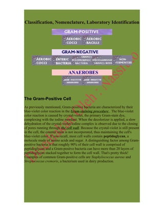
  • 1. Classification, Nomenclature, Laboratory Identification The Gram-Positive Cell As previously mentioned, Gram-positive bacteria are characterized by their blue-violet color reaction in the Gram-staining procedure . The blue-violet color reaction is caused by crystal-violet, the primary Gram-stain dye, complexing with the iodine mordant. When the decolorizer is applied, a slow dehydration of the crystal-violet/iodine complex is observed due to the closing of pores running through the cell wall. Because the crystal-violet is still present in the cell, the counter stain is not incorporated, thus maintaining the cell's blue-violet color. If you recall, most cell walls contain peptidoglycan, a molecule made of amino acids and sugar. A distinguishing factor among Gram- positive bacteria is that roughly 90% of their cell wall is comprised of peptidoglycan and a Gram-positve bacteria can have more than 20 layers of peptidoglycan stacked together to form the cell wall. That's pretty thick! Examples of common Gram-positive cells are Staphylococcus aureus and Streptococcus cremoris, a bacterium used in dairy production.
  • 2. GRAM-POSITIVE COCCI The Gram-positive cocci are grouped together based on their Gram-stain reaction, thick cell wall composition, and spherical shape. Most of the organisms in these groups are members of the Micrococcaceae family All of the organisms in these groups are non-endospore forming chemosynthetic heterotrophs. We will discuss only the clinically relevent bacteria from Micrococcus and Staphylococcus of the Micrococcaceae family. Streptococcus and Enterococcus(formerly a species of Streptococcus) are discussed as well because of the many diseases they inflict on humans. The chart below shows the paths to identification of the genera discussed. GRAM-POSITIVE RODS The Gram-positive rods in this section will be divided into three distinct varieties based upon their ability to produce endospores and their morphological appearance: • ENDOSPORE-FORMING o BACILLUS • REGULAR, NON-ENDOSPORE-FORMING
  • 3. o LACTOBACILLUS o LISTERIA o ERYSIPELOTHRIX • IRREGULAR, NON-ENDOSPORE-FORMING o CORYNEBACTERIUM These bacteria are ubiquitous in nature and most are aerobic or facultatively anaerobic. The Gram-Negative Cell Unlike Gram-positive bacteria, which asuume a violet color in Gram staining, Gram negative bacteria incorporate the counterstain rather than the primary stain. Because the cell wall of Gram(-) bacteria is high in lipid content and low in peptidoglycan content, the primary crystal-violet escapes from the cell when the decolorizer is added. This is because primary stains like to bind with peptidoglycan- something the G(-) cell has very little of. Gram(-) bacteria cause a lot of problems because many species are pathogenic. This pathogenic capability is usually associated with certain components of their cell walls, particularly the lipopolysaccharide (endotoxin) layer. Remember the Black Plague which wiped out a a third of the population of Europe? It was caused by the tiny G(-) rod, Yersinia pestis. Most enteric (bowel related) illnesses can also be attributed to this group of bacteria. Some of my favorites are Salmonella food poisoning. If you choose, you can directly access the diseases section of this tutorial to learn more. MISCELLANEOUS GRAM-NEGATIVE RODS VIBRIO CAMPYLOBACTER HELICOBACTER
  • 4. NEISSERIA The Neisseria genus consists of aerobic, non-spore-forming Gram-negative coccobacilli which inhabit the mucous membranes of many animals (and humans). These non-motile microbes require a moist environment and warm temperatures (human body temperature range) to achieve optimum growth. An important means of identification of Neisseria species is the oxidase test, for which all members test positive. Additionally, Neisseria grow well on chocolate agar containing antibiotics that inhibit growth of Gram-negative bacteria, Gram-positive bacteria, and molds. The two most clinically significant members of the genus Neisseria are N. gonorrhoeae and N. meningitidis. N. gonorrhoeae Infection by the diplococcoid bacterium N. gonorrhoeae is referred to as a gonococcal infection. Gonorrhea is transmitted between humans through intimate contact of the mucous membrane. This sexually transmitted organism can be carried by men and women for many years without any sign or symptoms. In infected males, the disease is characterized by a urethral discharge of pus and can eventually result in other complications such as prostatitis and periurethral abscess. The incubation period of the bacterium can last from a day to a week. Females with gonorrhea exhibit vaginal discharge, abdominal pain, and abnormal non-menstrual bleeding. Ironically, the widespread use of birth control devices such as the pill has actually increased the number of gonococcal infections in the United States. Use of the pill can lower the glycogen concentration of the vaginal membrane. This environmental change inhibits the growth of acid-producing bacteria, such as Lactobacillus, which are the natural flora of the vagina. The vaginal pH soon becomes less acidic and a variety of organisms are able to grow there. As with most other sexually transmitted diseases, gonorrhea is prevalent in young adult and homosexual populations. This disease may sound really bad, but it is treatable. N. gonorrheae is sensitive to ultraviolet radiation, drying, and antibiotics.
  • 5. Because chlamydia infection is often associated with a gonococcal infection, a regimen of ceftriaxone and doxycycline is used to kill both organisms. LABORATORY INDICATIONS: • Oxidase + • Glucose fermentative N. meningitidis It doesn't take a genius to figure out that N. meningitidis causes meningitis, inflammation of the membranes covering the central nervous system. The different strains of N. meningitidis are classified by their capsular polysaccharides. This bacterium is the second leading cause of meningitis in the United States. Early symptoms may include headache, fever, and vomiting. Death can quickly follow due to endotoxin shock or focal cerebral involvement. Infection doesn't always lead to death, however. The organism can often assume a carrier status with very few carriers actually developing the disease. Infected patients can be treated with penicillin, while rifampin may be used prophylactically as a means of preventing the disease state in carriers. ENTEROBACTERIACEAE Members of genera belonging to the Enterobacteriaceae family have earned a reputation placing them among the most pathogenic and most often encountered organisms in clinical microbiology. These large Gram- negative rods are usually associated with intestinal infections, but can be found in almost all natural habitats. They are the causative agents of such diseases as meningitis, bacillary dysentery, typhoid, and food poisoning. As well as being oxidase negative, all members of this family are glucose fermenters and nitrate reducers. In most cases, the pathogenicity of a particular enteric bacterium can be determined by its ability to metabolize lactose. Non-utilizers are usually pathogenic while the lactose utilizers are not. Because many different species in this family can cause similar symptoms, biochemical tests are crucial to the identification, diagnosis, and treatment of infection. We will discuss the twelve genera of the Enterobacteriaceae family which are most commonly encountered in the clinical laboratory: • ESCHERICHIA COLI
  • 6. • SHIGELLA • EDWARDSIELLA • SALMONELLA • CITROBACTER • KLEBSIELLA • ENTEROBACTER • SERRATIA • PROTEUS • MORGANELLA • PROVIDENCIA • YERSINIA
  • 7. Gram-staining Procedure Gram-staining is a four part procedure which uses certain dyes to make a bacterial cell stand out against against its background. The specimen should be mounted and heat fixed on a slide before you procede to stain it. The reagents you will need to successfully perform this operation are: • Crystal Violet (the Primary Stain) • Iodine Solution (the Mordant) • Decolorizer (ethanol is a good choice) • Safranin (the Counterstain) • Water (preferably in a squirt bottle) Before starting, make sure that all reagents, as well as the squirt-bottle of water, are easily accessible because you won't have time to go get them during the staining procedure. Also, make sure you are doing this near a sink because it can get really messy. Wear the appropriate lab attire. STEP 1: Place your slide on a slide holder or a rack. Flood (cover completely) the entire slide with crystal violet. Let the crystal violet stand for about 60 seconds. When the time has elapsed, wash your slide for 5 seconds with the water bottle. The specimen should appear blue-violet when observed with the naked eye. STEP 2: Now, flood your slide with the iodine solution. Let it stand about a minute as well. When time has expired, rinse the slide with water for 5 seconds and immediately procede to step three. At this point, the specimen should still be blue-violet.
  • 8. STEP 3: This step involves addition of the decolorizer, ethanol. Step 3 is somewhat subjective because using too much decolorizer could result in a false Gram (-) result. Likewise, not using enough decolorizer may yield a false Gram (+) results. To be safe, add the ethanol dropwise until the blue-violet color is no longer emitted from your specimen. As in the previous steps, rinse with the water for 5 seconds. STEP 4: The final step involves applying the counterstain, saffranin. Flood the slide with the dye as you did in steps 1 and 2. Let this stand for about a minute to allow the bacteria to incorporate the saffranin. Gram positive cells will incorporate little or no counterstain and will remain blue-violet in appearance. Gram negative bacteria, however, take on a pink color and are easily distinguishable from the Gram positives. Again, rinse with water for 5 seconds to remove any excess of dye. After you have completed steps 1 through 4, you should dry the slide with bibulous paper or allow it to air dry before viewing it under the microscope.
  • 9. STAPHYLOCOCCUS Clinically, the most important genus of the Micrococcaceae family is Staphylococcus. The Staphylococcus genus is classified into two major groups: aureus and non-aureus. S. aureus is a leading cause of soft tissue infections, as well as toxic shock syndrome (TSS) and scalded skin syndrome. It can be distinguished from other species of Staph by a positive result in a coagulase test(all other species are negative). The pathogenic effects of Staph are mainly asssociated with the toxins it produces. Most of these toxins are produced in the stationary phase of the bacterial growth curve. In fact, it is not uncommon for an infected site to contain no viable Staph cells. The S. aureus enterotoxin causes quick onset food poisoning which can lead to cramps and severe vomiting. Infection can be traced to contaminated meats which have not been fully cooked. These microbes also secrete leukocidin, a toxin which destroys white blood cells and leads to the formation of pus and acne. Particularly, S. aureus has been found to be the causative agent in such ailments as pneumonia, meningitis, boils, arthritis, and osteomyelitis (chronic bone infection). Most S. aureus are penicillin resistant, but vancomycin and nafcillin are known to be effective against most strains. Of the non-aureus species, S. epidermis is the most clinically significant. This bacterium is an opportunistic pathogen which is a normal resident of human skin. Those susceptible to infection by the bacterium are IV drug users, newborns, elderly, and those using catheters or other artificial appliances. Infection is easily treatable with vancomycin or rifampin. LABORATORY INDICATIONS: • Anaerobic glucose fermentation with acid production • Catalase + • Nitrate + • Coagulase +
  • 10. STREPTOCOCCUS The Streptococcus genus consists of Gram-positive, aerobic bacteria which appear as chains under microscopic observation. The organisms in this genus are characterized by a coccus appearance, a thick cell wall, and aerobic action on glucose. Four different classification systems exist for this important microorganism: CLINICAL • Pyogenic Streptococci • Oral Streptococci • Enteric Streptococci, HEMOLYSYS • alpha-hemolysis • beta-hemolysis • gamma-hemolysis SEROLOGICAL-Lancefield (A-H), (K-U) BIOCHEMICAL(physiological)
  • 11. GROUP A The first group in the Lancefield classification system includes only one species of Streptococcus, S. pyogenes. This particular opportunistic pathogen is responsable for about 90% of all cases of pharyngitis. A common form of pharyngitis is "Strep throat" which is characterized by inflamation and swelling of the throat, as well as development of pus-filled regions on the tonsils. Penicillin is usually administered to patients as soon as possible to quell the possibility of the infection spreading from the upper respiratory system into the lungs. Once in the lungs, the infection could give rise to pneumonia. Some cases also develop into rheumatic fever if left untreated. Other diseases linked to S. pyogenes are skin infections such as impetigo, cellulitis, and erysipelas. LABORATORY INDICATIONS: • Catalase - • Beta-hemolysis • Bacitracin sensitive GROUP B The B classification of Lansefield also includes only one bacterium, S. agalactiae. For years this bacterium has been the causative agent in mastitis in cows. Currently, it has been found to be a cause of sexually transmitted urogenital infections in females. Although infection is easily treated with penicillin, proper diagnosis is necessary for women nearing labor because the infection can easily spread to the child via the birth canal. LABORATORY INDICATIONS: • CAMP + • Beta-hemolysis GROUP D Type D Streptococcus is the next clinically important bacterium because of the multitude of diseases it is known to cause. Although many are harmless, the pathogenic strains cause complications of the human digestive tract. This group has recently been reclassified into two divisions: Enterococcus and non- Enterococcus. The Enterococci include E. faecalis, a cause of urinary tract infections, and E. faecium, a bacterium resistant to many common antibiotics. Diseases such as septicemia, endocarrditis, and appendicitis have also been attributed to group D Strep. Fecal matter from infected individuals is a source
  • 12. for isolation and identification techniques. Once identified, Group D Strep can be treated with ampicillin alone or in combination with gentamicin. LABORATORY INDICATIONS: • Hydrolysis of bile esculin (dark brown medium) -this indicates the ability of the bacteria to tolerate bile from the liver • Growth in high salt conc. OTHER IMPORTANT STREP Streptococcus pneumoniae Because its surface carbohydrate antigens do not correspond to a specific Lancefield group, S. pneumonia is discussed separately. Although not given a letter designation, S. pneumoniae can be considered a Pyogenic (pus- producing) strain of Strep. It can be distinguished from other Pyogenic bacteria by its high sensitivity to Optochin (no growth zone of inhibition). This bacterium causes pneumonia (obviously!), meningitis, and otitis media. It also demonstrates alpha-hemolytic growth on blood agar. Viridans Group The Viridans Streptococci, consisting of S. mutans and S. mitis, are alpha- hemolytic bacteria. These bacteria inhabit the mouth. In fact, a large percentage of tooth decay can be attributed to S. mutans. BACILLUS Bacillus represents a genus of Gram-positive bacteria which are ubiquitous in nature (soil, water, and airborne dust). Some species are natural flora in the human intestines. When grown on blood agar, Bacillus produces large, spreading, gray-white colonies with irregular margins. A unique characteristic of this bacterium is its ability to produce endospores when environmental conditions are stressful. The only other known spore-producing bacterium is Clostridium. Although most species of Bacillus are harmless saprophytes, two species are considered medically significant: B.anthracis and B. cereus.
  • 13. B. anthracis B. anthracis is the bacterium which causes anthrax in cows, sheep, and sometimes humans. Anthrax is transmitted to humans via direct contact with animal products or inhalation of endospores. Under the microscope, B. anthracis cells appear to have square ends and seem to be attached by a joint to other cells. The spores are best observed when the bacterium is cultered on artificial media. Sources of infection are usually industrial or agricultural and the infection is classified as one of three types: • CUTANEOUS INFECTION (95% of human cases) • INHALATION ANTHRAX (rare) • GASTROINTESTINAL ANTHRAX (very rare!) LABORATORY INDICATIONS: • Nonhemolytic (sheep blood agar) • Non-motile • Gel hydrolysis - • Catalase + B. cereus Unlike B. anthracis, B.cereus is a motile bacterium which can cause toxin- mediated food poisoning. It is known to inhabit many kinds of food including stew, cereal, and milk. Most recently, however, it has been found in fried rice. The two toxins released by the bacterium lead to vomiting and diarrhea, symptoms similar to those of Staphylococcus food poisoning. Because toxin production usually takes place after the infected foods are cooked, proper cold storage of food is recommended immediately after preparation. LABORATORY INDICATIONS: • Hemolytic (sheep blood agar) • Motile • Gel hydrolysis + • Glucose, maltose, & salicin fermentative • Catalase +
  • 14. Biochemical Tests, Media, Techniques CATALASE TEST Some bacteria and macrophages can reduce diatomic oxygen to hydrogen peroxide or superoxide. Both of these molecules are toxic to bacteria. Some bacteria, however, possess a defense mechanism which can minimize the harm done by the two compounds. These resistant bacteria use two enzymes to catalyze the conversion of hydrogen peroxide and superoxide back into diatomic oxygen and water. One of these enzymes is catalase and its presence can be detected by a simple test. The catalase test involves adding hydrogen peroxide to a culture sample or agar slant. If the bacteria in question produce catalase, they will convert the hydrogen peroxide and oxygen gas will be evolved. The evolution of gas causes bubbles to form and is indicative of a positive test.
  • 15. CITRATE TEST The citrate test is used to determine the ability of a bacterium to utilize citrate as its only source of carbon. Bacteria can break the conjugate base salt of citrate into organic acids and carbon dioxide. The carbon dioxide can combine with the sodium from the conjugate base salt to form a basic compound, sodium carbonate. A pH indicator in the medium detects the presence of this compound by turning blue (a positive test). COAGULASE TEST Like the mannitol salts agar, the coagulase test is another method for differienting between pathogenic and non- pathogenic strains of Staphylococcus. Bacteria that produce coagulase use it as a defense mechanism by clotting the areas of plasma around them, thereby enabling themselves to resist phagocytosis by the host's immune system. The sample in question is usually inoculated onto 0.5 ml of rabbit plasma and incubated at 37 degrees celsius for one to four hours. A positive test is denoted by a clot formation in the test tube after the allotted time.
  • 16. OPTOCHIN TEST The optochin test is a presumptive test that is used to identify strains of Streptococcus pneumoniae. Optochin (ethyl hydrocupreine) disks are placed on inoculated blood agar plates. Because S. pneumoniae is not optochin resistant, a zone of inhibition will develop around the disk where the bacteria have been lysed. This zone is typically 14mm from the disk or greater. OXIDASE TEST Cytochrome oxidase is an enzyme found in some bacteria that transfers electrons to oxygen, the final electron acceptor in some electron transport chains. Thus, the enzyme oxidizes reduced cytochrome c to make this transfer of energy. Presence of cytochrome oxidase can be detected through the use of an Oxidase Disk which acts as an electron donator to cytochrome oxidase. If the bacteria oxidize the disk (remove electrons) the disk will turn purple, indicating a positive test. No color change indicates a negative test.
  • 17. UREASE TEST Urease is an enzyme that breaks the carbon-nitrogen bond of amides to form carbon dioxide, ammonia, and water. Members of genus Proteus are known to produce urease. Urease can be detected by plating bacteria onto an amide containing medium, specifically urea. When urea is broken down, ammonia is released and the pH of the medium increases (becomes more basic). This pH change is detected by a pH indicator that turns pink in a basic environment. A pink medium indicates a positive test for urease. MEDIA CHOCOLATE AGAR Chocolate agar is a nutrient medium which is used in culturing fastidious organisms such as Haemophilus species and Neisseria. It is comprised of sheep blood that provides the X and V factors necessary for Haemophilus growth. Chocolate agar, however, does not reveal hemolysis data, so species
  • 18. differentiation among the members of Haemophilus must be performed in another manner. EOSIN METHYLENE BLUE (EMB) AGAR EMB agar is a differential medium used in identification and isolation of Gram- negative enteric rods. EMB agar also inhibits the growth of Gram-positive organisms. The differential basis of this medium involves two indicator dyes, eosin and methylene blue, that distinguish between lactose fermenting and non- lactose fermenting organisms. Lactose fermenters form colonies with dark centers and clear borders while the non-lactose fermenters form completely coloroless colonies. MACCONKEY AGAR MacConkey agar is probably the most popular solid differential medium in the world. It is mainly used in identification of lactose fermenting, Gram-negative enteric pathogens and for inhibiting growth of Gram-positive organisms. Bacterial colonies that can ferment lactose turn the medium red. This red color is due to the pH indicators response to the acidic environment created by
  • 19. fermenting lactose. Organisms that do not ferment lactose do not cause a color change. MANNITOL SALTS AGAR A common medium used for the isolation of pathogenic staphylococci is the Mannitol Salts Agar. The high salt concentration of this medium inhibits the growth of most other organisms. Pathogenic staphylococci not only grow on the medium, but they also produce acid from it. This acid production turns the pH indicator from red to yellow. Non-pathogenic staphylococci can grow on the medium but produce no acid from it. SMEAR PREPARATION The preparation of a smear is required for many laboratory procedures, including the Gram-stain. The purpose of making a smear is to fix the bacteria onto the slide and to prevent the sample from being lost during a staining procedure. A smear can be prepared from a solid or broth medium. Below are some guidelines for preparing a smear for a Gram-stain.
  • 20. 1. Place one needle of solid bacterial growth or two loops of liquid bacterial growth in the center of a clean slide. 2. If working from a solid medium, add one drop (and only one drop) of water to your specimen with a water bottle. If using a broth medium, do not add the water. 3. Now, with your inoculating loop, mix the specimen with the water completely and spread the mixture out to cover about half of the total slide area. 4. Place the slide on a slide warmer and wait for it to dry. The smear is now ready for the staining procedure.