
Recommended
More Related Content
What's hot
What's hot (20)
Viewers also liked
Viewers also liked (20)
Similar to Anatomy & Physiology
Similar to Anatomy & Physiology (20)
Recently uploaded
Recently uploaded (20)
Anatomy & Physiology
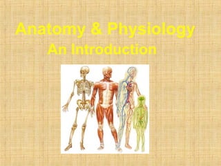
- 1. Anatomy & Physiology An Introduction
- 2. Anatomy - The study of the structure of the human body Physiology - The study of body function An Overview of Anatomy
- 3. Chemical level – atoms form molecules Cellular level – cells and their subunits Tissue level – a group of cells performing a common function Organ level – a discrete structure made up of more than one tissue Organ system – organs working together for a common purpose Organism – the result of all simpler levels working together Structural Organization
- 5. Forms external body covering Protects deeper tissues from injury Synthesizes vitamin D Site of cutaneous receptors (pain, pressure, etc.) and sweat and oil glands The Integumentary System
- 6. The Integumentary System • Integument is skin • Skin and its appendages make up the integumentary system • A fatty layer (hypodermis) lies deep to it • Two distinct regions – Epidermis – Dermis
- 7. Functions of skin • Protection – Cushions and insulates and is waterproof – Protects from chemicals, heat, cold, bacteria – Screens UV • Synthesizes vitamin D with UV • Regulates body heat • Prevents unnecessary water loss • Sensory reception (nerve endings)
- 8. Epidermis • Keratinized stratified squamous epithelium • Four types of cells – Keratinocytes – deepest, produce keratin (tough fibrous protein) – Melanocytes - make dark skin pigment melanin – Merkel cells – associated with sensory nerve endings – Langerhans cells – macrophage-like dendritic cells • Layers (from deep to superficial) – Stratum basale or germinativum – single row of cells attached to dermis; youngest cells – Stratum spinosum – spinyness is artifactual; tonofilaments (bundles of protein) resist tension – Stratum granulosum – layers of flattened keratinocytes producing keratin (hair and nails made of it also) – Stratum lucidum (only on palms and soles) – Stratum corneum – horny layer (cells dead, many layers thick)
- 9. Dermis • Strong, flexible connective tissue: your “hide” • Cells: fibroblasts, macrophages, mast cells, WBCs • Fiber types: collagen, elastic, reticular • Rich supply of nerves and vessels • Critical role in temperature regulation (the vessels) • Two layers (see next slides) – Papillary – areolar connective tissue; includes dermal papillae – Reticular – “reticulum” (network) of collagen and reticular fibers
- 11. Hypodermis • “Hypodermis” (Gk) = below the skin • “Subcutaneous” (Latin) = below the skin • Also called “superficial fascia” “fascia” (Latin) =band; in anatomy: sheet of connective tissue • Fatty tissue which stores fat and anchors skin (areolar tissue and adipose cells) • Different patterns of accumulation (male/female)
- 12. Skin color • Three skin pigments – Melanin: the most important – Carotene: from carrots and yellow vegies – Hemoglobin: the pink of light skin • Melanin in granules passes from melanocytes (same number in all races) to keratinocytes in stratum basale – Digested by lysosomes – Variations in color – Protection from UV light vs vitamin D?
- 13. Skin appendages • Derived from epidermis but extend into dermis • Include – Hair and hair follicles – Sebaceous (oil) glands – Sweat (sudoiferous) glands – Nails
- 15. Nails • Of hard keratin • Corresponds to hooves and claws • Grows from nail matrix
- 16. • Functions of hair – Warmth – less in man than other mammals – Sense light touch of the skin – Protection - scalp • Parts – Root imbedded in skin – Shaft projecting above skin surface • Make up of hair – hard keratin • Three concentric layers – Medulla (core) – Cortex (surrounds medulla) – Cuticle (single layers, overlapping)
- 17. • Types of hair – Vellus: fine, short hairs – Intermediate hairs – Terminal: longer, courser hair • Hair growth: averages 2 mm/week – Active: growing – Resting phase then shed • Hair loss – Thinning – age related – Male pattern baldness • Hair color – Amount of melanin for black or brown; distinct form of melanin for red – White: decreased melanin and air bubbles in the medulla – Genetically determined though influenced by hormones and environment
- 18. Sebaceous (oil) glands • Entire body except palms and soles • Produce sebum by holocrine secretion • Oils and lubricates
- 19. Disorders of the integumentary system • Burns – Threat to life • Catastrophic loss of body fluids • Dehydration and fatal circulatory shock • Infection – Types • First degree – epidermis: redness (e.g. sunburn) • Second degree – epidermis and upper dermis: blister • Third degree - full thickness • Infections • Skin cancer
- 20. SKELETON SYSTEM
- 21. – Protects and supports body organs – Provides a framework for muscles – Blood cells formed within bones – Stores minerals The Skeletal System
- 22. Bone- hardest tiddue in body consist of 80% organic and inorganic material and 20 % of water. Bone cells are called osteocytes. Func. of bones :- 1. provide framework to the body. 2. gives attachment to muscles and tendons 3. permit movement of the body as a whole and parts of the body by forming joints. 4. are site of blood cells development 5. provide reservoir for calcium.
- 23. Joints- site at which any 2 or more bone come together.Joint can be broadly classified as of 3 types :- 1. Fibrous or fixed joints-immovable, fibrous tissue between bones. e.g.between bones of the skull. 2. Cartilaginous or slightly movable joints- have a pad of cartilaginous between ends of bones making up the joints allowing very slight movement caused by compression of the pad. e.g joint between bones of vertebral column.
- 24. 3. Synovial or freely movable joints- These joints allows maximum movement and are classified according to the range of movement possible. a) ball and socket joint b) Hinge joint c)Gliding joint d) pivot joint e) Saddle joint
- 25. MUSCULAR SYSTEM
- 26. Allows manipulation of environment Locomotion Facial expression Maintains posture Produces heat The Muscular System
- 27. The Muscular System • The ability to move is an essential activity of the human body • ½ our body weight comes from muscles • Consists of over 600 individual muscles. • 3 purposes: – Body movement – Body shape – Body heat (maintain temp.)
- 28. The Muscular System • Body movements are determined by three types of muscles – Smooth (involuntary) – cannot be controlled by will. – Cardiac – control the contractions of the heart. – Skeletal (Voluntary) – can be controlled by will.
- 29. Function of Skeletal muscles • Attach to bones to provide voluntary movement – Tendons: strong, tough connective cords – Fascia: tough, sheet-like membrane • Produce heat and energy for the body • Help maintain posture • Protect internal organs • Called striated (striped) because they have striations of alternating light and dark band
- 30. Functions cont’d. • Fleshy body parts are made of skeletal muscles • Provide movements to the limbs, but contract quickly, fatigue easily and lack the ability to maintain contraction for long periods – Blinking eyes, talking, breathing, eating, dancing and writing all produced by these muscles
- 32. Function of Smooth Muscle • Called smooth muscle because they are unmarked by striations, small spindle shaped • Unattached to bones, act slowly, do not tire easily and can remain contracted for a long time • Not under conscious control so they are also called involuntary muscles • Found in walls of internal organs (intestines, bladder, stomach, uterus, blood vessels)
- 33. Function of cardiac muscle • Found only in the heart • Involuntary muscle • Requires a continuous supply of oxygen to function • Cardiac muscle cells begin to die after 30 seconds of oxygen cut-off • Striated and branched
- 35. Characteristics of Muscles • All muscles have 4 common characteristics – Excitability – ability to respond to a stimulus (ie: nerve impulse) – Contractibility – muscle fibers that are stimulated by nerves contract (become shorter) and causes movement – Extensibility – ability to be stretched – Elasticity – allows the muscle to return to its original shape after it has been stretched
- 36. • FIBROMYALGIA • Chronic, widespread pain in specific muscle site; numbness and tingling in arms or legs; headaches • Cause unknown • Treat symptoms – pain relief; stress reduction and muscle relaxers • MUSCULAR DYSTROPHY • Group of inherited diseases that cause chronic, progressive muscle atrophy resulting in total disability and early death • No cure • Treatment used to slow progression of disease
- 37. • MYASTENIA GRAVIS • Chronic condition where nerve impulses are not transmitted correctly leading to progressive muscular weakness and paralysis; affects respiratory muscles and can be fatal • Cause unknown • Treatment is supportive • MUSCLE SPASMS/CRAMPS • Sudden, painful involuntary muscle contractions • Caused from overexertion, low electrolytes or poor circulation • Treat by applying gentle pressure and stretching of the affected muscle
- 38. • STRAIN • Overstretching of a muscle or tendon frequently in legs, back or arms • Caused by sudden muscle exertion • Treated by resting, muscle relaxants, or pain medications, elevation of extremity and applying hot/cold compresses
- 39. NERVOUS SYSTEM
- 40. Fast-acting control system Responds to internal and external changes The Nervous System
- 41. Peripheral Nervous System • 3 kinds of neurons connect CNS to the body – sensory – motor – interneurons • Motor - CNS to muscles and organs • Sensory - sensory receptors to CNS • Interneurons: Connections Within CNS Spinal Cord Brain Nerves
- 42. Peripheral Nervous System Skeletal (Somatic) Sympathetic Parasympathetic Autonomic Peripheral Nervous System
- 43. Somatic System • Nerves to/from spinal cord – control muscle movements – somatosensory inputs • Both Voluntary and reflex movements • Skeletal Reflexes – simplest is spinal reflex arc Muscle Motor Neuron Interneuron Skin receptors Sensory Neuron Brain
- 44. Autonomic System • Two divisions: – sympathetic – Parasympatheitic • Control involuntary functions – heartbeat – blood pressure – respiration – perspiration – digestion • Can be influenced by thought and emotion
- 45. Sympathetic • “ Fight or flight” response • Release adrenaline and noradrenaline • Increases heart rate and blood pressure • Increases blood flow to skeletal muscles • Inhibits digestive functions CENTRAL NERVOUS SYSTEM Brain Spinal cord SYMPATHETIC Dilates pupil Stimulates salivation Relaxes bronchi Accelerates heartbeat Inhibits activity Stimulates glucose Secretion of adrenaline, nonadrenaline Relaxes bladder Stimulates ejaculation in male Sympathetic ganglia Salivary glands Lungs Heart Stomach Pancreas Liver Adrenal gland Kidney
- 46. Parasympathetic • “ Rest and digest ” system • Calms body to conserve and maintain energy • Lowers heartbeat, breathing rate, blood pressure CENTRAL NERVOUS SYSTEM Brain PARASYMPATHETIC Spinal cord Stimulates salivation Constricts bronchi Slows heartbeat Stimulates activity Contracts bladder Stimulates erection of sex organs Stimulates gallbladder Gallbladder Contracts pupil
- 47. Central Nervous System • Brain and Spinal Cord Spinal Cord Brain
- 48. Problems from lack of movement• Contractures – Tightening and shortening of a muscle resulting in a permanent flexing of a joint • Muscle atrophy – Muscles become weak and joints become stiff • Circulatory impairment – Blood clots and pressure ulcers can develop • Mineral loss – Especially calcium from the bones making bones brittle and easily to be fractured • Other problems – Poor appetite; constipation; urinary infections; respiratory problems; and pneumonia
- 49. • Left & Right sides are separate • Corpus Callosum : major pathway between hemispheres • Some functions are ‘lateralized’ – language on left – math, music on right • Lateralization is never 100% Brain has 2 Hemispheres Left Hemisphere Corpus Callosum Right Hemisphere
- 50. Each hemisphere is divided into 4 lobes Frontal Parietal Occipital Temporal
- 52. The Nervous System: Summary • Major structures of the nervous system – CNS: Brain & Spinal Cord – PNS: Somatic, Autonomic (Sympathetic & Parasympathetic) – Two hemispheres & 4 lobes • Organization – Afferent (sensory) neurons-towards CNS – Efferent (motor) neurons-away from CNS – Sympathetic-speeds up – Parasympathetic-slows down Central Nervous System Peripheral Nervous System
- 54. ENDOCRINE SYSTEM
- 55. Glands secrete hormones that regulate Growth Reproduction Nutrient use The Endocrine System
- 56. Endocrine Overview • Hormones- chemical messengers travel through body • Target cell or organ- organ or cells that a hormone affects
- 57. Anterior Pituitary Gland • Growth Hormone (GH)- affects growth of skeletal muscles and bones • Prolactin (PRL)- stimulates milk production after pregnancy • Gonadotropic- regulates hormone activity of sex organs • Also effects adrenal cortex and thyroid hormone release
- 58. Posterior Pituitary • Oxytocin- helps during pregnancy • Antidiuretic hormone (ADH)- inhibits urine production – Alcohol inhibits ADH causing increased output of urine
- 59. Thyroid Gland • Thyroid hormone – Controls the rate at which glucose is “burned” • Calcitonin – Decreases calcium levels in blood deposits on bone
- 60. Parathyroid Gland • Parathyroid hormone (PTH)- regulates Ca2+ in blood – If Ca2+ level drops bones is broken down
- 61. Adrenal Gland • Adrenal Cortex – Releases hormones that regulate mineral content in blood • Adrenal Medulla – Produces epinephrine and norepinephrine – Helps with stressful situations
- 62. Pancreas • Insulin- increases glucose uptake into cells and stores in liver • Glucagon- stimulates breakdown of glucose stores in the liver
- 63. Pineal Gland • Melatonin – Establishes body’s day/ night cycle
- 64. Thymus • Thymosin – “Programs” T cells
- 65. Gonads • Androgens (male)- sperm formation, development of male char • Estrogens (female)- development of female char
- 66. The Circulatory System The Heart, Blood Vessels, Blood Types
- 67. The Closed Circulatory System •Humans have a closed circulatory system, typical of all vertebrates, in which blood is confined to vessels and is distinct from the interstitial fluid. –The heart pumps blood into large vessels that branch into smaller ones leading into the organs. –Materials are exchanged by diffusion between the blood and the interstitial fluid bathing the cells.
- 68. The Cardiovascular System •Three Major Elements – Heart, Blood Vessels, & Blood –1. The Heart- cardiac muscle tissue –highly interconnected cells –four chambers •Right atrium •Right ventricle •Left atrium •Left ventricle
- 70. Pathway of the blood •Superior Vena Cava •Right Atrium •Tricuspid Valve •Right Ventricle •Pulmonary Semilunar Valve •Lungs •Pulmonary Vein •Bicuspid Valve •Left Ventricle •Aortic Semilunar Valve •Aorta •To the bodies organs & cells
- 71. The Cardiovascular System 2. Blood Vessels -A network of tubes –Arteriesarterioles move away from the heart •Elastic Fibers •Circular Smooth Muscle –Capillaries – where gas exchange takes place. •One cell thick •Serves the Respiratory System –VeinsVenules moves towards the heart •Skeletal Muscles contract to force blood back from legs •One way values •When they break - varicose veins form
- 72. The Cardiovascular System 3. The Blood A. Plasma Liquid portion of the blood. Contains clotting factors, hormones, antibodies, dissolved gases, nutrients and waste
- 73. The Cardiovascular System •The Blood B. Erythrocytes - Red Blood Cells –Carry hemoglobin and oxygen. Do not have a nucleus and live only about 120 days. –Can not repair themselves.
- 74. The Cardiovascular System •The Blood C. Leukocytes – White Blood cells –Fight infection and are formed in the bone marrow –Five types – neutrophils, lymphocytes, eosinophils, basophils, and monocytes.
- 75. The Cardiovascular System The Blood •D. Thrombocytes – Platelets. –These are cell fragment that are formed in the bone marrow from magakaryocytes. –Clot Blood by sticking together – via protein fibers called fibrin.
- 76. Disorders of the Circulatory System • Anemia - lack of iron in the blood, low RBC count • Leukemia - white blood cells proliferate wildly, causing anemia • Hemophilia - bleeder’s disease, due to lack of fibrinogen in thrombocytes • Heart Murmur - abnormal heart beat, caused by valve problems • Heart attack - blood vessels around the heart become blocked with plaque, also called myocardial infarction
- 77. Functions of the Heart • Generating blood pressure • Routing blood – Heart separates pulmonary and systemic circulations • Ensuring one-way blood flow – Heart valves ensure one-way flow • Regulating blood supply – Changes in contraction rate and force match blood delivery to changing metabolic needs
- 78. Size, Shape, Location of the Heart •Size of a closed fist •Shape –Apex: Blunt rounded point of cone –Base: Flat part at opposite of end of cone •Located in thoracic cavity in mediastinum
- 79. Heart Wall • Three layers of tissue – Epicardium: This serous membrane of smooth outer surface of heart – Myocardium: Middle layer composed of cardiac muscle cell and responsibility for heart contracting – Endocardium: Smooth inner surface of heart chambers
- 80. External Anatomy •Four chambers –2 atria –2 ventricles •Auricles •Major veins –Superior vena cava –Pulmonary veins •Major arteries –Aorta –Pulmonary trunk
- 81. Heart Valves •Atrioventricular –Tricuspid –Bicuspid or mitral •Semilunar –Aortic –Pulmonary •Prevent blood from flowing back
- 82. Blood Flow Through Heart
- 83. The Cardiovascular System Blood vessels transport blood Carries oxygen and carbon dioxide Also carries nutrients and wastes Heart pumps blood through blood vessels
- 84. The Cardiovascular System Blood vessels transport blood Carries oxygen and carbon dioxide Also carries nutrients and wastes Heart pumps blood through blood vessels
- 85. Conducting System of Heart
- 86. Cardiac Arrhythmias • Tachycardia: Heart rate in excess of 100bpm • Bradycardia: Heart rate less than 60 bpm • Sinus arrhythmia: Heart rate varies 5% during respiratory cycle and up to 30% during deep respiration • Premature atrial contractions: Occasional shortened intervals between one contraction and succeeding, frequently occurs in healthy people
- 87. Cardiac Cycle • Heart is two pumps that work together, right and left half • Repetitive contraction (systole) and relaxation (diastole) of heart chambers • Blood moves through circulatory system from areas of higher to lower pressure. – Contraction of heart produces the pressure
- 88. Heart Sounds • First heart sound or “lubb” – Atrioventricular valves and surrounding fluid vibrations as valves close at beginning of ventricular systole • Second heart sound or “dupp” – Results from closure of aortic and pulmonary semilunar valves at beginning of ventricular diastole, lasts longer • Third heart sound (occasional) – Caused by turbulent blood flow into ventricles and detected near end of first one-third of diastole
- 89. Location of Heart Valves
- 90. Mean Arterial Pressure (MAP) • Average blood pressure in aorta • MAP=CO x PR – CO is amount of blood pumped by heart per minute • CO=SV x HR – SV: Stroke volume of blood pumped during each heart beat – HR: Heart rate or number of times heart beats per minute • Cardiac reserve: Difference between CO at rest and maximum CO – PR is total resistance against which blood must be pumped
- 91. Picks up fluid leaked from blood vessels Disposes of debris in the lymphatic system Houses white blood cells (lymphocytes) Mounts attack against foreign substances in the body The Lymphatic System
- 93. Keeps blood supplied with oxygen Removes carbon dioxide Gas exchange occurs through walls of air sacs in the lungs The Respiratory System
- 94. Organization and Functions of the Respiratory System • Consists of an upper respiratory tract (nose to larynx) and a lower respiratory tract ( trachea onwards) . • Conducting portion transports air. - includes the nose, nasal cavity, pharynx, larynx, trachea, and progressively smaller airways, from the primary bronchi to the terminal bronchioles • Respiratory portion carries out gas exchange. - composed of small airways called respiratory bronchioles and alveolar ducts as well as air sacs called alveoli
- 96. Respiratory System Functions 1. supplies the body with oxygen and disposes of carbon dioxide 2. filters inspired air 3. produces sound 4. contains receptors for smell 5. rids the body of some excess water and heat 6. helps regulate blood pH
- 97. Breathing • Breathing (pulmonary ventilation). consists of two cyclic phases: • inhalation, also called inspiration - draws gases into the lungs. • exhalation, also called expiration - forces gases out of the lungs.
- 98. Upper Respiratory Tract • Composed of the nose and nasal cavity, paranasal sinuses, pharynx (throat), larynx. • All part of the conducting portion of the respiratory system.
- 99. Respiratory mucosa • A layer of pseudostratified ciliated columnar epithelial cells that secrete mucus • Found in nose, sinuses, pharynx, larynx and trachea • Mucus can trap contaminants – Cilia move mucus up towards mouth
- 100. Nose • Internal nares - opening to exterior • External nares opening to pharynx • Nasal conchae - folds in the mucous membrane that increase air turbulence and ensures that most air contacts the mucous membranes
- 101. Nose rich supply of capillaries warm the inspired air • olfactory mucosa – mucous membranes that contain smell receptors • respiratory mucosa – pseudostratified ciliated columnar epithelium containing goblet cells that secrete mucus which traps inhaled particles, • lysozyme kills bacteria and lymphocytes and • IgA antibodies that protect against bacteria
- 102. Nose provides and airway for respiration • moistens and warms entering air • filters and cleans inspired air • resonating chamber for speech detects odors in the air stream rhinoplasty: surgery to change shape of external nose
- 103. Paranasal Sinuses • Four bones of the skull contain paired air spaces called the paranasal sinuses - frontal, ethmoidal, sphenoidal, maxillary • Decrease skull bone weight • Warm, moisten and filter incoming air • Add resonance to voice. • Communicate with the nasal cavity by ducts. • Lined by pseudostratified ciliated columnar epithelium.
- 104. Pharynx • Common space used by both the respiratory and digestive systems. • Commonly called the throat. • Originates posterior to the nasal and oral cavities and extends inferiorly near the level of the bifurcation of the larynx and esophagus. • Common pathway for both air and food.
- 105. Nasopharynx • Superior-most region of the pharynx. Covered with pseudostratified ciliated columnar epithelium. • Located directly posterior to the nasal cavity and superior to the soft palate, which separates the oral cavity. • Normally, only air passes through. • Material from the oral cavity and oropharynx is typically blocked from entering the nasopharynx by the uvula of soft palate, which elevates when we swallow. • In the lateral walls of the nasopharynx, paired auditory/eustachian tubes connect the nasopharynx to the middle ear. • Posterior nasopharynx wall also houses a single pharyngeal tonsil (commonly called the adenoids).
- 106. Laryngopharynx • Inferior, narrowed region of the pharynx. • Extends inferiorly from the hyoid bone to the larynx and esophagus. • Terminates at the superior border of the esophagus and the epiglottis of the larynx. • Lined with a nonkeratinized stratified squamous epithelium. • Permits passage of both food and air.
- 107. Lower Respiratory Tract • Conducting airways (trachea, bronchi, up to terminal bronchioles). • Respiratory portion of the respiratory system (respiratory bronchioles, alveolar ducts, and alveoli).
- 108. Larynx • Voice box is a short, somewhat cylindrical airway ends in the trachea. • Prevents swallowed materials from entering the lower respiratory tract. • Conducts air into the lower respiratory tract. • Produces sounds. • Supported by a framework of nine pieces of cartilage (three individual pieces and three cartilage pairs) that are held in place by ligaments and muscles.
- 109. Larynx • Muscular walls aid in voice production and the swallowing reflex • Glottis – the superior opening of the larynx • Epiglottis – prevents food and drink from entering airway when swallowing • pseudostratified ciliated columnar epithelium
- 110. Conducting zone of lower respiratory tract
- 111. Trachea • A flexible tube also called windpipe. • Extends through the mediastinum and lies anterior to the esophagus and inferior to the larynx. • Anterior and lateral walls of the trachea supported by 15 to 20 C-shaped tracheal cartilages. • Cartilage rings reinforce and provide rigidity to the tracheal wall to ensure that the trachea remains open at all times • Posterior part of tube lined by trachealis muscle • Lined by ciliated pseudostratified columnar epithelium.
- 112. Respiratory Zone of Lower Respiratory Tract
- 113. Conduction vs. Respiratory zones • Most of the tubing in the lungs makes up conduction zone – Consists of nasal cavity to terminal bronchioles • The respiratory zone is where gas is exchanged – Consists of alveoli, alveolar sacs, alveolar ducts and respiratory bronchioles
- 114. Respiratory Bronchioles, Alveolar Ducts, and Alveoli • Lungs contain small saccular outpocketings called alveoli. • They have a thin wall specialized to promote diffusion of gases between the alveolus and the blood in the pulmonary capillaries. • Gas exchange can take place in the respiratory bronchioles and alveolar ducts as well as in the alveoli, each lung contains approximately 300 to 400 million alveoli. • The spongy nature of the lung is due to the packing of millions of alveoli together.
- 115. Gross Anatomy of the Lungs • Each lung has a conical shape. Its wide, concave base rests upon the muscular diaphragm. • Its superior region called the apex projects superiorly to a point that is slightly superior and posterior to the clavicle. • Both lungs are bordered by the thoracic wall anteriorly, laterally, and posteriorly, and supported by the rib cage. • Toward the midline, the lungs are separated from each other by the mediastinum. • The relatively broad, rounded surface in contact with the thoracic wall is called the costal surface of the lung.
- 116. Lungs Left lung • divided into 2 lobes by oblique fissure • smaller than the right lung • cardiac notch accommodates the heart Right • divided into 3 lobes by oblique and horizontal fissure • located more superiorly in the body due to liver on right side
- 117. Pleura and Pleural Cavities • The outer surface of each lung and the adjacent internal thoracic wall are lined by a serous membrane called pleura. • The outer surface of each lung is tightly covered by the visceral pleura. • while the internal thoracic walls, the lateral surfaces of the mediastinum, and the superior surface of the diaphragm are lined by the parietal pleura. • The parietal and visceral pleural layers are continuous at the hilus of each lung.
- 118. Blood supply of Lungs • pulmonary circulation - • bronchial circulation – bronchial arteries supply oxygenated blood to lungs, bronchial veins carry away deoxygenated blood from lung tissue superior vena cava • Response of two systems to hypoxia – pulmonary vessels undergo vasoconstriction bronchial vessels like all other systemic vessels undergo vasodilation
- 119. Respiratory events • Pulmonary ventilation = exchange of gases between lungs and atmosphere • External respiration = exchange of gases between alveoli and pulmonary capillaries • Internal respiration = exchange of gases between systemic capillaries and tissue cells
- 120. Two phases of pulmonary ventilation • Inspiration, or inhalation - a very active process that requires input of energy.The diaphragm, contracts, moving downward and flattening, when stimulated by phrenic nerves. • Expiration, or exhalation - a passive process that takes advantage of the recoil properties of elastic fiber. ・ The diaphragm relaxes.The elasticity of the lungs and the thoracic cage allows them to return to their normal size and shape.
- 121. DIGESTIVE SYSTEM
- 122. Breaks down food into absorbable units Indigestible foodstuffs eliminated as feces The Digestive System
- 123. Introduction There are four stages to food processing: 1.Ingestion: taking in food 2.Digestion: breaking down food into nutrients 3.Absorption: taking in nutrients by cells 4.Egestion: removing any leftover wastes
- 124. The Human Digestive System • Begins when food enters the mouth. • It is physically broken down by the teeth. • It is begun to be chemically broken down by amylase, an enzyme in saliva that breaks down carbohydrates.
- 125. The Human Digestive System • The tongue moves the food around until it forms a ball called a bolus. • The bolus is passed to the pharynx (throat) and the epiglottis makes sure the bolus passes into the esophagus and not down the windpipe!
- 126. The Human Digestive System • The bolus passes down the esophagus by peristalsis. • Peristalsis is a wave of muscular contractions that push the bolus down towards the stomach.
- 127. The Human Digestive System • To enter the stomach, the bolus must pass through the lower esophageal sphincter, a tight muscle that keeps stomach acid out of the esophagus.
- 128. The Human Digestive System • The stomach has folds called rugae and is a big muscular pouch which churns the bolus (Physical Digestion) and mixes it with gastric juice, a mixture of stomach acid, mucus and enzymes.
- 129. The Human Digestive System The acid kills off any invading bacteria or viruses. The enzymes help break down proteins and lipids. Chemical Digestion. The mucus protects the lining of the stomach from being eaten away by the acid.
- 130. The Human Digestive System The stomach does do some absorption too. Some medicines (i.e. aspirin), water and alcohol are all absorbed through the stomach. The digested bolus is now called chyme and it leaves the stomach by passing through the pyloric sphincter.
- 131. The Human Digestive System • Chyme is now in the small intestine. • The majority of absorption occurs here. • The liver and pancreas help the small intestine to maximize absorption. • The small intestine is broken down into three parts:
- 132. The Human Digestive System 1. Duodenum • Bile, produced in the liver but stored in the gall bladder, enters through the bile duct. It breaks down fats. • The pancreas secretes pancreatic juice to reduce the acidity of the chyme.
- 133. The Human Digestive System 2. Jejunum • The jejunum is where the majority of absorption takes place. • It has tiny fingerlike projections called villi lining it, which increase the surface area for absorbing nutrients.
- 134. The Human Digestive System • Each villi itself has tiny fingerlike projections called microvilli, which further increase the surface area for absorption.
- 135. The Human Digestive System 3. Ileum • The last portion of the small intestine is the ileum, which has fewer villi and basically compacts the leftovers to pass through the caecum into the large intestine.
- 136. The Human Digestive System • The large intestine (or colon) is used to absorb water from the waste material leftover and to produce vitamin K and some B vitamins using the helpful bacteria that live here.
- 137. The Human Digestive System • All leftover waste is compacted and stored at the end of the large intestine called the rectum. • When full, the anal sphincter loosens and the waste, called feces, passes out of the body through the anus.
- 138. Digestion and Homeostasis • The endocrine, nervous, digestive and circulatory systems all work together to control digestion. • Before we eat, smelling food releases saliva in our mouths and gastrin in our stomachs which prepares the body for a snack. The Hormone Gastrin
- 139. Digestion and Homeostasis A large meal activates receptors that churn the stomach and empty it faster. If the meal was high in fat, digestion is slowed, allowing time for the fat to be broken down. Hence why we feel fuller after eating a high fat meal.
- 140. THE IMMUNE SYSTEM Immune system has three main function:- 1.To fight against invading pathogens to defend the body. 2.Immune surveillance (inspection). 3.Get rid of worn- out body cells.
- 141. Bacteria and microbes cause several diseases in man. Normally our body is able to defend against most diseases causing microbes. Usually bacteria are engulfed and rapidly destroyed by the WBC. The resistance of a body to a disease is called immunity. The foreign microbes or toxins produced by them may act as antigens, which in turn cause formation of antibodies. Part of antibodies sometimes remain in the blood plasa and this seems to protect the individual from future attacks.
- 142. Immune system consists of white blood cells, lymphatics and some other organs involved in body like liver and spleen.
- 143. Overall function is to produce offspring Testes produce sperm and male sex hormones Ovaries produce eggs and female sex hormones Mammary glands produce milk Reproductive System
- 144. Eliminates nitrogenous wastes Regulates water, electrolyte, and acid- base balance The Urinary System
- 145. Thank you for your attention
Editor's Notes
- key words: peripheral nervous system
- key words: peripheral nervous system; skeletal nervous system; somatic nervous system; autonomic nervous system; sympathetic nervous system; parasympathetic nervous system
- keywords: sympathetic nervous system; fighlt or flight response
- key words: parasympathetic nervous system; rest and digest system
- key words: central nervous system; brain; spinal cord
- key words: left hemisphere; right hemisphere
- key words: cerebral cortex; lobes; frontal; parietal; temporal; occipital
- key words: cerebral cortex; lobes; frontal; parietal; temporal; occipital