
Recommended
More Related Content
What's hot
What's hot (20)
Viewers also liked
Viewers also liked (20)
Similar to Tongue ppt
Similar to Tongue ppt (20)
Recently uploaded
Recently uploaded (20)
Tongue ppt
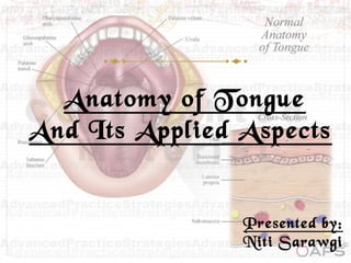
- 1. Anatomy of Tongue And Its Applied Aspects Presented by: Niti Sarawgi
- 2. Contents Introduction Development of tongue Anatomy of tongue Parts and surfaces of the tongue Muscles of the tongue Vascular supply of the tongue Lymphatic drainage of the tongue Innervation of the tongue Examination of the tongue Clinical considerations and diseases of the tongue Conclusion References
- 4. Development of tongue Starts to develop near the end of the fourth week Epithelium: Anterior 2/3: from 2 lingual swellings and one tuberculum impar, i.e., from first branchial arch supplied by lingual nerve (post-trematic) and chorda tympani (pre-trematic) Posterior 1/3: from the cranial half of the hypobranchial eminence, i.e., from the third arch supplied by glossopharyngeal nerve
- 5. Posterior most: from the fourth arch supplied by vagus nerve Muscles develop from the occipital myotomes which are supplied by hypoglossal nerve Connective tissue develops from local mesenchyme
- 7. Parts and surfaces of the tongue Oral Part • Apex • Dorsum part • Ventral part
- 8. Ventral surface The thin strip of tissue that runs vertically from the floor of the mouth to the undersurface of the tongue is called the lingual frenulum. It tends to limit the movement of the tongue. On either side of frenulum there is a prominence produced by deep lingual veins. more laterally there is a fold called plica fimbriata
- 9. Glands of BlandinNuhn Anterior lingual glands (also called apical glands) are deeply placed seromucous glands that are located near the tip of the tongue on each side of the frenulum linguae. They are found on the under surface of the apex of the tongue, and are covered by a bundle of muscular fibers derived from the Styloglossus and Longitudinalis inferior. They are between 12 to 25 mm. in length, and approximately 8 mm. wide, and each opens by three or four ducts on the under surface of the tongue's apex
- 10. Glands of VonEbner They are serous salivary glands Located adjacent to the moats surrounding the circumvalate and foliate pappilae Von Ebner's glands secrete lingual lipase This secretion flushes material from the moat to enable the taste buds to respond rapidly to changing stimuli Von Ebner's glands are innervated by cranial nerve IX, the glossopharyngeal nerve.
- 11. Gland of Weber They lie along the lateral border of the tongue These glands are pure mucous secreting glands. These open into the crypts of the lingual tonsils on the posterior tongue dorsum. Abscess formed due to accumulation of pus and fluids in this gland is called Peritonsillar Abscess
- 12. Lies behind the palatoglossal arches Forms the anterior wall of the oropharynx Devoid of papillae Underlying lymphoid nodules embedded in the submucosa collectively called as lingual tonsils Pharyngeal or Postsulcal Part Epiglottis Lingual tonsil Median epiglotic fold Lateral epiglotic fold valleculae Palatine tonsil
- 13. Muscles of the tongue Intrinsic muscles Superior longitudinal Inferior longitudinal Transverse Vertical Extrinsic muscles Styloglossus Hyoglossus Genioglossus Palatoglossus
- 15. GenioglossusGenioglossus Arises from superior genial tubercle above the origin of geniohyoid Hyoid bone Insertion : the fibres radiate widely to be inserted into the mucous membrane of the tongue; the lowest fibres passing down to the hyoid body
- 16. Action Protrusion Bilaterally –Central part depression Unilaterally – Diverges to the Opposite side
- 17. HyoglossusHyoglossus • Origin: greater cornu, front of body of hyoid bone • Insertion: side of the tongue between styloglossus and inferior longitudinal ActionsActions • Depresses the tongue
- 18. Chondroglossus A part of hyoglossus Separated from it by genioglossus Origin: medial side and base of lesser cornua Insertion: intrinsic musculature between hyoglossus and genioglossus
- 19. StyloglossusStyloglossus • Origin : styloid process near its apex • Insertion : longitudinal part into the inferior longitudinal muscles Oblique part into hyoglossus • ActionAction • Draws the tongue upwards and backward hyoglossushyoglossus styloglossus Inferior longitudinal muscles Styloid process
- 20. PalatoglossusPalatoglossus Origin: palatine aponeurosis of soft palate Insertion: side of the tongue ““more a part of soft palate thanmore a part of soft palate than the tonguethe tongue”” ActionAction: elevates the posterior part of the tongue Bilaterally- approximates the palatoglossal folds to constrict the isthmus of the fauces
- 21. MUSCLES ORIGIN INSERTION ACTION(S) Genioglossus Upper genial tubercle of mandible Upper fibres: tip of the tongue Middle fibres: dorsum Lower fibres: hyoid bone Upper fibres: retract the tip Middle fibres: depress the tongue Lower fibres: pull the posterior part forward (thus protrusion of the tongue from the mouth) Hyoglossus Greater cornu, front of lateral part of body of hyoid bone Side of tongue Depress the tongue Retracting the protruded tongue Styloglossus Tip, anterior surface of styloid process Side of tongue Pulls the tongue upwards and backwards during swallowing Palatoglossus Oral surface of palatine aponeurosis Side of tongue (junction of oral and pharygeal part) Pulls up root of tongue, approximates palatoglossal arches, closes oropharyngeal isthmus
- 23. Superior longitudinalSuperior longitudinal • Origin: submucous fibrous layer below the dorsum of the tongue and lingual septum • Insertion: extends to the lingual margin • ActionAction • Turns the apex and sides of the tongue upward to make the dorsum concave
- 24. Inferior longitudinalInferior longitudinal • Narrow band close to the inferior surface of the tongue • Origin: root of tongue and body of hyoid bone • Insertion: apex of tongue • ActionAction • Curls the tip inferiorly and shortens the tongue
- 25. TransverseTransverse • Origin: median fibrous septum • Insertion: fibrous tissue at the margins of tongue • ActionAction • Narrows and elongates the tongue
- 26. VerticalVertical • Origin: dorsum surface of the borders of the tongue • Insertion: ventral surface of the borders of the tongue • ActionAction • Flattens and broadens the tongue
- 27. Vascular supply of the tongue Lingual arteryLingual artery •A branch of external carotid artery(after passing deep to the hyoglossus muscles) •Divides into : •Dorsal lingual arteriesDorsal lingual arteries: supply posterior part •Deep lingual arteryDeep lingual artery : supplies the anterior part •Sublingual arterySublingual artery : supplies the sublingual gland and floor of the mouth
- 28. • Dorsal lingual vein-Dorsal lingual vein- drains the dorsum and sides of the tongue • Deep lingual veinsDeep lingual veins (Ranine veins) - drains the tip of the tongue and join sublingualsublingual veinsveins from sublingual salivary gland • All these veins terminate directly or indirectly into internalinternal jugular veinsjugular veins
- 29. Lymphatic drainageLymphatic drainage Lymph from one side (esp. of the posterior side), may reach the nodes of the both sides of the neck (in contrast to the blood supply which remains unilateral) Tip - drain to submental nodes or directly to deep cervical nodes Marginal lymphatics from the anterior part tend to drain to ipsilateral submandibular nodes or directly to inferior deep cervical nodes
- 30. Central lymphatics - drain to deep cervical nodes of either side Posterior part - drains directly and bilaterally to deep cervical nodes The deep cervical nodes usually involved: jugulodigastric and jugulo-omohyoid nodes All lymph from the tongue is believed to eventually drain through the jugulo-omohyoid node before reaching the thoracic duct or right lymphatic duct
- 32. Nerve Supply Motor: all muscles of the tongue (intrinsic and extrinsic) are supplied by hypoglossal nerve except palatoglossus which is supplied by pharyngeal plexus Sensory: anterior 2/3 of the tongue: general sensation: lingual nerve - branch of the mandibular nerve (with cell bodies in the trigeminal ganglion) taste: chorda tympani (with cell bodies in the geniculate ganglion of facial nerve) parasympathetic secretomotor fibres to the anterior lingual gland run in the chorda tympani from the superior salivary nucleus, and relay in the submandibular genglion
- 33. posterior 1/3 of the tongue: innervated by the glossopharyngeal nerve (both general sensation and taste), with cell bodies in the glossopharyngeal ganglia in the jugular foramen posterior most part of the tongue: innervated by the vagus nerve through the internal laryngeal branch (with cell bodies in the inferior vagal ganglion)
- 35. Mucous Membrane on Ventral Surface It is thin, smooth and loosely attached to the underlying Connective Tissue It is freely mobile and not raised into papillae because epithelium is closely adherent to underlying muscle by a thin lamina propria. It is covered with non- keratinized stratified squamous epithelium. .
- 36. Mucous Membrane On Dorsal Surface The dorsal surface Of the tongue is covered with a mucous membrane, which is firmly adherent to the underlying C.T. It is raised into small projections similar to the villi, but known as papillae (limited only to anterior 2/3ra of tongue). The stratified squamous epithelium covering the dorsal surface of the tongue is mostly keratinized
- 37. Papillae of tongue They are 4 varieties Filiform Fungiform Foliate Circumvallate
- 38. Filiform papillaFiliform papilla • Minute, conical, cylindrical projections which cover most of the presulcul dorsal area. • Increase the friction between the tongue and food • They bear many secondary papillae which are more pointed than those of vallate and fungiform papillae and covered with keratin
- 39. Fungiform papillaFungiform papilla Located mainly on the lingual margin Differ from filiform because are larger, rounded and deep red in colour Bears one or more taste buds on its apical surface These are mushroom shaped, more numerous near tip & margins of tongue but some of them scattered over the dorsum
- 40. Foliate papillaFoliate papilla Red leaf-like mucosal ridges Bilaterally at the sides of the tongue near sulcus terminalis Bear numerous taste buds
- 41. Circumvallate papillaCircumvallate papilla Large cylindrical structures 8 to 12 in number Form a ‘V’ shaped row in front of sulcus terminalis on the dorsal surface of the tongue The entire structure is covered with squamous epithelium, in both sulcal walls & taste buds around
- 43. Taste budsTaste buds • Present in relation to cirumvallate papilla, fungiform papillae and foliate papilla • Also present on the soft palate, the epiglottis, the palatoglossal arches, and the posterior wall of the oropharynx
- 44. Neuroepithelial taste cells or gustatory cells in taste buds: They are modified columnar elongated cells which act as receptors. They have darkly-stained' elongated central nuclei. The superficial part of these cells is provided with short hairs (hairlets or microvilli). These hairlets project into the taste pore. The base of the taste cells is surrounded by sensory nerve fibres, carry the impulses of taste sensation to the brain.
- 45. Supporting cells in taste buds : They are elongated columnar cells with dark cytoplasm but lightly-stained nuclei. They form the outer wall of the taste bud. They have long microvilli that extend from their surfaces into the taste pore. Basal cells are present at the base of the taste bud. They act as stem cells for renewal of taste cells and supporting cells.
- 46. Taste discrimination Gustatory receptors detect four main types of taste sensation Sweet: tip Sour: middle Salty: anterolateral Bitter: base However recent evidence indicates that all areas of tongue are responsive to all taste stimuli
- 47. Clinical examination of tongue • InspectionInspection • The tongue is examined for:The tongue is examined for: ColourColour Swelling Ulcer Coating Size variation Distribution of filiform and fungiform papilla Crenations Fissures Atrophy or hypertrophy of papilla
- 48. Frenal attachment and deviations as patient moves out the tongue
- 49. Palpation
- 50. Gently palpate the muscles of the tongue
- 51. Clinical considerations Injury to hypoglossal nerveInjury to hypoglossal nerve • Trauma like fractured mandible may injure hypoglossal nerve • Paralysis ,atrophy of one side of tongue • Tongue deviates to paralyzed side during protrusion due to action of unaffected genioglossus muscles • Others infranuclear lesion (i.e., in motor neuron disease and in syringobulbia): gradual atrophy and muscular twitchings of the affected half of the tongue observed supranuclear lesion (i.e., in pesudobulbar palsy): produce paralysis without palsy (tongue is stiff, small and moves sluggishly)
- 52. Paralysis of genioglossus muscleParalysis of genioglossus muscle • Muscle tends to fall backward, obstructing airway • Total relaxation of genioglossus occur during general anaesthesia so airway is inserted to prevent tongue from relapsing Sublingual absorption of drugsSublingual absorption of drugs • For quick absorption, pill or spray is put under the tongue where it dissolves and enter the lingual veins (nirtroglycerin in angina pectoris)
- 53. The presence of rich network of lymphatics and loose areolar tissue in the substance of tongue is responsible for enormous swelling of tongue in acute glossitis The undersurface of the tongue is a good site for observation of jaundice Carcinoma of Tongue is quite common. The affected side of the tongue is removed along with all the deep cervical lymph nodes Carcinoma of posterior 1/3 of the tongue is more dangerous due to bilateral lymphatic spread In unconscious patients , the tongue may fall and obstruct the airway. In grand mal epilepsy, the tongue is commonly bitten by the front incisors during the attack
- 54. Diseases of the tongue Inherited, congenital, and developmental anomalies Disorders of the lingual mucosa Diseases affecting the body of the tongue Malignant tumors of the tongue
- 55. Inherited, congenital, and developmental anomalies Variation in morphology Ankyloglossia Fissured tongue Macroglossia Hypoglossia Lingual thyroid and thyroglossal duct
- 57. • Tongue tie can be classified as: • Milder formMilder form: do not influence jaw development, tooth position or phonation • Severe formSevere form: exhibit Midline mandibular diastema, periodontal defects • Extreme formExtreme form: complete attachment of tongue to the floor of the mouth or alveolar gingiva
- 58. Microglossia (hypoglossia)Microglossia (hypoglossia) Uncommon developmental condition of unknown origin characterized by abnormally small tongue Entire tongue may be missing (aglossia) length of the mandibular arch will be smaller due to the smaller size of the tongue.
- 62. Proliferation of floor of pharyngeal wall 4th week Descends the neck anterior to trachea and larynx 7th week Pathophysiology of lingual thyroid
- 63. Disorders of lingual mucosa • Geographic tongueGeographic tongue • Hairy tongueHairy tongue • Nonkeratotic and keratotic white lesionsNonkeratotic and keratotic white lesions – Candidiasis – Leukoplakia, hairy leukoplakia • Nutritional defficiencies and hematologicNutritional defficiencies and hematologic abnormalitiesabnormalities – Vitamin B12 deficiency – Iron deficiency anemia • InfectionsInfections – Tertiary syphilis
- 64. Geographic tongueGeographic tongue:: • Psoriasiform mucositis of the dorsum of the tongue • Prevalence is 1% to 2% • Irregular reddish areas of depapillation • thinning of the dorsal tongue epithelium usually surrounded by a narrow zone of regenerating papillae -whiter than the surrounding tongue surface
- 66. CandidiasisCandidiasis (Moniliasis) • Most common intraoral oppertunistic fungal infection • Causative agent: Candida albicans • Factors determining the clinical evidence of candidiasis: Immune status of the host Oral mucosal enviroment Strains of Candida
- 68. Pernicious anemiaPernicious anemia • Most common forms of vitamin B12 deficiency Clinical featuresClinical features • Beefy red tongueBeefy red tongue • Erythematous areas on tip and margins • De-papilation • Candidal infection
- 69. Iron deficiency anemiaIron deficiency anemia • Most common form of anemia found in 50% females
- 70. PlummerVinson syndromePlummerVinson syndrome Also known as Paterson Kelly Syndrome • Clinical featuresClinical features • Microcytic hypochromic anemia • Smooth and sore tongue • Angular chelitis • Spoon shaped nails Disorders of lingual mucosaDisorders of lingual mucosa
- 71. Tertiary syphilis and Tertiary syphilis and interstitial glossitisinterstitial glossitis • Tongue may be affected by gumma formation • Non-ulcerating, irregular • indurations • Asymmetric pattern of grooves • Leukoplakia
- 72. Blandin and Nuhn mucocele The Blandin and Nuhn mucocele occurs exclusively on the anterior ventral surface of the tongue at the midline. Although the lesions may have clinical features similar to those of the mucocele, which is found elsewhere they tend to be more polypoid with a pedunculated base Because of repeated trauma against the lower teeth, the surface may be red and granular or white and keratotic.
- 74. Squamous cell carcinoma of the tongueSquamous cell carcinoma of the tongue Most common intraoral site 60% of lesions arise from the anterior 2/3rd of the tongue The affected side of the tongue is removed surgically. All the deep cervical lymph nodes are also removed, i.e. block dissection of neck. Unilateral block dissection of the neck should be efficacious for early carcinoma of the lateral border of the tongue but because of the bilateral lymphatic drainage bilateral dissection should be performed if the tip of the tongue, the frenulum ,or the dorsum of the tongue is involved.
- 75. CONCLUSIO N
- 76. • ReferencesReferences B.D Chaurasia(2006) Human Anatomy,Regional and Applied,Dissection. Henry Gray(2004),Gray's Anatomy . Neelima Anil Malik, Textbook of Oral and Maxillofacial Surgery. Frank H.Netter,MD. Atlas of human anatomy. William Henry Hollinshead. Anatomy for Surgeons: The head and neck T.W. Sadler ,Langman’s Medical Embryology Internet source.
Editor's Notes
- The tongue is muscular hydrostat on the floors of the mouths of most vertebrates which manipulates food for mastication. It is the primary organ of taste (gustation), as much of the upper surface of the tongue is covered in papillae and taste buds. It is sensitive and kept moist by saliva, and is richly supplied with nerves and blood vessels. In humans a secondary function of the tongue is phonetic articulation. it serves as a natural means of cleaning one's teeth. Also helps in maintaining equilibrium and development of proper occlusion It s avg length is 10 cm or 4inches from the oropharynx
- A third median swelling is formed from the posterior part of 4th arch- epiglottis
- Special sensory innervation- chorda tympani branch of facial nerve posterior-glossopharyngeal
- Arch 1- anterior 2/3rd foramen caecum – the site from which the thyroid diverticulum grows down in an embryoi Arch 2- initial contribution is lost Arch 3- posterior 1/3 (pharyngeal) Arch 4- epiglottis n adjacent structures
- Oral part placed in the floor of the mouth.just infront of the palatoglossal arch . APEX is the tip of the tongue forms the anterior free end which at rest lies behind the upper incisor teeth. DORSUM PART is a curved upper surface with each margin shows 4 to 5 vertical folds named foliate papillae and the superior surface is covered with papillae which make it rough. Ventral surface consists of a plexus of veins that makes it extremely vascular and a lingual frenum that attaches the tongue to the floor
- Ventral surface is smooth purplish and reflected onto oral floor and gums
- It is named after
- beginning the process of lipid hydrolysis in the mouth
- Posterior most part of the tongue is connected to the epiglottis by 3 folds of mucous membrane –medial n lateral epiglottic fold On either side of the median folds there is a depression called as vallecula Lateral folds separate the vallecula from the piriform fossa
- Tongue is divided into two halfes by a median fibrous septum . Each half consists EXTRINSIC – ATTACHED TO THE BONE INTRINSIC- WITHIN THE TONGUE WHOLLY NOT ATTACHED TO THE BONE. Alter the shape of the tongue
- Root – tonsillar and ascending pharyngeal arteries
- Lymph from the posterior third – superior deep cervical lymphnodes on both sides Lymph from the medial part of the anterior two third – inferior deep cervical lymph nodes Lymph from the lateral parts of the anterior two thirds - submandibular lymph nodes
- The pharyngeal plexus is a plexus of nerves formed by: • The pharyngeal branch of the vagus, which includes the cranial root of the accessory. This provides the motor supply to the muscles except for the tensor palati which is supplied by the mandibular division of the trigeminal. • The glossopharyngeal nerve, which provides the sensory supply to the pharynx. • Branches from the sympathetic trunk.
- MUCOUS MEMBRANE of tongue (covering both the surfaces) is formed of stratified squamous epithelium. The superficial cells of the mucous membrane of the tongue are continually shed off and are replaced by new cells
- The sense of taste is dependent on scattered groups of sensory cells, the taste buds Limited to the pre sulcul part of the tongue Produce its characteristic roughness Increase the area of the contact between the tongue and the contents of the mouth
- The distribution of taste was basically published by a PHD student Dr.hanig in his thesis. His mapping had a very rough picture of the taste distribution without any concrete data but it began to be passed down the generations. A few scientists tried correcting it nd finally 1974 virginia collins set it right n alsofound taste buds in other locations 5 taste – umami as found in 1901 – japaneses scientist ikeda – taste of sea vegetable , soy sauce , ripe tomato or monosodium glutamate
- Color variation – white: leukoplakia, oral thrush , lichen planus : purple- vitamin b2 def; blue-copd , poisoning , cyanosis Red- scarlet fever , vitamin deficiency , kawasaki syndrome ; yellow-preceds black hairytongue, jaundice Black-hairy Swelling- food allergies, yellow fever , anaphylaxis , hereditary angioedema , glossitis Ulcer-cancer , lp , behcets Coating – lp , oral thrush, leukoplakia
- Wrap the tip of tongue with gauge piece With warm mirror – base of tongue and valate papila Guide the tongue to right- retract the cheek to left- examine foliate papilla and entire border of tongue Repeat the same for the other side Tell patient to touch palate with tip of the tongue- examine ventral surface
- Block dissection of neck cz recurrence of malignancy occurs in lymph nodes This can be prevented by making the patient lie on one side head down or mechanically keeping it out Epilepsy – prevented by putting a mouth gag at the beginning of the seizure
- after the formation of tongue massive cell degeneration occurs between the tongue and the floor of the mouth and the only part attaching them is the lingual frenum . Congenital shortness of the lingual frenum or the frenal attachment that extends nearly to the tip of the tongue binding the tongue to the floor of the mouth and restricting its extension
- Rx – frenulectomy
- Mostly associated with other malformations – hands n feet , clefts Severe dentoskeletal malocclusion can result
- A relatively common condition characterized by an increase in the size of tongue. Either true or psuedo Characteristic – indentations on the lateral margins Syndrome- downs , beckwith – wiedemann Surgical intervention to get normal size and function – mastication , articulation , speech
- Scrotal or lingua plicata Characterized by grooves that vary in depth dorsal and lateral aspects of the tongue Reported prevalence is 2% to 21%. Lil clinical significance except – accumulation of food debris n micro-organism
- Lingual thyroid is an anomalous condition in which follicles of thyroid tissue are found in the substance of the tongue When the thyroid anlage fail to migrate to its desired position.- around foramen caecum Mainly was observed in females – hormonal imblance –puberty , pregnancy symptoms – dysphagia , dysphonia , fullness in throat Rx- determine that the actual thyroid is present , drugshormone therapy - excision
- Benign migratory glossitis or Wandering rash—stress Rx-topical or systemic for symptomatic lesion
- Lingua nigra /villosa Hypertrophy of the filliform papilla of the dorsum of the tongue Results from failure of normal desquamation of tongue papillae and epithelium Presipitating factors-poor oral hygiene , tobacco Hiv Cf- halitosis -food debris rx- surgical – electrodessication, co2 laser
- Etiology – aids , immunocompr.nutritional def , radiation therapy etc Primary and secondary P-acute chronic candida associated and keratinized lesions superinfected with candia Diagnosis- pas or methanamine silver . Rx-Antifungal agenst
- ‘A predominantly white lesion of the oral mucosa that cannot be characterized as any other definable lesions; some leukoplakia will transform into camcer- axell t , 1996 Patients with oral leukoplakia are 5 times at risk of developing oral cancer Homogenous and non homogenous , smoking ,
- Most common oral cancer – SCC , other kaposis sarcoma
- Etiology – oncogenes – mutation genetic ; premalignant lesions , tobacco , alcohol, HPV Survival – 5yrs- us 63% Cf- non healing ulcer more that 14 days, small , painless, pale or, white or red patch: burning sensation and pain when tumour advances …………difficulty in tongue movement , swallowing , pain and parasthesia. Dignosis – clinical examination , biopsy ,
- Tongue – versatile organ with a variety of functions Richly suppled by vessels and nerve , most common site for oral cancer As a oral and maxillofacial surgeon it is very imp to understand the atomy and clinical considerations so as to preserve max function following a surgery