Human System System Of Reproductive Female

There will be scientific program,pre and post congress workshops covering vast topics like Repeated IVF failures,Endometriosis,Stimulation Protocols-Review & new strategies,Oocyte,Hands on Laparascopic suturing and Operative hysteroscopy,Advanced Reproductive techniques,Rise & fall of Metformin,Fitness for Fertility,Letrozole in infertility and ART,Recent Advances in ART,Ovarian Pathology,Monitoring Ovarian Function,Antagonist,Oocyte Cryo banking,Unexplained Infertility,Ovulation Induction,Embryology,Cyro Preservation& Vitrification,Oocyte Retrieval,IVF lite,Ovarian Imaging,Ovarian Tumor,Egg donation,Oocyte Donation,GnRH antagonist in IUI,Repeated IVF failures Incharge,Endometriosis,Reproductive Endocrinology,Oocyte Incharge,Reproductive Surgery,Androlgy for the gynecologist and more.
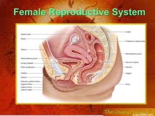
![The Female Reproductive System ,[object Object]](data:image/gif;base64,R0lGODlhAQABAIAAAAAAAP///yH5BAEAAAAALAAAAAABAAEAAAIBRAA7)
Recommended
More Related Content
What's hot
What's hot (20)
Viewers also liked
Viewers also liked (20)
Similar to Human System System Of Reproductive Female
Similar to Human System System Of Reproductive Female (20)
Recently uploaded
Recently uploaded (20)
Human System System Of Reproductive Female
- 5. Uterus
- 6. Vagina
- 9. Copulatory organ (external genitalia):
- 11. Functions: 1-Exocrine function: produce ova2-Endocrine function: secrete estrogen & progesterone hormones.
- 12. Location and Description Each ovary is oval (almond) shaped, measuring 3×2×1 cm. ovarian fossa: The ovary usually lies against the lateral wall of the pelvis in a depression called the ovarian fossa, bounded by the external iliac vessels above and by the obturator nerve, the ureter & the internal iliac vessels, behind. The position of the ovary is variable.
- 13. The ovary has:1)2 ends: Upper (tubal) end:is directed up & laterally & attached to: Ovarian fimbria of the Fallopian tube. Suspensory (infundibulo-pelvic) ligament of the ovary, which is a peritoneal fold that forms the upper lateral part of the broad ligament. It transmits the ovarian vessels & nerves from the side wall of the pelvis to the broad ligament. Lower (uterine) end:is directed down & medially. It is attached to the upper lateral angle of the uterus by the ligament of the ovary. 2) 2 surfaces: Lateral surface:related to the parietal peritoneum of the lateral pelvic wall & obturator nerve and vessels (in the floor of the fossa). Medial surface:related to fimbriated end of Fallopian tube. 3) 2 borders: Posterior border:free. Anterior border:attached to the upper lateral part of broad ligament by mesovarium (which transmits the ovarian nerves & vessels to the hilum of the ovary).
- 17. The ovarian artery arises from the abdominal aorta at the level of the first lumbar vertebra.
- 18. Veins
- 20. The lymph vessels of the ovary follow the ovarian artery and drain into the para-aortic nodes
- 21. Nerve Supply:
- 22. Parasympathetic: From pelvic splanchnic nerves (S 2, 3, 4).
- 23. Sympathetic: from T 10, 11.
- 25. Parts Of The Uterine Tube From Medial To Lateral Intramural (interstitial) part: It is the shortest (1 cm) and narrowest part. It passes through the wall of the superoateral angle of the uterus to open into the uterine cavity. Isthmus: It is narrow and 2 cm in length . Ampulla: It is the longest (5 cm), thin-walled, tortuous and widestpart. It is the site of fertilization. Infundibulum (fimbriated end): It is 2cm in length and funnel-shaped. It pierces the broad ligament to open into the peritoneal cavity near the ovary. Its margins carry fimbria which spread over the medial surface of the ovary.
- 26. Tubal ligation: A simple and effective method of birth control is to surgically ligate the uterine tubes, preventing spermatozoa from reaching ova. Conduct of the ovum in the uterine tube to the uterine cavity is helped by: ciliary movement of mucosal lining & peristaltic movement of the tube
- 29. Location:
- 30. It is located in the central part of the pelvis:
- 31. Anterior to the rectum
- 32. posterosuperior to the bladder. Dimension:measuring 3×2×1 inches
- 35. Anteriorly:it is not covered by peritoneum and related to the U.B.
- 36. Posteriorly:it is covered by peritoneum of Douglas pouch which separates it from the rectum.
- 37. Laterally:it gives attachment to the broad ligament andis related to the ureter and uterine vesselsjust below the root of the broad ligament.
- 40. Posteriorly: The body of the uterus is related posteriorly to the rectouterine pouch (pouch of Douglas) with coils of ileum or sigmoid colon within it.
- 44. Lymphatic drainage of the uterus
- 46. Peritoneal Covering Of The Uterus The peritoneum reflected from the rectum to the upper part of the vagina, forming recto-uterine pouch, then cover the posterior surface of the uterus, fundus, anterior surface of the body of the uterus ,which is reflected at the isthmus on the upper surface of U.B. forming the utero-vesical pouch. So anterior surface of the cervix and vagina have no peritoneal covering
- 51. 2) 4 borders:
- 52. upper free border: its medial 4/5 surroundsFallopian tubeits lateral 1/5 forms thesuspensory ligament of the ovary
- 53. lower border:rests on the pelvic floor
- 54. medial border:attached to the side of the uterus
- 56. Positions Of The Uterus Normal position antevertedanteflexed: the long axis of the uterus is bent forward on the long axis of the vagina.This position is referred to as anteversion (90 degree) of the uterus . the long axis of the body of the uterus is bent forward at the level of the internal os with the long axis of the cervix. This position is termed anteflexion (170 degree) of the uterus .
- 57. Abnormal position: retroverted, retroflexed the fundus and body of the uterus arebent backward on the vagina so that they lie in the rectouterine pouch (pouch of Douglas). In this situation, the uterus is said to be retroverted. If the body of the uterus is, in addition, bent backward on the cervix, it is said to be retroflexed. Leads to back pain.
- 58. Supporting Factors Of Uterus The uterus is supported mainly by: A)muscles 1) the tone of the Pelvic diaphragm (pelvic floor) muscles: levator ani muscles and coccygeus muscle. resisting downward push of uterus during increased intra-abdominal pressure. 2)Urogenital diaphragm:the muscles of the deep perineal pouch. 3)Perineal body: is a fibromuscular body between the vagina & anal canal; receiving the insertions of all perineal muscles. Thus, maintains the integrity of the pelvic floor.
- 59. Levator Ani Muscle They form a broad muscular sheet stretching across the pelvic cavity, they support the pelvic viscera and resist the intra-abdominal pressure transmitted downward through the pelvis. The medial edges of the anterior parts of the levatorani muscles are attached to the cervix of the uterus by the pelvic fascia. It is incomplete anteriorly to allow passage of urethra and vagina in female. Action: it supports and maintains the pelvic viscera in position.
- 60. Some of the fibers of levator ani are inserted into a fibromuscular structure called the perineal body This structure is important in maintaining the integrity of the pelvic floor; The perineal body lies in the perineum between the vagina and the anal canal. It thus supports the vagina and, indirectly, the uterus.
- 61. and the condensations of pelvic fascia, which form three important B)ligaments. The Transverse Cervical, Pubocervical, and Sacrocervical Ligaments These three ligaments are attached to the cervix and the vault of the vagina and play an important part in supporting the uterus and keeping the cervix in its correct position
- 62. Ligaments of the cervix: Transverse cervical (Mackenrodt’s ) cardinal ligament: It is the main supporting factor of the uterus. It a fan-shaped ligament, which is formed of condensed extraperitoneal tissue between the side wall of the pelvis and side of cervix & vagina. Pubo-cervical ligament: It is a condensation of extraperitoneal tissue, which extends from the front of cervix & upper part of vagina to the back of the pubis, around the sides of the urethra. Utero-sacral (sacrocervical) ligament: It is a condensation of extraperitoneal tissue, which extends from the back of the cervix to the front of 2nd & 3rd pieces of sacrum, around the sides of the rectum.
- 64. Uterine Prolapse Damage to the levatoresani muscles or ligaments of the cervix during childbirth or general poor body muscular tone may result in downward displacement of the uterus called uterine prolapse. In advanced cases, the cervix descends the length of the vagina and may protrude through the orifice. if the perineal body is damaged during childbirth, prolapse of the pelvic viscera may occur. Caesarean section: opening the abdomen, when normal child birth is not possible. Hysterectomy: removal of uterus in case of cancer.
- 66. It is copulatory organ in female & serve as a passage for menstrual flow & child birth.
- 67. Its upper part surrounding the cervix forming
- 68. Anterior,
- 69. posterior , right lateral and left lateral fornices.
- 70. It is 8 cm long
- 71. vaginal opening may bepartially covered by the hymen
- 72. Relations Anterior wall: (7 cm) Not covered by peritoneum Its upper 1/3 is pierced by the cervix. Its middle 1/3 is related to the base of U.B. Its lower 1/3 is related to the urethra. Posterior wall: (9 cm) Its upper1/4 is covered by peritoneum which is reflected to the rectum to form the recto-vaginal (Douglas pouch) which contains coils of ileum Its middle 2/4 related to rectum. Its lower 1/4 is related to perineal body and anal canal. Lateral relations (from above downwards): Upper part: uterine artery & ureter. Middle part:levatorani (sphincter vaginae). Lower part :greater vestibular gland (in the perineum)
- 73. Fornices of vagina: These are 4 pouches formed by the upper part of vagina around the vaginal part of cervix (2 lateral, 1 anterior & 1 posterior) The posterior fornix is the deepest one & the only one covered by peritoneum Lateral one related to uterine artery & ureter.
- 77. the levatores ani muscles
- 78. and the transverse cervical, pubocervical, and sacrocervical ligaments. These structures are attached to the vaginal wall by pelvic fascia.
- 79. The middle part of the vagina is supported by the urogenital diaphragm.