Atlas on bethesda system for reporting cervical cytology
•
111 likes•51,768 views

This is an atlas with more nearly 100 images, authentic taken from NCI web atlas. Useful to understand and report pap smears. The subject has been presented in a way which will help students reproduce in exams.

Recommended
More Related Content
What's hot
What's hot (20)
Similar to Atlas on bethesda system for reporting cervical cytology
Similar to Atlas on bethesda system for reporting cervical cytology (20)
More from Ashish Jawarkar
More from Ashish Jawarkar (20)
Recently uploaded
Recently uploaded (20)
Atlas on bethesda system for reporting cervical cytology
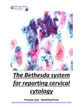
- 1. 1 The Bethesda system for reporting cervical cytology Preview only - Download from http://www.scribd.com/doc/182313900/Atlas-on-bethesda-system-for-reporting-cervical-cytology
- 2. 2 Systemic Pathology Notes The Bethesda system for reporting cervical cytology (2001) * Dr. Ashish V. Jawarkar (M.D.) Consultant Pathologist Vadodara Overview: 1. General points 2. Reporting format 3. Causes of false negative pap tests 4. Description of each category of reporting a. Satisfactory for evaluation b. Unsatisfactory for evaluation c. NILM i. Cells present 1. Superficial and intermediate squamous cells 2. Benign endometrial cells ii. Cellular changes 1. Reactive changes 2. Radiation induced changes 3. Hyperkeratosis 4. Parakeratosis 5. Cytolysis 6. atrophy iii. organisms 1. T. Vaginalis 2. Leptrotrichia 3. Candida spp. 4. Bacterial vaginosis 5. Actinomyces israelli 6. H. simplex virus d. Squamous cell abnormalities i. Atypical squmous cells 1. ASC-US 2. ASC-H 3. LSIL 4. HSIL ii. Invasive squamous cell carcinoma e. Glandular cell abnormalities i. Atypical Glandular cells 1. AGC-NOS 2. AGC-N 3. AEC-NOS 4. AEC-N ii. Adenocarcinoma in situ iii. Adenocarcinoma 1. Endocervical adenocarcinoma 2. Endometrial adenocarcinoma Preview only - Download from http://www.scribd.com/doc/182313900/Atlas-on-bethesda-system-for-reporting-cervical-cytology
- 3. 3 * Based on: ( Bethesda atlas (Solomon D., Nayar R. (editors). The Bethesda System for Reporting Cervical Cytology, Second Edition. New York: Springer-Verlag, 2004. * General Points 1. The system was developed in 1988 for reporting cervical/vaginal cytology, to provide uniform guidelines for reporting and reviewing gynaecologic Papaniculou smears. 2. It was subsequently modified in 1991 and 2001. 3. Most effective cancer prevention test available till date. It has become the index to which all other cancer screening tests are compared. 4. Yearly screening is estimated to reduce a woman’s risk of getting carcinoma by 93%. 5. One of the numerous advances made in cervical cytology in the last decade has been introduction of liquid based cytology. This has improved sensitivity of the technique, reduced the number of specimen with obscuring blood and inflammation and increased LSIL:ASC ratio. Also HPV testing can be directly performed on LBC specimen. *The Bethesda system 2001 classification (reporting format) 1.Specimen type 2.General Categorization 3.Interpretation/result 4. Suggestions 1. Satisfactory for evaluation *describe presence or absence of transformation zone component (*not required in 2001 system) 2.Unsatisfactory for evaluation a. Specimen rejected b. Specimen not processed c. Specimen processed but unsatisfactory 1. NILM 2. Epithelial cell abnormality 3. Others 1. Negative for intraepithelial lesion/malignancy Organisms 1.T. Vaginalis 2.Fungal organisms suggestive of candida spp. 3.Shift in bacterial flora s/o bacterial vaginosis 4.Bacterial morphology consistent with actinomyces 5.cellular changes c/w H. simplex Other non neoplastic findings 1.Reactive cellular changes 2.Atrophy 3.Glandular status post hysterectomy Other findings Endometrial cells in women more than 40 years of age 2. Epithelial cell abnormalities Squamous cells Atypical squamous cells (ASC-US, ASC-H) LSIL HSIL Invasive squamous cell carcinoma Glandular cells Atypical endocervical cells 1.Endocervical cells NOS 2.Endocervical cells favor neoplasia Atypical endometrial cells 1.Endometrial cells NOS Atypical glandular cells 1.glandular cells NOS 2.Glandular cells favor neoplasia Adenocarcinoma in situ Adenocarcinoma 3. Other malignant neoplasms (specify) (if any) Preview only - Download from http://www.scribd.com/doc/182313900/Atlas-on-bethesda-system-for-reporting-cervical-cytology
- 4. 4 Satisfactory for evaluation LBC smear Adequate squamous cellularity This image depicts the approximate cellular density of an adequate ThinPrep specimen which significantly exceeds the minimum criteria. It is to be used as a guide in assessing squamous cellularity of ThinPrep specimens Satisfactory for evaluation LBC smear Borderline adequate squamous cellularity Slides with borderline or low cellularity should be assessed by estimation of average cell number in at least 10 fields. At 40X, there were approximately 11 cells per field when 10 microscopic fields along a diameter were evaluated for squamous cellularity. This would give an estimated total cell count between 5,000 - 10, 000. Laboratories may choose to append a quality indicator statement such as "borderline or low squamous cellularity" when specimens meet minimal criteria, but have only 5,000-20,000 cells. *Not satisfactory for evaluation 1. <8000 squamous cells on conventional smears, <5000 squamous cells on LBC 2. >75% cells are obscured by blood or inflammation 3. If a single cell is atypical, it should not be reported as unsatisfactory Preview only - Download from http://www.scribd.com/doc/182313900/Atlas-on-bethesda-system-for-reporting-cervical-cytology
- 5. 5 Unsatisfactory for evaluation Conventional smear 75 cells in this field, smear should be reported as unsatisfactory if all fields have these many or less cells Unsatisfactory for evaluation Conventional smear If 50 - 75% of the slide has this appearance, obscuring inflammation should be mentioned in the quality indicators section of the report Preview only - Download from http://www.scribd.com/doc/182313900/Atlas-on-bethesda-system-for-reporting-cervical-cytology
- 6. 6 Metaplastic squamous cells LBC smear Normal polygonal squamous metaplastic cells with round to oval nuclei and bland chromatin pattern. On liquid based preparations cells may appear more rounded, and nuclei may appear smaller. This would be interpreted as "NILM” *Negative for intraepithelial lesion or malignancy (NILM) CELLS PRESENT: 1. Superficial squamous cells Predominate at the time of ovulation when Estrogen is high Intermediate squamous cells Predominate in late luteal phase when progesterone is high 2. Benign endometrial cells a. Can be seen during menses b. Any time during 1st half of menstrual cycle c. Benign appearing endometrial cells in a post menopausal patient should be looked at carefully, may be associated with endometrial polyps/hyperplasia or hormonal alterations. 3. Benign Glandular cells Preview only - Download from http://www.scribd.com/doc/182313900/Atlas-on-bethesda-system-for-reporting-cervical-cytology
- 7. 7 There are generally two types of squamous cells seen on Pap Tests – superficial and intermediate cell types. The superficial cells are the largest of the three and have small pyknotic (degenerative) nuclei and cytoplasm that generally stains eosinophilic. The intermediate squamous cells are similar in appearance but are slightly smaller in size and have larger, clearly structured, round nuclei with cytoplasm that usually stains basophilic. Both cell types are polygonal in shape. The intermediate cell type is the most common cell type seen. Endometrial cells Conventional smear Exodus pattern is characterized by a double contour pattern of external glandular epithelium with internal stromal cells. The stroma in this group is slightly eccentric. Nuclear features are easily appreciated, despite this being a conventional Pap smear. The nuclei have delicate even chromatin. Histiocytes are often seen in association with the double contour fragments of exodus. Preview only - Download from http://www.scribd.com/doc/182313900/Atlas-on-bethesda-system-for-reporting-cervical-cytology
- 8. 8 CELLULAR CHANGES: 1. Reactive/reparative changes – a. Mild nuclear enlargement b. Prominent nucleoli c. Abundant cytoplasm d. Mitoses No increase in N:C ratio May be associated with – Inflammation (repair) A case of uterine prolapse (inflammation/repair changes) Conventional smear Flat monolayer sheets with distinct cytoplasmic outlines, streaming nuclear polarity, prominent nucleolus in almost every cell. 2. Radiation induced changes – a. Quite enlarged b. Frequently multinucleated c. Cytoplasmic vacuolation d. Cellular degeneration Preview only - Download from http://www.scribd.com/doc/182313900/Atlas-on-bethesda-system-for-reporting-cervical-cytology
- 9. 9 Radiation therapy induced changes Conventional smear Enlarged nuclei with abundant polychromatic cytoplasm with vacuolization. Mild nuclear hyperchromasia without coarse chromatin, prominent nucleoli (coexisting repair). Note multinucleation (upper right corner insert). Reactive changes associated with radiation therapy LBC smear Abundant cytoplasm. Smudgy chromatin with mild nuclear enlargement 3. Hyperkeratosis – a. Anucleated, orange staining squamous cells b. Can be seen in women with uterine prolapsed Preview only - Download from http://www.scribd.com/doc/182313900/Atlas-on-bethesda-system-for-reporting-cervical-cytology
- 10. 10 T. Vaginalis LBC smear Good example of a flagella. Flagella are usually not seen in conventional Pap smears. Also seen in this image (left lower inset) is a "kite shaped" trichomonad- another finding noted in liquid based preparations. Reactive squamous cells associated with T. Vaginalis Conventional smear Minimal nuclear enlargement, cytoplasmic polychromasia Preview only - Download from http://www.scribd.com/doc/182313900/Atlas-on-bethesda-system-for-reporting-cervical-cytology
- 11. 11 a. Leptotrichia 1. Large filamentous bacteria that appear as gray hair like structures 2. They coexist with T. Vaginalis Leptothrix with T. Vaginalis LBC smear The finding of Trichomonas and leptothrix together has been referred to as "spaghetti and meatballs" The leptothrix should be distinguishable from Doderlein bacilli, that are normally seen. When leptothrix are seen, one should search for the possible presence of trichomonads. In liquid based preparations, the leptothrix organisms may tend to clump (as seen in this image) as opposed to conventional smears. Leptothrix with T. Vaginalis Conventional smear Preview only - Download from http://www.scribd.com/doc/182313900/Atlas-on-bethesda-system-for-reporting-cervical-cytology
- 12. 12 ASC-US LBC smear 1. ATYPICAL SQUAMOUS CELLS – CANNOT EXCLUDE HSIL (ASC-H) Cytologic changes are suggestive of but not enough for diagnosis of HSIL Criteria: 1. Resemble parabasal/basal cells 2. Nuclei are hyperchromatic and have irregular margins ASC-H Conventional smears Less mature squamous cells/metaplastic cells with polygonal shape, and slightly enlarged nuclei with occasional nuclear contour irregularities Preview only - Download from http://www.scribd.com/doc/182313900/Atlas-on-bethesda-system-for-reporting-cervical-cytology
- 13. 13 ASC-H LBC smear Metaplastic cells with increased N:C ratios and nuclear contour irregularities Preview only - Download from http://www.scribd.com/doc/182313900/Atlas-on-bethesda-system-for-reporting-cervical-cytology
- 14. 14 SQUAMOUS INTRAEPITHELIAL LESION (LOW GRADE (LSIL) AND HIGH GRADE (HSIL)) Bethesda CIN WHO Cell type Arrangement No. of abnormal cells Nuclear size Hyperchromasia N:C ratio Koilocytosis LSIL CIN I Mild Dysplasia Superficial or intermediate Singly or in sheets + 4-6 times nomal + + +++ HSIL CIN II Moderate dysplasia Parabasal Severe dysplasia Basal CIN III Carninoma in situ (CIS)* Basal/spindle/pleomorphic Singly or in sheets ++ Singly or in sheets +++ Singly or in sheets or syncitia ++++ ++ + + Overall size of nucleus/cell is not as much as LSIL but N:C ratio is greatly increased ++ +++ ++++ ++ +++ ++++ ++ +/+/- *CIS consists of three types of cells 1. Small basal cell type – cells similar to severe dysplasia but demonstrate even lesser cytoplasm and higher N:C ratio 2. Large cell non keratinizing type – syncitial like cell sheets in which individual cell membranes are difficult to identify 3. Large cell keratinizing type – pleomorphic highly atypical tadpole like cells, thich orangophilic cytoplasm LSIL Conventional Smear Nuclear enlargement and hyperchromasia is of sufficient degree for the interpretation of LSIL. Demonstration of HPV cytopathic effect is not necessary for an interpretation of LSIL, if required nuclear changes are present. Preview only - Download from http://www.scribd.com/doc/182313900/Atlas-on-bethesda-system-for-reporting-cervical-cytology
- 15. 15 Endometrial adenocarcinoma Conventional Smear Cluster of small cells with enlarged round or oval nuclei, small nucleoli and vacuolated cytoplasm in a background of "watery" diathesis. Endometrial adenocarcinoma LBC smear Papillary cluster with large cells and prominent nucleoli. Preview only - Download from http://www.scribd.com/doc/182313900/Atlas-on-bethesda-system-for-reporting-cervical-cytology
- 16. 16 This is only a preview Download entire document from http://www.scribd.com/doc/182313900/Atlas-on-bethesda-system-for-reporting-cervical-cytology Preview only - Download from http://www.scribd.com/doc/182313900/Atlas-on-bethesda-system-for-reporting-cervical-cytology