9.Congenital heart disease
•Download as PPT, PDF•
10 likes•1,216 views

congenital heart disease

Recommended
More Related Content
What's hot
What's hot (20)
Similar to 9.Congenital heart disease
Similar to 9.Congenital heart disease (20)
More from PNK SINGH
More from PNK SINGH (20)
Recently uploaded
Recently uploaded (20)
9.Congenital heart disease
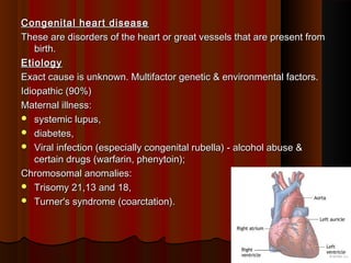
- 1. Congenital heart diseaseCongenital heart disease These are disorders of the heart or great vessels that are present fromThese are disorders of the heart or great vessels that are present from birth.birth. EtiologyEtiology Exact cause is unknown. Multifactor genetic & environmental factors.Exact cause is unknown. Multifactor genetic & environmental factors. Idiopathic (90%)Idiopathic (90%) Maternal illness:Maternal illness: systemic lupus,systemic lupus, diabetes,diabetes, Viral infection (especially congenital rubella) - alcohol abuse &Viral infection (especially congenital rubella) - alcohol abuse & certain drugs (warfarin, phenytoin);certain drugs (warfarin, phenytoin); Chromosomal anomalies:Chromosomal anomalies: Trisomy 21,13 and 18,Trisomy 21,13 and 18, Turner's syndrome (coarctation).Turner's syndrome (coarctation). 11
- 2. 22
- 3. 33
- 4. 44
- 5. INCIDENCE AND RELATIVE FREQUENCY OF CONGENITALINCIDENCE AND RELATIVE FREQUENCY OF CONGENITAL CARDIAC MALFORMATIONSCARDIAC MALFORMATIONS Lesion defects % of all CHDLesion defects % of all CHD Ventricular septal defect 30Ventricular septal defect 30 Atrial septal defect 10Atrial septal defect 10 Patent ductus arteriosus 10 (where in the ductusPatent ductus arteriosus 10 (where in the ductus arteriosus fails to close after birth so poor wait gain,inc. work ofarteriosus fails to close after birth so poor wait gain,inc. work of breathing)breathing) Pulmonary stenosis 7Pulmonary stenosis 7 Coarctation of aorta 7Coarctation of aorta 7 Aortic stenosis 6Aortic stenosis 6 Tetralogy of Fallot 6(usually a right to leftTetralogy of Fallot 6(usually a right to left shunt ,in which higher resistance to right ventricular outflow resultsshunt ,in which higher resistance to right ventricular outflow results in more severe cyanosis symptoms.)in more severe cyanosis symptoms.) Complete transposition of great arteries 4(is a group of congenitalComplete transposition of great arteries 4(is a group of congenital heart disease involving an abnormal spatial arrangement of any ofheart disease involving an abnormal spatial arrangement of any of vessels .sup&inf.,pulmonary artery&vein)vessels .sup&inf.,pulmonary artery&vein) Others 20Others 20 55
- 6. CHD classified broadly into 3 major categories: CHD classified broadly into 3 major categories: ShuntsShunts Left -to-right shuntLeft -to-right shunt Right -to-left shuntRight -to-left shunt ObstructionObstruction ShuntsShunts Abnormal communication between chambers &/or blood vessels.Abnormal communication between chambers &/or blood vessels. Abnormal channels permit the flow of blood from left to right or theAbnormal channels permit the flow of blood from left to right or the reverse, depending on pressure relationships.reverse, depending on pressure relationships. 66
- 7. Rt – Lt shuntRt – Lt shunt When blood from the rt side enters the left sideWhen blood from the rt side enters the left side Cyanosis results because there is diminished pulmonary blood flowCyanosis results because there is diminished pulmonary blood flow and poorly oxygenated blood enters the systemic circulation (calledand poorly oxygenated blood enters the systemic circulation (called cyanotic congenital heart disease, the most important examples ofcyanotic congenital heart disease, the most important examples of which arewhich are Tetralogy of Fallot,Tetralogy of Fallot, Transposition of the great arteries,Transposition of the great arteries, Persistent truncus arteriosus(rare type ,in which single b/d vesselPersistent truncus arteriosus(rare type ,in which single b/d vessel comes out of right to left ventricals(pulmonary artery&arota)),comes out of right to left ventricals(pulmonary artery&arota)), Tricuspid atresia(valve is missing or abnormally developed ),Tricuspid atresia(valve is missing or abnormally developed ), 77
- 8. Left -to-right shuntsLeft -to-right shunts Increase pulmonary blood flow and are not initially associated withIncrease pulmonary blood flow and are not initially associated with cyanosis.cyanosis. it causes pulmonary hypertension, this increases pressure in theit causes pulmonary hypertension, this increases pressure in the right heart more than that of the left.right heart more than that of the left. As, a result , thereby reversing the shunt to right-to-left withAs, a result , thereby reversing the shunt to right-to-left with unoxygenated blood in the systemic circulation occurs.unoxygenated blood in the systemic circulation occurs. Late cyanotic congenital heart disease or Eisenmenger syndromeLate cyanotic congenital heart disease or Eisenmenger syndrome ASD,ASD, VSD,VSD, Patent ductus arteriosus [PDA]Patent ductus arteriosus [PDA] 88
- 9. Comparison of Left Versus Right Shunt Congenital DiseaseComparison of Left Versus Right Shunt Congenital Disease 99
- 10. TETRALOGY OF FALLOTTETRALOGY OF FALLOT The four components of the tetralogy areThe four components of the tetralogy are Pulmonary stenosis,Pulmonary stenosis, Overriding of the ventricular septal defect by the aorta,Overriding of the ventricular septal defect by the aorta, Ventricular septal defect andVentricular septal defect and Right ventricular hypertrophy. (boot shaped)Right ventricular hypertrophy. (boot shaped) The combination results in elevated right ventricular pressure and right-to-The combination results in elevated right ventricular pressure and right-to- left shunting of cyanotic blood across the ventricular septal defect.left shunting of cyanotic blood across the ventricular septal defect. 1010
- 11. 1111
- 12. Coarctation of the aortaCoarctation of the aorta Definition: segmental narrowing of the aortaDefinition: segmental narrowing of the aorta Preductal coarctation (infantile-type)Preductal coarctation (infantile-type) Severe narrowing of aorta proximal to the ductus arteriosisSevere narrowing of aorta proximal to the ductus arteriosis Usually associated with a patent ductus arteriosis (PDA)Usually associated with a patent ductus arteriosis (PDA) Right ventricular hypertrophyRight ventricular hypertrophy Cyanosis of the lower part of the bodyCyanosis of the lower part of the body 1212
- 13. Postductal coarctation (adult-type)Postductal coarctation (adult-type) Narrowing of the aorta distal to the ductus arteriosisNarrowing of the aorta distal to the ductus arteriosis Collateral circulation via the internal mammary(thoratic) andCollateral circulation via the internal mammary(thoratic) and intercostal arteriesintercostal arteries There is hypertension in upper extremity & low blood pressure inThere is hypertension in upper extremity & low blood pressure in lower extremitylower extremity Chest x-ray: notching of the ribs(it is a radiological sign,where theChest x-ray: notching of the ribs(it is a radiological sign,where the surface of rib is deformed.)surface of rib is deformed.) 1313
- 14. ComplicationsComplications Congestive heart failureCongestive heart failure Intracerebral hemorrhageIntracerebral hemorrhage Dissecting aortic aneurysmDissecting aortic aneurysm 1414
- 15. PERSISTENT DUCTUS ARTERIOSUS or PATENT DUCTUSPERSISTENT DUCTUS ARTERIOSUS or PATENT DUCTUS AETERIOSUS (PDA)AETERIOSUS (PDA) Ductus arteriosis remains open after birth.Ductus arteriosis remains open after birth. Direct communication between the aorta and pulmonary arteryDirect communication between the aorta and pulmonary artery During fetal life, before the lungs begin to function, most of the bloodDuring fetal life, before the lungs begin to function, most of the blood from the pulmonary artery passes through the ductus arteriosus intofrom the pulmonary artery passes through the ductus arteriosus into the aorta.the aorta. Associated with prematurity and congenital rubella infections.Associated with prematurity and congenital rubella infections. 1515
- 16. Pathophysiology of PDAPathophysiology of PDA Since the pressure in the aorta is higher than that in the pulmonarySince the pressure in the aorta is higher than that in the pulmonary artery (PA), there will be a continuous arteriovenous shunt,artery (PA), there will be a continuous arteriovenous shunt, Volume of which depends on the size of the ductus.Volume of which depends on the size of the ductus. As much as 50% of the left ventricular output may be recirculatedAs much as 50% of the left ventricular output may be recirculated through the lungs, with a consequent increase in the work of thethrough the lungs, with a consequent increase in the work of the heart.heart. 1616
- 17. 1717
- 18. 1818
- 19. Atrial Septal DefectAtrial Septal Defect There is an abnormal opening in the atrial septumThere is an abnormal opening in the atrial septum Blood flows left to rightBlood flows left to right Patent foramen ovale fails to closePatent foramen ovale fails to close Right heart becomes dilatedRight heart becomes dilated Too much blood to the lungsToo much blood to the lungs 1919
- 20. Ventricular Septal DefectVentricular Septal Defect Abnormal opening in the ventricular septumAbnormal opening in the ventricular septum Left to right shunt – majorityLeft to right shunt – majority Dilated right heart – too much blood to lungs – increase inDilated right heart – too much blood to lungs – increase in pulmonary pressurepulmonary pressure Smaller defects can close spontaneouslySmaller defects can close spontaneously 2020
- 22. Tricuspid atresia(absence or abnormal narrowing opening)Tricuspid atresia(absence or abnormal narrowing opening) Absent tricuspid orifice,Absent tricuspid orifice, Hypoplastic(undevpt.of tissue & organs) RV, RA to LA shun VSDHypoplastic(undevpt.of tissue & organs) RV, RA to LA shun VSD shunt, other anomalies.shunt, other anomalies. Transposition of the great vesselsTransposition of the great vessels • Aorta arises from the morphological RV, pulmonary artery from LVAorta arises from the morphological RV, pulmonary artery from LV • Shunt via atria, ductus and possibly VSDShunt via atria, ductus and possibly VSD 2222
- 23. Pulmonary atresiaPulmonary atresia Pulmonary valve atretic and pulmonary artery hypoplasticPulmonary valve atretic and pulmonary artery hypoplastic RA to LA shunt, pulmonary flow via ductus.RA to LA shunt, pulmonary flow via ductus. Ebstein's anomaly(is a congenital malformation of the heartEbstein's anomaly(is a congenital malformation of the heart that is characterized by apical displacement of the septalthat is characterized by apical displacement of the septal and posterior tricuspid valves leaflets)and posterior tricuspid valves leaflets) Tricuspid valve is dysplastic(abnormal growth) and displaced into RV,Tricuspid valve is dysplastic(abnormal growth) and displaced into RV, Right ventricle 'atrialised‘,(valves slightly displaced)Right ventricle 'atrialised‘,(valves slightly displaced) RA to LA shunt,RA to LA shunt, 2323
- 24. Eisenmenger SyndromeEisenmenger Syndrome Defined as the process in which a left-to-right shunt causes increasedDefined as the process in which a left-to-right shunt causes increased flow through the pulmonary vasculature,flow through the pulmonary vasculature, Causing pulmonary hypertension, which in turn, causes increasedCausing pulmonary hypertension, which in turn, causes increased pressures in the right side of the heartpressures in the right side of the heart and reversal of the shunt into a right-to-left shunt.and reversal of the shunt into a right-to-left shunt. 2424
- 25. Clinical effects of long standing CyanosisClinical effects of long standing Cyanosis Clubbing of the tips of fingers & toesClubbing of the tips of fingers & toes PolycythaemiaPolycythaemia Cardiac dilatation &/or hypertrophyCardiac dilatation &/or hypertrophy Pulmonary HTN developsPulmonary HTN develops 2525