1. Paulo N. Rocha Effector mechanisms in transplant
Troy J. Plumb
Steven D. Crowley
rejection
Thomas M. Coffman
Authors’ addresses Summary: Antigens, provided by the allograft, trigger the activation
Paulo N. Rocha, Troy J. Plumb, Steven D. Crowley, and proliferation of allospecific T cells. As a consequence of this
Thomas M. Coffman, response, effector elements are generated that mediate graft injury
Duke University and Durham VA Medical and are responsible for the clinical manifestations of allograft rejection.
Centers, Durham, NC, USA. Donor-specific CD8þ cytotoxic T lymphocytes play a major role in this
process. Likewise, CD4þ T cells mediate delayed-type hypersensitivity
Correspondence to: responses via the production of soluble mediators that function to
Thomas M. Coffman, MD further activate and guide immune cells to the site of injury. In
Chief, Division of Nephrology addition, these mediators may directly alter graft function by modulat-
Box 3014, Duke University Medical Center ing vascular tone and permeability or by promoting platelet aggrega-
Durham, NC 27705 tion. Allospecific CD4þ T cells also promote B-cell maturation and
USA differentiation into antibody-secreting plasma cells via CD40–CD40
Tel.: þ1 919 286 6947 ligand interactions. Alloantibodies that are produced by these B cells
Fax: þ1 919 286 6879 exert most of their detrimental effects on the graft by activating the
E-mail: coffm002@mc.duke.edu complement cascade. Alternatively, antibodies can bind Fc receptors on
natural killer cells or macrophages and cause target cell lysis via antibody-
dependent cell-mediated cytotoxicity. In this review, we discuss these
major effector pathways, focusing on their role in the pathogenesis
of allograft rejection.
Introduction
Injury and destruction of an organ transplant during rejection
is carried out by effector elements generated as part of the
immune response to alloantigens on the graft. These effector
responses are redundant and can cause precise, antigen-specific
cell injury or can affect the physiological functions of the
graft through the non-specific actions of inflammatory medi-
ators. Immune effector pathways are shaped by the differen-
tiation and maturation processes of alloantigen-specific T and
B lymphocytes that are described elsewhere. In this article, we
review three major effector elements that are important in
Immunological Reviews 2003 the pathogenesis of allograft rejection: the cytotoxic T-cell
Vol. 196: 51–64
Printed in Denmark. All rights reserved response, delayed-type hypersensitivity, and antibodies and
complement. We focus on recent advances in understanding
Copyright ß Blackwell Munksgaard 2003
Immunological Reviews
the role of these effector mechanisms in organ transplant
0105-2896 rejection.
51
2. Rocha et al Á Effector mechanisms in transplant rejection
Lymphocyte-medicated cytotoxicity receptor expressed on T lymphocytes], a newly recognized
member of the tumor necrosis factor (TNF) family expressed
Generation of antigen-specific cytotoxic T lymphocytes (CTLs)
on activated T cells, can also influence CTL activation in the
is a major immunological effector mechanism in allograft
alloimmune response (13–15). These alternative pathways
rejection. Much of the current understanding of CTLs derives
may assume a more important role after blockade of CD40/
from in vitro studies exploring the cellular immune response to
CD40L and/or CD28/B7.
alloantigens, infectious agents, and tumors. This work has
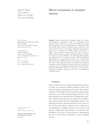
In the 1–3 days following activation of CTL precursors, form-
been summarized in several recent reviews (1–4). The role
ation of cytotoxic granules containing perforin and granzymes
of CTLs in rejection has long been a popular theme in trans-
can be detected. When the target cell is identified and engaged
plantation research. For example, Strom and associates (5)
through specific interactions between the T-cell receptor (TCR)
showed that donor-specific CTLs could be eluted from reject-
and CD8 on the CTL and MHC class I on the target cell, these
ing human renal allografts. Later, Rosenberg et al. (6) showed
granules fuse with the effector cell membrane and extrude the
that adoptive transfer of CD8þ effector T cells was sufficient to
granule contents into the immunological synapse (Fig. 1) (4).
induce rejection of major histocompatibility complex (MHC)
Along with perforin and granzymes, the cytotoxic granules
class I-mismatched skin grafts in mice. Additional complexity
also contain serglycin, calreticulin, Fas ligand (FasL), and
of the role of CTLs in rejection was suggested by studies of
granulysin. In the presence of calcium, perforins assemble
Wood and Morris (7), showing that donor-specific CTLs could
into polyperforins and insert into the target cell membrane
also be isolated from long-surviving, ‘accepted’ rat renal allo-
to facilitate the uptake of granzyme B (GB) by an uncertain
grafts. Today, it seems clear that CD8þ CTLs play a key role in
mechanism into the target cell cytoplasm, where GB mediates
rejection of organ transplants, but this role may be accentuated
apoptosis (16). This chain of events leading to the insertion of
in certain circumstances.
perforin and the delivery of GB into the target cell is known as
The CD8þ CTL is primed and activated by recognition of
the granzyme exocytosis (GE) pathway. The final common
donor MHC class I antigens. These antigens also serve as the
pathway for these cytolytic processes is triggering of apoptosis
targets for the mature cytolytic effector. In the prevailing view,
in the target cell.
recipient CD8þ T cells are primed by direct presentation of
Upon entry into the target cell, GB can trigger apoptosis
donor antigens by ‘passenger’ antigen-presenting cells (APCs)
through several pathways, including direct cleavage of procas-
from the graft (1). Although donor APCs are the major path-
pase-3 and indirect activation of procaspase-9 through a
ways for CTL induction, Kreisel et al. (8) reported a novel
complex pathway (17, 18). In this pathway, GB acting through
mechanism of direct antigen presentation by activated donor
vascular endothelial cells. This pathway can induce responder
CD8þ cells with an effector phenotype that are sufficient to
Caspases
cause acute rejection (AR). Although the role of class I MHC
antigens is unequivocal, the requirement for costimulatory Target cell
molecules in the allogeneic priming of CTLs is controversial.
After cardiac transplantation, blockade of CD40–CD40 ligand Fas MHC I Ca++
(CD40L) interactions has been variously reported to impair
(9) or to have no effect (10) on activation of allospecific CD8þ Fas pathway GE pathway
CD8 TCR CD3
T cells. Moreover, the resistance to tolerance induction by FasL
costimulatory blockade has been attributed to the development
of CD8þ CTL effectors (11). Some of the variability in results Cytotoxic T cell
in this area might reflect differences in experimental condi-
tions, contributions of other pathways to provide help in the
Perforin
form of cytokines, or genetic background of animals used in Granzyme B Cytotoxic granule
Serglycin
the studies (12). Moreover, less traditional costimulatory Calreticulin
Fas ligand Polyperforins
molecules, such as leukocyte function-associated antigen-1 Granulysin
(LFA-1), membrane lymphotoxin, or LIGHT [homologous to
Fig. 1. Mechanisms of cytotoxic T lymphocyte-induced graft damage.
lymphotoxins, inducible expression, competes with herpes sim- FasL, Fas ligand; GE, granzyme exocytosis pathway; MHC, major
plex virus (HSV) glycoprotein D for HSV entry mediator, a histocompatibility complex; TCR, T-cell receptor.
52 Immunological Reviews 196/2003
3. Rocha et al Á Effector mechanisms in transplant rejection
BCL-2 interacting domain (BID) and BCL-2 homologous Conversely, some studies show that rejection can proceed in
antagonist/killer (BAK) induces the release from the mito- the absence of perforin. Diamond and Gill (31) employed a
chondrion of cytochrome C and second mitochondria-derived strategy of adoptively transferring primed CD8þ cells from
activator of caspase/direct inhibitor of apoptosis protein perforin- or FasL-deficient mice into severe combined immuno-
(IAP)-binding protein with low isoelectric point to form an deficiency disease (SCID) murine recipients of pancreatic
‘apoptosome’ with procaspase-9. In the apoptosome, procas- islet allografts to show that in vivo allograft rejection did not
pase-9 is cleaved to caspase-9, which then cleaves procaspase-3. depend on perforin or FasL. The ability of graft rejection to
Caspase-3 inactivates the inhibitor of caspase-activated DNAse proceed in the absence of perforin highlights the importance of
(ICAD) leading to DNA fragmentation (19). Recent studies the cation-independent mannose 6-phosphate receptor (CI-
support the relevance of caspase-3 and -9 to transplantation MPR) that mediates the uptake of GB by target cells and allows
physiology. For example, caspase-3 staining correlated with apoptosis in a perforin-independent manner. Blockade of GB
rejection in human cardiac allograft specimens (20). In addition, interactions with CI-MPR prevents GB uptake and apoptosis in
zinc chloride, an inhibitor of caspase-3, reduced apoptosis in rat target cells. Donor H-2k MPR– cells injected under the kidney
cardiac allografts and prolonged allograft survival (21). Finally, a capsule of BALB/c H-2d recipients are not rejected. Thus,
T-cell-specific tyrosine–kinase inhibitor, tautomycetin induced expression of the CI-MPR and its interaction with GB are
recipient T-cell apoptosis and prolonged rat cardiac allograft essential for in vivo allogeneic cell rejection (32).
survival by the phosphorylation of T-cell-specific residues lead- The molecules that choreograph apoptosis and lymphocyte
ing to the cleavage downstream of caspase-3 and caspase-9 (22). cytolysis are generally considered to be rejection-promoting
The caspase cascades therefore might represent novel targets for effector molecules. However, in other circumstances, they can
immunosuppressive intervention, if they can be pinpointed be protective. For example, it has been suggested that perforin
with adequate specificity and without untoward side effects. from donor cells can down regulate the alloimmune response
Along with the GE pathway, CD8þ CTLs can also utilize the by inducing apoptosis of recipient immune cells (33). In this
Fas-dependent pathway to induce cytolysis and apoptosis paradigm, the donor perforin may overwhelm the recipient
(23). In the Fas-dependent pathway, FasL is either packaged T-cell-expressed cathepsin B that normally serves to protect the
into cytotoxic granules with perforins and granzymes or is recipient effector cells from the actions of perforin. Similarly,
trafficked directly to the activated effector cell surface for it has been shown that expression of FasL confers immune
binding to the target cell. Binding of FasL on the effector cell privilege to the allograft by inducing Fas-dependent apoptosis
to Fas on the target cell membrane triggers apoptosis ultim- in the recipient immune effector cells. For instance, murine
ately through the same caspase effector mechanisms as in the testis tissue transplanted under the kidney capsule of an allo-
GE pathway. Expression of Fas and FasL can be detected in geneic recipient survives indefinitely due to the expression of
rejecting allografts in humans, but their presence is not always FasL on Sertoli cells (34). In a more recent series of human
specific for rejection (24, 25). CD4þ effector cells may also pre-implantation renal allograft biopsies, FasL expression was
eliminate cells expressing MHC class II antigen through a inversely correlated to subsequent AR (35). In another study,
Fas-dependent or Fas-independent mechanism (26). over-expression of FasL on a thyroid allograft prevented rejec-
Although early studies with perforin- or Fas-deficient mice tion and resulted in suppressed donor-specific CTL activity
demonstrated the importance of both the GE- and Fas-depend- (36). Finally, murine recipients of allogeneic bone marrow
ent pathways to cell-mediated cytotoxicity (27, 28), the fol- that had undergone vector-mediated transduction with FasL
lowing studies suggest that the GE pathway plays the had enhanced short-term engraftment relative to controls
dominant role in apoptosis induction in allograft rejection. (37). Despite these promising studies, a beneficial role for
Krupnick et al. (29) showed that in vitro CTL-mediated killing donor FasL in human transplantation has yet to be exploited.
of donor vascular endothelial cells deficient in Fas and FasL
proceeded with only mild impairment, indicating that the GE
Natural killer cells
pathway is the dominant contributor to cytotoxicity in this
system. Similarly, co-incubation of graft-infiltrating T cells The natural killer (NK) cell is a large granular lymphocyte that
from human renal allografts undergoing rejection with a GE acts as part of the innate immune system to kill virally and
pathway inhibitor (concanamycin A) substantially reduced in parasitically infected cells (38). NK cells also provide surveil-
vitro lysis and apoptosis of proximal tubular epithelial cells, lance in preventing the growth of some tumors. The NK cell
whereas incubation with a Fas inhibitor did not (30). does not rearrange TCR or immunoglobulin (Ig) genes to
Immunological Reviews 196/2003 53
4. Rocha et al Á Effector mechanisms in transplant rejection
facilitate binding to a specific antigen. Rather, the NK cell be quite low, even in florid rejection. Thus, the CTL pathway
expresses numerous activating (i.e. NKp46, NKp44, and appears to be one of several mechanisms that contribute to
NKp30) and inhibitory receptors (i.e. Ly49 class), the ligation transplant injury. Its relative contribution varies depending on
of which regulate NK-cell activation (39, 40). The ligands for immunosuppression, the type of graft, and the nature of MHC
these receptors are not all well described, but in the prevailing disparity between donor and recipient. Interestingly, compon-
theory, self-MHC class I molecules bind to Ly49 receptors to ents of the CTL system might actually be protective in some
provide an overriding inhibitory signal that prevents NK-cell circumstances.
activation (40). Thus, the absence of self-MHC class I mol-
ecules on a cell due to down regulation by infection or due to
Delayed-type hypersensitivity
allogeneic phenotype can result in its lysis by an NK cell. NK
cells lyse targets solely through the GE pathway with perforin- Another major effector limb of the T-cell response to an organ
containing granules that are preformed during development graft is the DTH response. DTH is primarily mediated by
rather than upon activation, as in the case of T lymphocytes. alloantigen-specific CD4þ T-helper 1 (Th1) cells. After trig-
However, similar to T lymphocytes, GB in the NK cell is gering by alloantigen, these Th1 cells secrete cytokines, such as
required for the induction of rapid apoptosis in the target interferon-g (IFN-g) and TNF (45). These cytokines have
cell (41). multiple pro-inflammatory actions, including activation of
Although early research implicated NK cells in the rejection monocytes and macrophages that are a prominent component
of bone marrow allografts (42), more recent studies have also of the cellular infiltrate in allograft rejection. This activation
suggested a role for NK cells in solid organ transplant rejec- causes a further amplification of cytokine and chemokine
tion. For instance, Ogura and associates (43) reported that production, along with generation of proteolytic enzymes,
transplantation of rat livers into CD8þ T-cell-depleted recipi- nitric oxide, and other soluble factors that perpetuate and
ents resulted in rejection and intra-graft expression of GB and shape the local inflammatory response. These factors also
FasL similar to that of unmanipulated allograft recipient con- directly impact the physiological functioning of the graft
trols. Although these authors demonstrate, in a separate study, through effects on vascular tone, permeability, and integrity.
infiltration of the liver allografts by recipient NK cells, their Finally, soluble mediators of the DTH response act in an
findings do not preclude the contribution to rejection of antigen-independent fashion to promote chemotaxis and
another effector arm of the recipient’s immune system, such further activation of immune cells.
as CD4þ effector cells or an antibody-mediated response.
Slightly more provocative is a murine cardiac allograft model
DTH and rejection
in which the removal of the CD28-costimulatory signal did not
afford long-term graft acceptance unless accompanied by Adoptive transfer experiments have suggested that an allospe-
depletion of recipient NK-receptor-bearing cells (44). In this cific DTH response alone is sufficient to mediate skin graft
study therefore NK cells were sufficient to mediate solid allo- rejection. Dalloul and associates (46) showed that adoptive
graft rejection in the absence of T-cell costimulation. Deple- transfer of CD4þ lymphocytes from CD8–/– mice could induce
tion of NK cells alone did not prevent allograft rejection. Taken rejection of MHC class I- or II-disparate skin grafts in SCID
together, these studies suggest that NK cells might play a role mice. In this circumstance, skin grafts were rejected in the
in the alloimmune response, but their importance in a host absence of a detectable CTL response. Likewise, Valujskikh et al.
with normal T-cell function has not been clearly demon- (47) showed that transfer of a Th1 alloreactive cell line that
strated. recognizes donor MHC peptides via the indirect pathway was
Generation of allospecific CTLs represents an immune effector sufficient to cause rejection of skin grafts in SCID mice. In this
pathway that can deliver precise, antigen-specific cell kill- circumstance, direct, donor-specific CTL responses were not
ing. The capacity of this system in rejection is reflected by the possible. Histologically, these grafts had a predominant
identification of apoptotic cells in biopsies of rejecting allo- macrophage infiltrate consistent with a DTH-type effector
grafts and the ability of adoptive transfer of CD8þ CTLs to mechanism, and donor antigens elicited a typical DTH
cause transplant rejection. However, the requirements for this response when injected subcutaneously (47).
pathway are not absolute, as depletion of CD8þ cells might The intensity of the donor-specific DTH response can be
have little effect on the course of rejection. Moreover, the assessed in transplanted animals in vivo by injecting donor
relative number of apoptotic cells in rejecting allografts may splenocytes or splenocyte lysates into either the ear or footpad
54 Immunological Reviews 196/2003
5. Rocha et al Á Effector mechanisms in transplant rejection
of the recipient (48). The reaction is characterized by a typical suppressed by these mechanisms and that attenuation of
DTH response with exudates, edema, and an intense cellular the intra-graft DTH response might contribute to long-term
infiltrate. The intensity of response is proportional to the graft survival.
degree of edema formation assessed as thickness of the pinnae Whereas Th1 cells are associated with the production and
or footpad. In rodent transplant models, as well in humans release of pro-inflammatory cytokines, TNF and IFN-g, with
with organ transplants, graft loss is associated with a vigorous subsequent activation of macrophages, Th2-type cells produce
systemic DTH response. By contrast, long-term graft accept- cytokines such as IL-4, IL-5, IL-10, and IL-13. Although the
ance is characterized by a blunted or absent DTH response Th2 cytokine profile generally inhibits cell-mediated immun-
(49–51). ity and the DTH response, this cytokine profile can induce
Bickerstaff and associates (52) have studied the regulation of antibody-mediated rejection (discussed elsewhere in this
this systemic DTH response in mice that have accepted heart article), and recent evidence suggests that IL-4 and IL-5
allografts after treatment with gallium nitrate. These studies promote eosinophil-mediated rejection. Eosinophils are
indicate that the DTH response, assessed by injecting alloanti- recruited to the graft by IL-4, IL-5, and IL-13 released by
gen subcutaneously, is suppressed by the actions of tissue Th2 cells. Upon recruitment and activation, eosinophils
growth factor-b (TGF-b) and interleukin (IL)-10. Injecting elaborate substances such as leukotrienes (LTs) (discussed
neutralizing antibodies to TGF-b and IL-10 along with the below), superoxides, major basic protein, eosinophil cationic
donor antigen can restore the DTH response. Similar blunting protein, and eosinophil peroxidase. In several models, Th2-
of DTH responses is observed in mouse recipients of dominant alloresponsive T cells can mediate allograft rejection.
spontaneously accepted kidney allografts. However, in this Histologic examination of many of these models reveals an
circumstance, suppression of DTH is mediated primarily by intense infiltrate of eosinophils (54). Using IFN-g and IL-2
TGF-b (49). Enhanced activity of TGF-b in recipients of long- double knockout (KO) mice, Zand et al. (55) demonstrated
surviving allografts appears to be due to activation of TGF-b that these animals rapidly rejected cardiac allografts, and the
by the protease plasmin rather than to enhanced production of intra-graft cytokine profile was characteristic of a Th2
TGF-b. In this regard, DTH responses can be restored in response. Le Moine et al. (56, 57) evaluated the role of
cardiac allograft acceptors by co-injecting antigen with eosinophils in a model of chronic skin allograft rejection. In
antibodies against tissue-specific plasminogen activator (53). this model, MHC class II-disparate skin grafts were applied
VanBuskirk et al. (51) used a ‘trans vivo’ model to measure after generalized T-cell depletion. The skin grafts survived for
DTH responses in human transplant recipients. Peripheral more than 60 days before they were eventually rejected, and
blood mononuclear cells (PBMCs) were harvested from three there was skewing toward a Th2 cytokine profile in the reject-
human transplant recipients (two kidney, one liver), who had ing grafts with marked increases in IL-4 and IL-5 but not IFN-g.
well-functioning allografts despite discontinuing their imm- Histologically, these grafts had predominant eosinophil
unosuppressive therapy. PBMCs from the patients were then infiltrates, and they developed an impressive obliterative arter-
co-injected with donor antigen into the footpad of SCID mice. iolopathy. When neutralizing antibodies to IL-4 were admin-
Similar to the rodent models, all three patients with long- istered, both graft vasculopathy and eosinophil infiltration
surviving allografts had suppressed DTH responses to donor were abolished. Using neutralizing antibodies to IL-5 or IL-5
antigens. By contrast, the response to tetanus toxoid was gene KO mice, eosinophil graft infiltration was inhibited, but
intact. However, when tetanus toxoid was injected with vasculopathy was unaffected. These studies suggest a critical
alloantigen, the DTH response was blunted, suggesting active role for eosinophils in settings where rejection is mediated by
suppression of bystander antigen responses. As in the mouse a predominant Th2-type response. It is worth noting that
experiments, antibodies against either TGF-b or IL-10 rescued significant eosinophil infiltrates are sometimes seen in severe
DTH responsiveness. Thus, the immunoregulatory cytokines allograft rejection in humans (58, 59).
TGF-b and IL-10 play an important role in the impaired DTH As discussed above, the DTH response is perpetuated and
response that is associated with allograft acceptance (51). shaped by soluble inflammatory mediators. These mediators
Furthermore, these studies suggest that inhibition of the act in several capacities to promote and amplify the inflamma-
DTH response by ‘acceptors’ is an active process that is tory response to an allograft. They recruit immune cells to the
mediated, at least in part, by the anti-inflammatory cytokines, graft and can promote the activation and differentiation of
TGF-b and IL-10. It is attractive to speculate that DTH antigen-specific T cells. Finally, they directly affect the physio-
responses in the long-surviving allografts might also be logical functions of the allograft through effects on vascular
Immunological Reviews 196/2003 55
6. Rocha et al Á Effector mechanisms in transplant rejection
tone and integrity. Among the wide range of inflammatory reduced in splenocytes or isolated T cells from mice lacking
mediators that contribute to the pathogenesis of allograft thromboxane prostanoid (TP) receptors. In addition, survival
rejection, lipid mediators generated by the metabolism of of cardiac allografts was prolonged in TP–/– recipients treated
arachidonic acid (AA) pathway play a prominent role (60). with sub-therapeutic doses of cyclosporine compared to
Eicosanoids are generated by the enzymatic metabolism of cyclosporine-treated wildtype controls. Similarly, survival of
AA. In the first step of these metabolic pathways, AA is elabor- kidney allografts transplanted into TP–/– animals is likewise
ated from membrane-bound phospholipids through the significantly prolonged compared to wildtype controls (our
actions of phospholipases. AA can then be further metabolized unpublished observation). Thus, the COX metabolite, TXA2,
to a variety of biologically active products including prosta- acting via the TP receptor promotes allograft rejection.
noids, LTs, P450 metabolites (HETEs and EETs), and the iso- Compared to the pro-inflammatory actions of TXA2, PGE2
prostanes. Most eicosanoid products are rapidly metabolized tends to inhibit or suppress immune responses. In transplant-
and, therefore, must work in an autocrine or paracrine fash- ation models, administration of PGE analogs inhibits rejection
ion. Among the eicosanoids, roles for prostanoids and LTs in and prolongs survival (60). The diverse biological actions of
the alloimmune response have been most clearly defined (60). PGE2 are mediated via four distinct GPCRs, the E prostanoid
These systems provide a prototypical example of the role of (EP) receptors (EP1,2,3,4) (70). EP receptor isoforms are
soluble mediators in rejection and, therefore, are reviewed in expressed on immune cells including macrophages and
some detail below. T cells. However, until recently, the precise EP receptor iso-
forms mediating the immunosuppressive actions of PGE2 were
not known. To address this question, we examined responses
Prostanoids and rejection
to PGE2 in splenocytes and purified T cells from mice lacking
Prostanoids are generated from AA by the cyclooxygenase each of the individual four EP receptors. These studies indi-
(COX) pathway (61). There are two isoforms of COX that cated that the Gs-coupled EP2 receptor mediates the inhibitory
have identical biochemical functions, but regulation and pat- effects of PGE2 upon T cells, whereas both the EP2 and EP4
terns of their expression are quite different. COX-1 is consti- receptors regulate macrophage functions (71). As the clinical
tutively expressed in most nucleated cells, whereas COX-2 use of PG analogs has been hindered by their lack of potency
expression is markedly up regulated in response to injury or and specificity, the identification of the relevant immuno-
inflammation. These enzymes are the targets of widely used modulatory receptors may facilitate exploring this pathway as a
conventional non-steroidal anti-inflammatory drugs (NSAIDs), therapeutic target.
which inhibit both COX isoforms, and coxibs, which are select-
ive for COX-2. The actions of the prostanoids, including effects
Leukotrienes in rejection
on inflammation and immunity, are mediated via G protein-
coupled receptors (GPCRs) (62). Although there is evidence LTs are another class of AA metabolites that contribute to the
suggesting a role for various prostanoids in transplantation, inflammatory response to an allograft. In this pathway, AA is
the actions of prostaglandin E2 (PGE2) and thromboxane (TX) metabolized to LTA4 via the actions of 5-lipoxygenase (5-LO).
A2 have been most thoroughly characterized (60). LTA4 can be hydrolyzed to form LTB4, or can be conjugated
A role for the prostanoid TXA2 in rejection was first sug- with glutathione to form LTC4. LTC4 can be further metab-
gested by Foegh and associates (63), who reported elevated TX olized to LTD4 and LTE4 by extracellular metabolism. LTC4 and
metabolites in the urine of patients with rejecting kidney its metabolites are collectively referred to as the cysteinyl-
transplants. These findings were subsequently confirmed in leukotrienes (CysLTs) and were previously known as the
animal models of rejection (64–66). TX is a potent vasocon- slow reacting substance of anaphylaxis (72).
strictor, and as a hemodynamic mediator, it can have a detri-
mental effect on allograft function (64, 67). However, TXA2
Leukotriene B4
may contribute to rejection by influencing cellular immune
responses (68, 69). LTB4 is primarily synthesized by neutrophils and macrophages
Recently, we have demonstrated that TXA2, acting through and is a potent chemotactic and chemokinetic factor for neu-
its receptor TP, directly influences cellular immune responses trophils. There are two receptors for LTB4: BLT1 and BLT2.
(Thomas et al., manuscript submitted). Proliferative responses These GPCRs are found in highest concentrations on leuko-
to alloantigens or anti-CD3 antibody were significantly cytes. BLT1 expression is highest in monocytes, whereas BLT2
56 Immunological Reviews 196/2003
7. Rocha et al Á Effector mechanisms in transplant rejection
is most highly expressed in lymphocytes (73). LTB4 increases number of circulating lymphocytes, accompanied by seques-
leukocyte adhesion to endothelial cells and extravasation into tration of lymphocytes in peripheral lymph nodes, mesenteric
tissues (74). LTB4 promotes the production of pro-inflamma- lymph nodes, and Peyer’s patches (88). Honig and associates
tory cytokines by T cells and monocytes, such as IL-1, IL-2, (85) found that FTY720 enhances CCL19- and CCL21-induced
and IFN-g (75–77). In addition, LTB4 upregulates the chemotaxis by activating the multidrug transporters, Abcb1
expression of integrins such as CD11b (78). In a mouse and Abcc1, thereby promoting peripheral lymphocyte seques-
heterotopic heart transplant model, Weringer et al. (79) tration. One function of Abcc1 is to transport LTC4 to the
demonstrated a clear role for LTB4 in allograft rejection, as extracellular space. In the setting of FTY720 administration,
mice treated with an LTB4 antagonist had significantly pro- 5-LO deficiency or inhibition renders T cells unresponsive to
longed graft survival. These findings were associated with a CCL19 and CCL21; however, the addition of exogenous CysLT
marked reduction in cellular staining for CD11b and a delayed (LTD4) restores responsiveness (85). These studies demon-
peak in graft reactive serum IgG levels (79). strate a clear role for CysLTs in lymphocyte migration, and
The CysLTs are primarily synthesized by eosinophils, mast they suggest a mechanism whereby altering CysLT release
cells, and macrophages. They stimulate smooth muscle con- results in sequestration of lymphocytes in peripheral lymph
traction, contributing to bronchiolar and arteriolar constric- nodes. In this circumstance, FTY720 appears to impair the
tion, and increase vessel permeability, promoting plasma alloimmune response by augmenting the release of CysLTs.
extravasation. The actions of the CysLTs are mediated by two
receptors, CysLT1 and CysLT2. These GPCRs are expressed in a
Antibodies and complement
wide variety of tissues and cell types, including the spleen,
lungs, eosinophils, and monocytes/macrophages (80). CysLTs Generation of CTL and DTH are the principal effector limbs of
may also contribute to allograft rejection as LTC4 levels are the T-cell response to an allograft. The third major element
enhanced in rejecting rat kidneys correlating with the devel- contributing to graft injury and rejection is the development
opment of cellular infiltrates, and administration of a CysLT of an alloantibody response to the transplant. Although anti-
receptor antagonist decreases vascular rejection (81, 82). body production is ultimately a B-cell function, the contribu-
Consistent with the apparent benefits of inhibiting indi- tion of T cells cannot be overlooked. As will be discussed
vidual LT receptors discussed above, global inhibition of LT below, B cells require help from alloreactive CD4þ T cells to
synthesis using 5-LO inhibitors improves function and pro- grow, differentiate, and secrete antibodies. The binding of
longs allograft survival in various models of transplantation, alloantibodies to ABO or MHC antigens expressed on endothe-
including kidney, heart, and pancreas allografts. In a rat kidney lial cells triggers a complex response involving the comple-
transplant model, inhibition of 5-LO improved allograft sur- ment and coagulation pathways that activate and recruit
vival, diminished MHC class II expression, and preserved inflammatory cells, ultimately resulting in graft injury. Allo-
allograft morphology (81). Despite the beneficial effects antibodies can also mediate antibody-dependent cellular cyto-
observed with pharmacological inhibition of 5-LO, transplant toxicity (ADCC). In this case, NK cells or macrophages bind to
outcomes in mice with targeted deletion of the 5lo gene were the Fc region of antibody molecules promoting lysis of target
quite different. In a mouse model of kidney transplantation, cells. The cross-linking of Fc receptors on NK cells triggers
allografts transplanted into 5-LO-deficient recipients had sig- perforin/granzyme-mediated cytotoxicity, whereas in macro-
nificantly reduced survival (83). Similarly, 5-LO deficiency phages this cross-linking promotes the release of mediators
accelerated the course and severity of autoimmune disease in such as nitric oxide (NO), TNF-a, and reactive oxygen species.
MRL-lpr mice, consistent with an unexpected role for 5-LO to The actions of alloantibodies and complement to promote
ameliorate immune injury (84). graft injury produce distinct clinical manifestations in hyper-
Recent studies by Honig and associates (85) support the acute, acute humoral, and chronic rejection. In addition, there
view that 5-LO products can inhibit the immune response. are some circumstances, in which, antibodies and complement
These studies suggest that LTC4 contributes to the efficacy of may have beneficial effects.
FTY720, a sphingosine-derived immunosuppressant.
Although its mechanism of action is not clearly understood,
Hyperacute rejection
FTY720 prolongs survival in heart and skin grafts without
impairing T-cell and B-cell activation (86, 87). Following Hyperacute allograft rejection (HAR) is the classic and most
administration of FTY720, there is a marked reduction in the exuberant example of antibody-mediated rejection. In this
Immunological Reviews 196/2003 57
8. Rocha et al Á Effector mechanisms in transplant rejection
process, large quantities of preformed antibodies against ABO interstitial hemorrhage, and severe injury to endothelial cells;
or MHC bind these antigens on endothelial cells and activate immunostaining reveals Ig and complement deposits along
the classic complement pathway (Fig. 2). endothelial surfaces of graft blood vessels. Given the lack of
The split products of the early complement components, effective treatment, HAR almost invariably leads to allograft loss.
such as C3a and C5a, function as anaphylatoxins attracting With the advent of blood typing and T-cell cross-match
inflammatory cells and platelets to the target area. The late testing, this type of rejection has become a rare event in the
complement components, namely C5b-9, form the membrane clinical arena. However, HAR remains a major barrier to
attack complex (MAC) that, in turn, activates and damages the xenotransplantation. For example, when a pig organ is trans-
endothelium (89, 90). Activated endothelial cells produce IL-8 planted into primates, ‘xenoreactive natural antibodies’ bind
and monocyte chemotactic protein-1 (MCP-1) to recruit neu- the carbohydrate galactose-a-1,3-galactose (Gala1, 3Gal)
trophils and monocytes to the site of injury (91). Other expressed in pig endothelial cells and cause HAR (94). More-
inflammatory cytokines, such as IL-1, are also secreted and over, xenografts appear to be particularly susceptible to
function to upregulate the expression of tissue factor in complement-mediated injury, because porcine complement
endothelial cells. There is release of preformed von Willebrand regulatory proteins fail to dampen the activation of human
factor (vWF) and P selectin from cytoplasmic Weibel-Palade complement on xenogeneic cells (95). In fact, much effort has
bodies to the surface of endothelial cells; vWF promotes the been concentrated on generating transgenic pigs that either do
formation and stabilization of the platelet plug, whereas P not synthesize Gala1,3Gal (96) or that express human com-
selectin, a member of the family of adhesion molecules, regu- plement regulatory proteins as potential organ donors for
lates interactions between endothelial cells and leukocytes humans (97). Although these recent advances of research on
(92). P selectin interacts with its natural ligand, P-selectin xenotransplantation are still far from translating into clinical
glycoprotein ligand-1 (PSGL-1), present on neutrophils and practice, they have undoubtedly provided invaluable insights
monocytes and mediates the adherence of these cells to the into the pathogenetic roles of antibodies and complement in
endothelium for subsequent extravasation into the tissue (93). HAR (98).
In essence, the actions of complement and inflammatory medi-
ators transform the endothelium from a protective barrier
between the blood and extravascular tissues into a pro-
Acute rejection
coagulant, chemoattractive, and adhesive interface that promotes
inflammation. Once endothelial cells are damaged, the under- In contrast to HAR, the role of antibodies in acute allograft
lying matrix is exposed, and there is release of tissue factor rejection is controversial. Acute rejection (AR) is typically
into the circulation, which binds factor VIIa and activates the viewed as a T-cell-mediated process. Mice lacking T cells
extrinsic coagulation cascade. The end result of these processes cannot reject fully mismatched allografts, and reconstitution
is widespread intra-vascular thrombosis, hemorrhage, and tis- of these animals with T cells restores the rejection process.
sue injury manifested grossly by a mottled and cyanotic graft. Moreover, T cells can be readily seen in histologic sections as
Pathologically, HAR is characterized by obstruction of small the principal components infiltrating the allograft during AR.
vessels (including glomerular capillaries) by platelet thrombi, Current anti-rejection protocols directly target T cells and are
ADCC Complement cascade
C3a
+ Coagulation
cascade
+
MØ C1q IL-8
NK Platelets
Fc MCP-1 VIIa
Fc P selectin +
TF IL-1
MAC TF vWF Fig. 2. Mechanisms of antibody-induced
graft damage. ADCC, antibody-dependent
cellular cytotoxicity; MCP-1, monocyte
chemotactic protein-1; vWF, von Willebrand
Activated endothelial cells factor.
58 Immunological Reviews 196/2003
9. Rocha et al Á Effector mechanisms in transplant rejection
able to prevent or treat the vast majority of AR episodes. On Rocha et al. (106) compared the outcome of 16 patients
the other hand, B cells and antibodies do not appear to be with AHR treated with PP þ IVIG with that of 43 patients
essential for graft rejection, as agammaglobulinemic mice are with acute cellular rejection diagnosed and treated during
able to reject the first and second set skin transplants at control the study period. The one-year graft survival by Kaplan
rate (99). Finally, antibody and complement deposition are Meier analysis was 81% in the AHR and 84% in the
not typical findings in most AR biopsies. ACR group (P ¼ NS) (106). Prospective trials comparing
Over the last decade, however, repeated clinical observa- PP þ IVIG with other regimens are needed. Given the low
tions have suggested a central role for antibodies in at least a incidence of humoral rejection, a multicenter approach will
subset of patients with AR. These patients are typically pre- likely be required to recruit the necessary number of
sensitized and present with severe allograft dysfunction early patients for such studies.
after transplant that is resistant to anti-T-cell therapy (100). There is a series of studies in animal models supporting an
The histologic features of antibody-mediated acute humoral important contribution of antibodies to the pathogenesis of
rejection (AHR) are distinct from those of typical acute cellular AR. Brandle et al. (107) used B-cell-deficient mice to show that
rejection (ACR). In AHR, neutrophils constitute a large pro- donor-specific antibodies contribute to the pathogenesis of
portion of the cellular infiltrate, which appears to preferen- acute allograft rejection. In a model of cardiac allograft rejec-
tially target the peritubular capillaries. Sensitive flow tion in the mouse using subtherapeutic doses of cyclosporine
cytometry techniques can detect circulating donor-specific to dampen cellular immune responses, graft survival was sig-
alloantibodies (DSAs) in the majority of AHR cases. nificantly prolonged in B-cell-deficient mice compared to
A recent study showed that 95% of allograft biopsies from controls (107). Using a different donor-recipient strain com-
recipients with DSAs at the time of rejection had positive bination, Wasowska et al. (91) documented prolonged
staining for the complement split product C4d (101). It has cardiac allograft survival in Ig KO mice, despite an intact
been suggested that staining for C4d might have some advan- cellular immune response. When complement-activating anti-
tages as a marker of complement activation, as C4d remains bodies to donor antigens were passively transferred to Ig KO
covalently bound to the endothelium, serving as a footprint of mice 10 days after transplant, AR was rapidly restored. Trans-
complement activation by alloantibodies (102). Initial studies fer of antibodies at earlier time points led to a slower onset of
depicted C4d staining as a very sensitive and specific diagnos- rejection, suggesting a significant interaction between anti-
tic tool that could reliably distinguish AHR from cellular bodies and the cellular components of the immune response
rejection or calcineurin toxicity (102, 103). However, emer- (91). This interaction might occur in several ways. First,
ging data have raised questions about the sensitivity and spe- antibodies can coat donor endothelial cells and bind recipient
cificity of this assay in the diagnosis of AHR. For example, macrophages and NK cells to promote target cell lysis
Bohmig et al. (104) showed that a substantial proportion of via ADCC. Second, it is well documented that B cells require
patients with AHR (defined by AR and circulating DSAs T-cell help for antibody production (108). T-cell and
detected by flow cytometry) had negative C4d staining, result- B-cell interactions via CD40L–CD40 and CD28–B7 pathways
ing in a sensitivity of 31%. Moreover, Nickeleit (105) are essential for B-cell growth, differentiation, and Ig-class
demonstrated C4d positivity in 40–50% of biopsies with switching (109). Interruption of these pathways inhibits
histologic signs of acute cellular rejection as well as in some both cellular and humoral responses prolonging allograft
cases that did not require any anti-rejection therapy. There- survival (110, 111).
fore, treatment decisions should probably not be made based The apparent actions of antibody to enhance T-cell
solely on C4d-staining results. Rather, clinicians should use responses to an allograft might also involve activation of
C4d staining in conjunction with the clinical presentation, complement. In this regard, Pratt and associates (112) have
flow cytometry results, and histology before deciding whether provided clear evidence of collaboration between complement
antibodies or T cells should be the primary targets of anti- components and cellular immunity during allograft rejection.
rejection therapy. The authors transplanted wildtype or C3-gene-disrupted
Historically, AHR was associated with poor allograft (C3–/–) kidneys into MHC-mismatched recipients and showed
prognosis, but recent studies have suggested that regimens that recipients of C3–/– grafts experienced long-term graft
that target antibody removal and resynthesis such as plas- survival; in contrast, wildtype kidneys were rapidly rejected
mapheresis with intravenous Ig (PP þ IVIG), immuno- with a mean graft survival of 12.5 days. When wildtype
adsorption, or rituximab, might improve clinical outcomes. kidneys were transplanted into C3–/– recipients, there was
Immunological Reviews 196/2003 59
10. Rocha et al Á Effector mechanisms in transplant rejection
only a mild prolongation in graft survival (16.2 Æ 1.2, mean In an elegant study using a murine model of cardiac trans-
Æ SEM) indicating that locally synthesized C3 had a greater plantation, Hancock et al. (111) demonstrated that adminis-
impact on rejection than circulating C3. T cells were isolated tration of anti-CD4 monoclonal antibody induced long-term
from recipients of wildtype or C3–/– grafts and were graft survival but did not prevent pathological findings of
re-challenged with donor antigen in vitro. In these experiments, chronic rejection. By contrast, when anti-CD40L antibody
proliferative responses to donor antigens were lower, if the was administered to disrupt the interactions between B and T
recipient had received a C3–/– kidney. Taken together, these cells, allograft survival was indefinite and histologic evidence
findings suggest that C3 produced locally by the rejecting renal of CR, including transplant arteriosclerosis, was abolished.
allograft augments the alloimmune response by apparently This treatment also led to increased vascular expression of
contributing to T-cell priming. ‘protective’ genes such as heme oxygenase-1 (HO-1), Bcl-xL,
These data reinforce the notion that the immune response to and A20. When these genes were induced in endothelial cells
an allograft comprises cellular and humoral components that by in vitro culture in the presence of Th2 cytokines prior to
interact to produce graft injury. Although the central role of alloantibody exposure, there was marked protection against
T cells during acute allograft rejection remains unquestioned, endothelial cell activation, as evidenced by decreased E-selectin
significant contributions from antibodies and complement expression. In vivo treatment with anti-CD4 antibody along
are being slowly uncovered. Moreover, there are important with agents that induce HO-1 (metalloporphyrins) resulted
interactions between these components of the immune system in almost complete protection from CR and significantly
during the alloimmune response. Therapeutic interventions decreased intra-graft apoptosis (111). These data are in keep-
aimed at interrupting each of these effector pathways or their ing with recent findings by Plissonnier et al. (115), which
interactions could have a major influence on the fate of organ indicate that apoptosis might be an important mechanism of
transplants. antibody-mediated injury during CR. The authors transplanted
MHC-mismatched aortic grafts into rats that had been pre-
sensitized against donor antigens by skin transplant and
Chronic rejection
demonstrated that alloantibodies induced apoptosis of graft
The pathogenesis of chronic rejection (CR) remains incom- vascular cells in vitro and in vivo (115).
pletely understood. However, there are data that suggest key Production of alloantibodies in association with episodes of
roles for antibodies and complement in the pathogenesis of CR. AR has been long recognized. The pathogenetic role of these
Russell and Ley (113) performed cardiac transplants between antibodies, however, remains in question. Recent evidence
inbred mice, where the recipients were depleted of CD4þ and suggests that the development of anti-MHC class II alloantibod-
CD8þ T cells. This maneuver was sufficient to induce long- ies after transplantation may be a risk factor for CR, indepen-
term graft survival but did not prevent the development of dent of AR (116). In a recent report, all kidney transplant
obstructive coronary lesions typical of CR. The authors showed failures due to CR were preceded by the development of
that transplants between strains that produced antibodies to anti-human leukocyte antigen (HLA) antibodies (117).
donor cells (B10.A to B10.BR) developed more intense cor- Mauiyyedi et al. (118) tested the role of C4d antibody staining
onary vasculopathy than those in the reverse combination, in in kidney biopsies of patients with chronic allograft nephro-
which antibodies were not detected. In the latter strain com- pathy (CAN) and showed that 23/38 CAN biopsies (61%)
bination, the severity of the coronary lesions could be had peritubullar capillaries (PTC) staining for C4d, compared
increased in a dose-dependent fashion by the administration with 1 of 46 (2%) of controls (P < 0.001). The authors
of antibodies against donor antigens. Similarly, continuing concluded that antibodies play a pivotal role in the pathogenesis
injections of antidonor antibodies were sufficient to of 61% of cases of CAN and suggested classifying this subset of
induce striking coronary lesions in SCID recipients of patients under the label of ‘chronic humoral rejection’. Subse-
heart allografts (113). Further evidence that antibodies are quent studies by Theruvath (119) and Regele (120), however,
required for the full pathologic expression of CR stems from have shown much lower rates of C4d positivity (13 and 34%,
more recent experiments from the same group, in which respectively) among biopsies performed for CAN. Moreover,
B-cell-deficient mice failed to develop the typical arterial Nickeleit et al. (105) suggested that C4d is a marker of active
lesions of CR (114). Others have shown that transferring AR and not CR. Given the inconsistency of these observations, it
antibodies specific to donor antigen into Ig-deficient mice is quite clear that more studies are needed before a role for C4d,
can restore these lesions (111). as marker of humoral immunity during CR, can be established.
60 Immunological Reviews 196/2003
11. Rocha et al Á Effector mechanisms in transplant rejection
Potential beneficial effects of antibody and complement in prolonging graft survival independent of the strain combina-
transplantation tion. This finding is in stark contrast with the results obtained
with a single injection of antibodies after transplantation
The data presented so far depict antibodies and complement
(121). The mechanisms of antibody-mediated graft enhance-
as important effectors of allograft damage during rejection.
ment are unclear; proposed mechanisms involve the produc-
However, there is evidence suggesting that, in special circum-
tion of anti-idiotypic or blocking antibodies. Administration
stances, both antibodies and complement might have benefi-
of F(ab0 )2 fragments alone does not promote enhancement of
cial effects on graft survival. Earlier studies indicated that
graft survival, suggesting that this process is Fc dependent (122).
transfer of cytotoxic alloantibodies might have in vivo effects
This finding is further supported by the fact that non-opsonizing
that are both damaging and protective to the allograft.
IgM antibodies are unable to induce enhancement (123).
Oluwole and coworkers (121) demonstrated that the donor-
Sohn and coworkers (124) recently showed that the devel-
recipient strain combination and the timing of administration
opment of antigen-specific tolerance after intra-ocular
are crucial in determining the outcome of antibody transfer. For
(immune privileged site) injection is dependent on the com-
example, when ACI rats (‘low responders’) are the recipients
plement fragment iC3b binding to APCs. Ligation of iC3b to its
of Wistar Furth hearts, transfer of antibodies leads to enhance-
receptor on APCs resulted in the sequential production of TGF-
ment of graft survival, but when the donor-recipient combin-
b and IL-10, which is essential for the induction of tolerance
ation is reversed and Wistar Furth rats (‘high responders’)
in this model (124). The extent to which this elegant observa-
become the recipients, antibody transfer results in HAR. More-
tion extends to the development of tolerance to alloantigens
over, the transfer of 1 mL of alloantibodies for several days
still needs to be elucidated.
prior to and on the day of transplantation is very effective in
References
1. Arakelov A, Lakkis FG. The alloimmune 10. Jones ND, et al. CD40-CD40 ligand- 17. Sutton VR, et al. Initiation of apoptosis by
response and effector mechanisms of allograft independent activation of CD8þ T cells can granzyme B requires direct cleavage of bid,
rejection. Semin Nephrol 2000;20:95–102. trigger allograft rejection. J Immunol but not direct granzyme B-mediated caspase
2. Barry M, Bleackley RC. Cytotoxic T 2000;165:1111–1118. activation. J Exp Med 2000;192:1403–1414.
lymphocytes: all roads lead to death. Nat Rev 11. Adams AB, et al. Heterologous immunity 18. Wang GQ, et al. Resistance to granzyme B-
Immunol 2002;2:401–409. provides a potent barrier to transplantation mediated cytochrome c release in Bak-
3. Krammer PH. CD950 s deadly mission in the tolerance. J Clin Invest 2003;111: deficient cells. J Exp Med 2001;194:
immune system. Nature 2000;407:789–795. 1887–1895. 1325–1337.
4. Russell JH, Ley TJ. Lymphocyte-mediated 12. Williams MA, et al. Genetic characterization 19. Wolf BB, Schuler M, Echeverri F, Green DR.
cytotoxicity. Annu Rev Immunol of strain differences in the ability to mediate Caspase-3 is the primary activator of
2002;20:323–370. CD40/CD28-independent rejection of skin apoptotic DNA fragmentation via DNA
5. Strom TB, Tilney NL, Carpenter CB, Busch GJ. allografts. J Immunol 2000;165:6849–6857. fragmentation factor-45/inhibitor of caspase-
Identity and cytotoxic capacity of cells 13. Scheu S, Alferink J, Potzel T, Barchet W, activated DNase inactivation. J Biol Chem
infiltrating renal allografts. N Engl J Med Kalinke U, Pfeffer K. Targeted disruption of 1999;274:30651–30656.
1975;292:1257–1263. LIGHT causes defects in costimulatory T cell 20. Narula J, et al. Annexin-V imaging for
6. Rosenberg AS, Mizuochi T, Sharrow SO, activation and reveals cooperation with noninvasive detection of cardiac allograft
Singer A. Phenotype, specificity, and function lymphotoxin beta in mesenteric lymph node rejection. Nat Med 2001;7:1347–1352.
of T cell subsets and T cell interactions genesis. J Exp Med 2002;195:1613–1624. 21. Kown MH, et al. Zinc-mediated reduction of
involved in skin allograft rejection. J Exp Med 14. Guo Z, et al. Cutting edge: membrane apoptosis in cardiac allografts. Circulation
1987;165:1296–1315. lymphotoxin regulates CD8(þ) T cell- 2000;102:228–232.
7. Dallman MJ, Wood KJ, Morris PJ. Specific mediated intestinal allograft rejection. J 22. Shim JH, et al. Immunosuppressive effects of
cytotoxic T cells are found in the nonrejected Immunol 2001;167:4796–4800. tautomycetin in vivo and in vitro via T cell-
kidneys of blood-transfused rats. J Exp Med 15. Corbascio M, et al. Anti-lymphocyte specific apoptosis induction. Proc Natl Acad
1987;165:566–571. function-associated antigen-1 monoclonal Sci USA 2002;99:10617–10622.
8. Kreisel D, et al. Mouse vascular endothelium antibody inhibits CD40 ligand-independent 23. Ju ST, Cui H, Panka DJ, Ettinger R, Marshak-
activates CD8þ T lymphocytes in a B7- immune responses and prevents chronic Rothstein A. Participation of target Fas
dependent fashion. J Immunol vasculopathy in CD40 ligand-deficient mice. protein in apoptosis pathway induced by
2002;169:6154–6161. Transplantation 2002;74:35–41. CD4þ Th1 and CD8þ cytotoxic T cells. Proc
9. Zhai Y, Meng L, Busuttil RW, Sayegh MH, 16. Metkar SS, et al. Cytotoxic cell granule- Natl Acad Sci USA 1994;91:4185–4189.
Kupiec-Weglinski JW. Activation of mediated apoptosis: perforin delivers 24. Oh SI, et al. Correlation of Fas and Fas ligand
alloreactive CD8þ T cells operates via CD4- granzyme B-serglycin complexes into target expression with rejection status of
dependent and CD4-independent cells without plasma membrane pore transplanted heart in human. Transplantation
mechanisms and is CD154 blockade sensitive. formation. Immunity 2002;16:417–428. 2001;71:906–909.
J Immunol 2003;170:3024–3028.
Immunological Reviews 196/2003 61
12. Rocha et al Á Effector mechanisms in transplant rejection
25. Xu B, et al. Apoptosis in chronic rejection of and rejection of bone-marrow grafts. reactive delayed-type hypersensitivity
human cardiac allografts. Transplantation Immunol Rev 1997;155:29–40. responses in cardiac allograft acceptor mice. J
2001;71:1137–1146. 41. Shresta S, MacIvor DM, Heusel JW, Russell JH, Immunol 2000;164:5132–5139.
26. Williams NS, Engelhard VH. Identification of Ley TJ. Natural killer and lymphokine- 54. Le Moine A, Goldman M, Abramowicz D.
a population of CD4þ CTL that utilizes a activated killer cells require granzyme B for the Multiple pathways to allograft rejection.
perforin- rather than a Fas ligand-dependent rapid induction of apoptosis in susceptible Transplantation 2002;73:1373–1381.
cytotoxic mechanism. J Immunol target cells. Proc Natl Acad Sci USA 55. Zand MS, et al. Interleukin-2 and interferon-
1996;156:153–159. 1995;92:5679–5683. gamma double knockout mice reject
27. Kagi D, et al. Fas and perforin pathways as 42. Murphy WJ, Kumar V, Bennett M. Rejection heterotopic cardiac allografts. Transplantation
major mechanisms of T cell-mediated of bone marrow allografts by mice with 2000;70:1378–1381.
cytotoxicity. Science 1994;265:528–530. severe combined immune deficiency (SCID). 56. Le Moine A, et al. IL-5 mediates eosinophilic
28. Kagi D, et al. Cytotoxicity mediated by T cells Evidence that natural killer cells can mediate rejection of MHC class II-disparate skin
and natural killer cells is greatly impaired in the specificity of marrow graft rejection. J allografts in mice. J Immunol
perforin-deficient mice. Nature Exp Med 1987;165:1212–1217. 1999;163:3778–3784.
1994;369:31–37. 43. Ogura Y, et al. Apoptosis and allograft 57. Le Moine A, et al. Critical roles for IL-4, IL-5,
29. Krupnick AS, et al. Mechanism of T cell- rejection in the absence of CD8þ T cells. and eosinophils in chronic skin allograft
mediated endothelial apoptosis. Transplantation 2001;71:1827–1834. rejection. J Clin Invest 1999;103:
Transplantation 2002;74:871–876. 44. Maier S, et al. Inhibition of natural killer cells 1659–1667.
30. Wever PC, et al. Mechanisms of lymphocyte- results in acceptance of cardiac allografts in 58. Almirall J, Campistol JM, Sole M, Andreu J,
mediated cytotoxicity in acute renal allograft CD28-/- mice. Nat Med 2001;7:557–562. Revert L. Blood and graft eosinophilia as a
rejection. Transplantation 1998;66:259–264. 45. Gallin J. Inflammation. In: Paul WE, ed. rejection index in kidney transplant. Nephron
31. Diamond AS, Gill RG. An essential Fundamental Immunolgy. New York: Raven 1993;65:304–309.
contribution by IFN-gamma to CD8þ T cell- Press, 1993:1015–1032. 59. Martinez OM, et al. Evidence for a
mediated rejection of pancreatic 46. Dalloul AH, Chmouzis E, Ngo K, nonclassical pathway of graft rejection
islet allografts. J Immunol 2000;165: Fung-Leung WP. Adoptively transferred involving interleukin 5 and eosinophils.
247–255. CD4þ lymphocytes from CD8 -/- mice are Transplantation 1993;55:909–918.
32. Motyka B, et al. Mannose 6-phosphate/ sufficient to mediate the rejection of MHC 60. Tilley SL, Coffman TM, Koller BH. Mixed
insulin-like growth factor II receptor is a class II or class I disparate skin grafts. messages: modulation of inflammation and
death receptor for granzyme B during J Immunol 1996;156:4114–4119. immune responses by prostaglandins and
cytotoxic T cell-induced apoptosis. Cell 47. Valujskikh A, Matesic D, Gilliam A, Anthony D, thromboxanes. J Clin Invest 2001;108:
2000;103:491–500. Haqqi TM, Heeger PS. T cells reactive to a 15–23.
33. Bose A, Inoue Y, Kokko KE, Lakkis FG. single immunodominant self-restricted 61. Rocca B, FitzGerald GA. Cyclooxygenases and
Cutting edge: perforin down-regulates CD4 allopeptide induce skin graft rejection in mice. prostaglandins: shaping up the immune
and CD8 T cell-mediated immune responses J Clin Invest 1998;101:1398–1407. response. Int Immunopharmacol
to a transplanted organ. J Immunol 48. Sirak J, Orosz CG, Wakely E, VanBuskirk AM. 2002;2:603–630.
2003;170:1611–1614. Alloreactive delayed-type hypersensitivity in 62. Funk CD. Prostaglandins and leukotrienes:
34. Bellgrau D, Gold D, Selawry H, Moore J, graft recipients: complexity of responses and advances in eicosanoid biology. Science
Franzusoff A, Duke RC. A role for CD95 divergence from acute rejection. 2001;294:1871–1875.
ligand in preventing graft rejection. Nature Transplantation 1997;63:1300–1307. 63. Foegh ML, et al. Urine i-TXB2 in renal
1995;377:630–632. 49. Bickerstaff AA, Wang JJ, Pelletier RP, Orosz CG. allograft rejection. Lancet 1981;2:431–434.
35. Porter CJ, Ronan JE, Cassidy MJ. Fas-fas- Murine renal allografts: spontaneous 64. Coffman TM, Yarger WE, Klotman PE.
ligand antigen expression and its relationship acceptance is associated with regulated Functional role of thromboxane production
to increased apoptosis in acute renal T cell-mediated immunity. J Immunol by acutely rejecting renal allografts in rats. J
transplant rejection. Transplantation 2001;167:4821–4827. Clin Invest 1985;75:1242–1248.
2000;69:1091–1094. 50. Orosz CG, Wakely E, Sedmak DD, Bergese SD, 65. Khirabadi BS, Foegh ML, Ramwell PW. Urine
36. Tourneur L, et al. Transgenic expression of VanBuskirk AM. Prolonged murine cardiac immunoreactive thromboxane B2 in rat
CD95 ligand on thyroid follicular cells allograft acceptance: characteristics of cardiac allograft rejection. Transplantation
confers immune privilege upon thyroid persistent active alloimmunity after treatment 1985;39:6–8.
allografts. J Immunol 2001;167:1338–1346. with gallium nitrate versus anti-CD4 66. Tannenbaum JS, Anderson CB, Sicard GA,
37. Whartenby KA, et al. Transduction of donor monoclonal antibody. Transplantation McKeel DW, Etheredge EE. Prostaglandin
hematopoietic stem-progenitor cells with Fas 1997;63:1109–1117. synthesis associated with renal allograft
ligand enhanced short-term engraftment in a 51. VanBuskirk AM, et al. Human allograft rejection in the dog. Transplantation
murine model of allogeneic bone marrow acceptance is associated with immune 1984;37:438–443.
transplantation. Blood 2002;100: regulation. J Clin Invest 2000;106:145–155. 67. Mangino MJ, Anderson CB, Deschryver K,
3147–3154. 52. Bickerstaff AA, VanBuskirk AM, Wakely E, Turk J. Arachidonate lipoxygenase products
38. Trinchieri G. Biology of natural killer cells. Orosz CG. Transforming growth factor-beta and renal allograft rejection in dogs.
Adv Immunol 1989;47:187–376. and interleukin-10 subvert alloreactive Transplantation 1987;44:805–808.
39. Hsieh CL, Obara H, Ogura Y, Martinez OM, delayed type hypersensitivity in cardiac 68. Leung KH, Mihich E. Prostaglandin
Krams SM. NK cells and transplantation. allograft acceptor mice. Transplantation modulation of development of cell-mediated
Transpl Immunol 2002;9:111–114. 2000;69:1517–1520. immunity in culture. Nature 1980;288:
40. George T, et al. Allorecognition by murine 53. Bickerstaff AA, Xia D, Pelletier RP, Orosz CG. 597–600.
natural killer cells: lysis of T-lymphoblasts Mechanisms of graft acceptance: evidence 69. Caughey GE, Pouliot M, Cleland LG, James MJ.
that plasminogen activator controls donor- Regulation of tumor necrosis factor-alpha and
62 Immunological Reviews 196/2003
13. Rocha et al Á Effector mechanisms in transplant rejection
IL-1 beta synthesis by thromboxane A2 in transplantation in the rat. Transplant Proc primate cardiac xenografts from humoral
nonadherent human monocytes. J Immunol 1987;19:3116–3118. injury. Nat Med 1995;1:423–427.
1997;158:351–358. 83. Goulet JL, et al. Deficiency of 96. Phelps CJ, et al. Production of alpha
70. Narumiya S, FitzGerald GA. Genetic and 5-lipoxygenase accelerates renal allograft 1,3-galactosyltransferase-deficient pigs.
pharmacological analysis of prostanoid rejection in mice. J Immunol Science 2003;299:411–414.
receptor function. J Clin Invest 2001;167:6631–6636. 97. Cozzi E, White DJ. The generation of
2001;108:25–30. 84. Goulet JL, et al. Deficiency of transgenic pigs as potential organ donors for
71. Nataraj C, et al. Receptors for prostaglandin 5-lipoxygenase abolishes sex-related humans. Nat Med 1995;1:964–966.
E(2) that regulate cellular immune survival differences in MRL-lpr/lpr mice. 98. Lin SS, et al. The role of antibodies in acute
responses in the mouse. J Clin Invest J Immunol 1999;163:359–366. vascular rejection of pig-to-baboon cardiac
2001;108: 1229–1235. 85. Honig SM, et al. FTY720 stimulates transplants. J Clin Invest 1998;101:
72. Haeggstrom JZ, Wetterholm A. Enzymes multidrug transporter- and cysteinyl 1745–1756.
and receptors in the leukotriene cascade. leukotriene-dependent T cell chemotaxis to 99. Cerny A, Ramseier H, Bazin H,
Cell Mol Life Sci 2002;59:742–753. lymph nodes. J Clin Invest 2003;111: Zinkernagel RM. Unimpaired first-set and
73. Yokomizo T, Izumi T, Shimizu T. 627–637. second-set skin graft rejection in
Co-expression of two LTB4 receptors in 86. Pinschewer DD, Ochsenbein AF, Odermatt agammaglobulinemic mice. Transplantation
human mononuclear cells. Life Sci 2001;68: B, Brinkmann V, Hengartner H, Zinkernagel 1988;45:1111–1113.
2207–2212. RM. FTY720 immunosuppression impairs 100. Sayegh MH, Colvin RB. Case record of the
74. Lewis RA, Austen KF, Soberman RJ. effector T cell peripheral homing without Massachusetts General Hospital. Weekly
Leukotrienes and other products of the affecting induction, expansion, and memory. clinicopathological exercises. Case 8–2003.
5-lipoxygenase pathway. Biochemistry and J Immunol 2000;164:5761–5770. A 35-year-old man with early dysfunction
relation to pathobiology in human diseases. 87. Yanagawa Y, Masubuchi Y, Chiba K. of a second renal transplant. N Engl J Med
N Engl J Med 1990;323:645–655. FTY720, a novel immunosuppressant, 2003;348:1033–1044.
75. Rola-Pleszczynski M. Differential effects of induces sequestration of circulating mature 101. Crespo M, et al. Acute humoral rejection in
leukotriene B4 on T4þ and T8þ lymphocytes by acceleration of lymphocyte renal allograft recipients. I. Incidence,
lymphocyte phenotype and homing in rats III. Increase in frequency of serology and clinical characteristics.
immunoregulatory functions. J Immunol CD62L-positive T cells in Peyer’s patches by Transplantation 2001;71:652–658.
1985;135:1357–1360. FTY720-induced lymphocyte homing. 102. Collins AB, et al. Complement activation in
76. Rola-Pleszczynski M, Bouvrette L, Gingras D, Immunology 1998;95:591–594. acute humoral renal allograft rejection:
Girard M. Identification of interferon-gamma 88. Chiba K, et al. FTY720, a novel diagnostic significance of C4d deposits in
as the lymphokine that mediates leukotriene immunosuppressant, induces sequestration peritubular capillaries. J Am Soc Nephrol
B4-induced immunoregulation. J Immunol of circulating mature lymphocytes by 1999;10:2208–2214.
1987;139:513–517. acceleration of lymphocyte homing in rats. 103. Mauiyyedi S, et al. Acute humoral rejection
77. Rola-Pleszczynski M, Lemaire I. I. FTY720 selectively decreases the number in kidney transplantation. II. Morphology,
Leukotrienes augment interleukin 1 of circulating mature lymphocytes by immunopathology, and pathologic
production by human monocytes. acceleration of lymphocyte homing. classification. J Am Soc Nephrol
J Immunol 1985;135: 3958–3961. J Immunol 1998;160:5037–5044. 2002;13:779–787.
78. Showell HJ, et al. The in vitro and in vivo 89. Saadi S, Platt JL. Transient perturbation of 104. Bohmig GA, et al. Capillary C4d deposition
pharmacologic activity of the potent and endothelial integrity induced by natural in kidney allografts: a specific marker of
selective leukotriene B4 receptor antagonist antibodies and complement. J Exp Med alloantibody-dependent graft injury. J Am
CP-105696. J Pharmacol Exp Ther 1995;181:21–31. Soc Nephrol 2002;13:1091–1099.
1995;273:176–184. 90. Nakashima S, Qian Z, Rahimi S, 105. Nickeleit V, Zeiler M, Gudat F, Thiel G,
79. Weringer EJ, Perry BD, Sawyer PS, Gilman SC, Wasowska BA, Baldwin WM III. Membrane Mihatsch MJ. Detection of the complement
Showell HJ. Antagonizing leukotriene B4 attack complex contributes to destruction of degradation product C4d in renal allografts:
receptors delays cardiac allograft rejection in vascular integrity in acute lung allograft diagnostic and therapeutic implications.
mice. Transplantation 1999;67:808–815. rejection. J Immunol 2002;169:4620–4627. J Am Soc Nephrol 2002;13:242–251.
80. Ogasawara H, et al. Characterization of 91. Wasowska BA, et al. Passive transfer of 106. Rocha PN, et al. Beneficial effect of
mouse cysteinyl leukotriene receptors alloantibodies restores acute cardiac plasmapheresis and intravenous
mCysLT1 and mCysLT2: differential rejection in IgKO mice. Transplantation immunoglobulin on renal allograft survival
pharmacological properties and tissue 2001;71: 727–736. of patients with acute humoral rejection.
distribution. J Biol Chem 2002;277: 92. Lasky LA. Selectins: interpreters of cell- Transplantation 2003;75:1490–1495.
18763–18768. specific carbohydrate information during 107. Brandle D, Joergensen J, Zenke G, Burki K,
81. Spurney RF, Ibrahim S, Butterly D, inflammation. Science 1992;258:964–969. Hof RP. Contribution of donor-specific
Klotman PE, Sanfilippo F, Coffman TM. 93. McEver RP, Cummings RD. Role of PSGL-1 antibodies to acute allograft rejection:
Leukotrienes in renal transplant rejection in binding to selectins in leukocyte evidence from B cell-deficient mice.
rats. Distinct roles for leukotriene B4 and recruitment. J Clin Invest 1997;100: Transplantation 1998;65:1489–1493.
peptidoleukotrienes in the pathogenesis of S97–S103. 108. Steele DJ, et al. Two levels of help for B cell
allograft injury. J Immunol 1994;152: 94. Parker W, Bruno D, Holzknecht ZE, Platt JL. alloantibody production. J Exp Med
867–876. Characterization and affinity isolation of 1996;183:699–703.
82. Coffman TM, Kolbeck P, Sanflippo F, xenoreactive human natural antibodies. 109. Lumsden JM, Williams JA, Hodes RJ.
Klotman P. Renal allograft function, J Immunol 1994;153:3791–3803. Differential requirements for expression of
arachidonic acid metabolism, and systemic 95. McCurry KR, et al. Human complement CD80/86 and CD40 on B cells for
cellular immunity after kidney regulatory proteins protect swine-to-
Immunological Reviews 196/2003 63
14. Rocha et al Á Effector mechanisms in transplant rejection
T-dependent antibody responses in vivo. 115. Plissonnier D, et al. Involvement of glomerular capillaries: a contribution of
J Immunol 2003;170:781–787. antibody-dependent apoptosis in graft humoral immunity to chronic allograft
110. Sayegh MH, Turka LA. The role of T-cell rejection. Transplantation 2000;69: rejection. J Am Soc Nephrol 2002;13:
costimulatory activation pathways in 2601–2608. 2371–2380.
transplant rejection. N Engl J Med 116. Pelletier RP, Hennessy PK, Adams PW, 121. Oluwole SF, Tezuka K, Wasfie T, Stegall MD,
1998;338:1813–1821. VanBuskirk AM, Ferguson RM, Orosz CG. Reemtsma K, Hardy MA. Humoral
111. Hancock WW, Buelow R, Sayegh MH, Clinical significance of MHC-reactive immunity in allograft rejection. The role of
Turka LA. Antibody-induced transplant alloantibodies that develop after kidney or cytotoxic alloantibody in hyperacute
arteriosclerosis is prevented by graft kidney-pancreas transplantation. Am J rejection and enhancement of rat cardiac
expression of anti-oxidant and anti- Transplant 2002;2:134–141. allografts. Transplantation 1989;48:
apoptotic genes. Nat Med 1998;4: 117. Lee PC, et al. All chronic rejection failures of 751–755.
1392–1396. kidney transplants were preceded by the 122. Capel PJ, Tamboer WP, de Waal RM,
112. Pratt JR, Basheer SA, Sacks SH. Local development of HLA antibodies. Jansen JL, Koene RA. Passive enhancement of
synthesis of complement component C3 Transplantation 2002;74:1192–1194. mouse skin allografts by alloantibodies is Fc
regulates acute renal transplant rejection. 118. Mauiyyedi S, et al. Chronic humoral dependent. J Immunol 1979;122:421–425.
Nat Med 2002;8:582–587. rejection: identification of antibody- 123. Lems SP, Tamboer WP, Capel PJ, Koene RA.
113. Russell PS, Chase CM, Winn HJ, Colvin RB. mediated chronic renal allograft rejection by Effects of IgG and IgM alloantibodies in the
Coronary atherosclerosis in transplanted C4d deposits in peritubular capillaries. J Am enhancement of mouse skin allografts and
mouse hearts. II. Importance of humoral Soc Nephrol 2001;12:574–582. the relation with their opsonizing capacity
immunity. J Immunol 1994;152: 119. Theruvath TP, et al. Control of antidonor in vivo. J Immunol 1981;127:665–669.
5135–5141. antibody production with tacrolimus and 124. Sohn JH, Bora PS, Suk HJ, Molina H,
114. Russell PS, Chase CM, Colvin RB. mycophenolate mofetil in renal allograft Kaplan HJ, Bora NS. Tolerance is dependent
Alloantibody- and T cell-mediated recipients with chronic rejection. on complement C3 fragment iC3b binding
immunity in the pathogenesis of transplant Transplantation 2001;72:77–83. to antigen-presenting cells. Nat Med
arteriosclerosis: lack of progression to 120. Regele H, et al. Capillary deposition of 2003;9:206–212.
sclerotic lesions in B cell-deficient mice. complement split product C4d in renal
Transplantation 1997;64:1531–1536. allografts is associated with basement
membrane injury in peritubular and
64 Immunological Reviews 196/2003

![Rocha et al Á Effector mechanisms in transplant rejection
Lymphocyte-medicated cytotoxicity receptor expressed on T lymphocytes], a newly recognized
member of the tumor necrosis factor (TNF) family expressed
Generation of antigen-specific cytotoxic T lymphocytes (CTLs)
on activated T cells, can also influence CTL activation in the
is a major immunological effector mechanism in allograft
alloimmune response (13–15). These alternative pathways
rejection. Much of the current understanding of CTLs derives
may assume a more important role after blockade of CD40/
from in vitro studies exploring the cellular immune response to
CD40L and/or CD28/B7.
alloantigens, infectious agents, and tumors. This work has
In the 1–3 days following activation of CTL precursors, form-
been summarized in several recent reviews (1–4). The role
ation of cytotoxic granules containing perforin and granzymes
of CTLs in rejection has long been a popular theme in trans-
can be detected. When the target cell is identified and engaged
plantation research. For example, Strom and associates (5)
through specific interactions between the T-cell receptor (TCR)
showed that donor-specific CTLs could be eluted from reject-
and CD8 on the CTL and MHC class I on the target cell, these
ing human renal allografts. Later, Rosenberg et al. (6) showed
granules fuse with the effector cell membrane and extrude the
that adoptive transfer of CD8þ effector T cells was sufficient to
granule contents into the immunological synapse (Fig. 1) (4).
induce rejection of major histocompatibility complex (MHC)
Along with perforin and granzymes, the cytotoxic granules
class I-mismatched skin grafts in mice. Additional complexity
also contain serglycin, calreticulin, Fas ligand (FasL), and
of the role of CTLs in rejection was suggested by studies of
granulysin. In the presence of calcium, perforins assemble
Wood and Morris (7), showing that donor-specific CTLs could
into polyperforins and insert into the target cell membrane
also be isolated from long-surviving, ‘accepted’ rat renal allo-
to facilitate the uptake of granzyme B (GB) by an uncertain
grafts. Today, it seems clear that CD8þ CTLs play a key role in
mechanism into the target cell cytoplasm, where GB mediates
rejection of organ transplants, but this role may be accentuated
apoptosis (16). This chain of events leading to the insertion of
in certain circumstances.
perforin and the delivery of GB into the target cell is known as
The CD8þ CTL is primed and activated by recognition of
the granzyme exocytosis (GE) pathway. The final common
donor MHC class I antigens. These antigens also serve as the
pathway for these cytolytic processes is triggering of apoptosis
targets for the mature cytolytic effector. In the prevailing view,
in the target cell.
recipient CD8þ T cells are primed by direct presentation of
Upon entry into the target cell, GB can trigger apoptosis
donor antigens by ‘passenger’ antigen-presenting cells (APCs)
through several pathways, including direct cleavage of procas-
from the graft (1). Although donor APCs are the major path-
pase-3 and indirect activation of procaspase-9 through a
ways for CTL induction, Kreisel et al. (8) reported a novel
complex pathway (17, 18). In this pathway, GB acting through
mechanism of direct antigen presentation by activated donor
vascular endothelial cells. This pathway can induce responder
CD8þ cells with an effector phenotype that are sufficient to
Caspases
cause acute rejection (AR). Although the role of class I MHC
antigens is unequivocal, the requirement for costimulatory Target cell
molecules in the allogeneic priming of CTLs is controversial.
After cardiac transplantation, blockade of CD40–CD40 ligand Fas MHC I Ca++
(CD40L) interactions has been variously reported to impair
(9) or to have no effect (10) on activation of allospecific CD8þ Fas pathway GE pathway
CD8 TCR CD3
T cells. Moreover, the resistance to tolerance induction by FasL
costimulatory blockade has been attributed to the development
of CD8þ CTL effectors (11). Some of the variability in results Cytotoxic T cell
in this area might reflect differences in experimental condi-
tions, contributions of other pathways to provide help in the
Perforin
form of cytokines, or genetic background of animals used in Granzyme B Cytotoxic granule
Serglycin
the studies (12). Moreover, less traditional costimulatory Calreticulin
Fas ligand Polyperforins
molecules, such as leukocyte function-associated antigen-1 Granulysin
(LFA-1), membrane lymphotoxin, or LIGHT [homologous to
Fig. 1. Mechanisms of cytotoxic T lymphocyte-induced graft damage.
lymphotoxins, inducible expression, competes with herpes sim- FasL, Fas ligand; GE, granzyme exocytosis pathway; MHC, major
plex virus (HSV) glycoprotein D for HSV entry mediator, a histocompatibility complex; TCR, T-cell receptor.
52 Immunological Reviews 196/2003](data:image/gif;base64,R0lGODlhAQABAIAAAAAAAP///yH5BAEAAAAALAAAAAABAAEAAAIBRAA7)