Thoracic Outlet Syndrome: Anatomy, Symptoms, Diagnostic Evaluation and Surgical Treatment
•
34 likes•28,495 views

Thoracic outlet syndrome, cervical outlet syndrome.

Recommended
More Related Content
What's hot
What's hot (20)
Viewers also liked
Viewers also liked (8)
Similar to Thoracic Outlet Syndrome: Anatomy, Symptoms, Diagnostic Evaluation and Surgical Treatment
Similar to Thoracic Outlet Syndrome: Anatomy, Symptoms, Diagnostic Evaluation and Surgical Treatment (20)
More from Povilas Pauliukas
More from Povilas Pauliukas (20)
Recently uploaded
Recently uploaded (20)
Thoracic Outlet Syndrome: Anatomy, Symptoms, Diagnostic Evaluation and Surgical Treatment
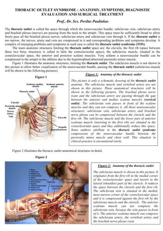
- 1. THORACIC OUTLET SYNDROME : ANATOMY, SYMPTOMS, DIAGNOSTIC EVALUATION AND SURGICAL TREATMENT Prof., Dr. Scs. Povilas Pauliukas The thoracic outlet is called the space through which the neurovascular bundle: subclavian vein, subclavian artery and brachial plexus (nerves) are passing from the neck to the armpit. This space must be sufficiently broad to allow freely pass all the brachial plexus nerves, subclavian artery and subclavian vein through it. If the thoracic outlet is too narrow, the nerves, artery and vein are compressed in it and corresponding problems and symptoms develop. A complex of emerging problems and symptoms in such case is called the thoracic outlet syndrome (TOS). The main anatomic structures limiting the thoracic outlet space are: the clavicle, the first rib (space between these two bony structures is called in latin the costoclavicular space), the subclavius muscle, situated in the costoclavicular space, the anterior and posterior scalene muscles. Very seldom a neurovascular bundle can be compressed in the armpit in the athletes due to the hypertrophied abnormal pectoralis minor muscle. Figure 1 illustrates the anatomic structures, limiting the thoracic outlet. The subclavius muscle is not shown in this picture to allow better visualization of the neurovascular bundle, passing the thoracic outlet (subclavius muscle will be shown in the following pictures). Figure 1 Figure 2 illustrates the thoracic outlet anatomical structures in detail. Figure 2 Figure 1: Anatomy of the thoracic outlet This picture is only a schematic drawing of the thoracic outlet anatomy. The subclavius muscle and vertebral artery are not shown in this picture. These anatomical structures will be shown in the following pictures. The brachial plexus nerve roots and the subclavian artery are passing through the gap between the anterior and medius scalene muscles (cervical outlet). The subclavian vein passes in front of the scalene muscles and they can not compress it. All three neurovascular structures: subclavian vein, subclavian artery and brachial nerve plexus can be compressed between the clavicle and the first rib. The subclavius muscle and the lower part of anterior scalenus muscle (inserting to the first rib) are situated in the costoclavicular space and they additionally reduce this space. Some authors attribute to the thoracic outlet syndrome compression of the neurovascular bundle between the pectoralis minor muscle and chest wall. This situation in clinical practice is encountered rarely. Figure 2: Anatomy of the thoracic outlet The subclavius muscle is shown in this picture. It originates from the first rib in the medial corner of the costoclavicular space and inserts to the lateral (shoulder) part of the clavicle. It reduces the space between the clavicle and the first rib. The subclavian vein is situated in the medial, most narrow corner of the costoclavicular space and it is compressed against the first rib by the subclavius muscle and the clavicle. The anterior scalenus muscle can not compress the subclavian vein, because the vein passes in front of it. The anterior scalenus muscle can compress the subclavian artery, the vertebral artery and the brachial nerve plexus roots.
- 2. All three scalene muscles are depicted in figure 3. Figure 3 When the gap between the anterior and medius scalene muscles (spatium interscalenum) is too narrow or absent for passing of the subclavian artery and brachial plexus, they are compressed by these two muscles, especially when the muscles contract and squeeze the nerves and arteries. The vertebral artery does not pass the gap between the scalene muscles (spatium interscalenum), but it is situated very close to the anterior scalenus muscle and in cases of brachial nerve plexus compression with the abnormal scalene muscle, abnormally inserting to the vertebral column or to the first rib, it is compressed by this muscle together with the brachial plexus. That’s, why many patients with symptoms of brachial nerve plexus roots compression (neurogenic thoracic outlet syndrome) have symptoms of vertebrobasilar insufficiency (insufficiency of blood flow in vertebrobasilar region of the brain, supplied with the blood by vertebral arteries). When the vertebral artery is compressed by the scalenus anterior muscle against the vertebral column, it is compressed at the entrance into the cervical outlet (spatium interscalenum) and it is wise to attribute such compression of vertebral arteries to the cervical outlet syndrome, especially due to the fact, that scalenectomy cures both problems at once: the neurogenic symptoms and symptoms of vertebrobasilar insufficiency. Normally, the vertebral artery enters its bony vertebral canal (canalis transversarius) at the sixth cervical vertebra. The anterior scalenus and longus colli muscles attach to the transverse process of the sixth cervical vertebra just above the vertebral artery entrance into the bony canal, leaving the free triangle space in the neck for the vertebral artery to pass from its origin on the subclavian artery to the entrance of the vertebral bony canal at the transverse process of the 6-th cervical vertebra (Figure 4). Normally, the vertebral artery is not compressed against the vertebrae by the scalenus anterior muscle, the longus colli muscle and between them. However, when the anterior scalenus muscle abnormally attaches to the transverse process of the 7-th cervical vertebra, the vertebral artery is entrapped between this muscle and the transverse process of the 7-th cervical vertebra. The same situation arises when the vertebral artery enters bony vertebral canal abnormally at higher level: at the transverse process of the 5-th, 4-th, or even 3-rd cervical vertebrae. In these cases, the vertebral artery is compressed against the transverse processes of the 6-th cervical vertebra and above situated vertebrae until the vertebral artery enters the bony canal. Therefore, the deviation of the vertebral artery from its normal course in the neck, or deviation of the scalene muscles anatomy from normal, creates the conflict between the anterior scalene muscle and the vertebral artery, causing compression of vertebral artery and symptoms of vertebrobasilar insufficiency. Due to the fact, that scalene muscles anomalies typically are bilateral, symmetrical, as a rule, both vertebral arteries are compressed by the anterior scalene muscle in cases of cervical outlet syndrome, which is also typically bilateral, on both sides. Another very important pathogenetic mechanism of impaired blood flow through the vertebral arteries and of vertebrobasilar insufficiency is the spasm of vertebral arteries. Vertebral arteries are arteries of muscle type, not elastic: they have smooth muscles in their walls and can contract. They react to compression by contraction of their walls and reduce their lumen sometimes twice or even more times. Duplex scanning of vertebral arteries in such cases reveals narrow, spastic, with diminished blood flow, vertebral arteries. A spasm of vertebral arteries causes the paroxysm of vertebrobasilar insufficiency, sometimes even a vertebrobasilar stroke. Clinical manifestations of such spastic paroxysm of vertebral Figure 3: Cervical outlet (spatium interscalenum) All three scalene muscles, the brachial nerve plexus, the subclavian artery, the vertebral artery and the subclavian vein are depicted in this picture. The anterior and medius scalene muscles originate from the first rib. The posterior scalenus muscle originates from the second rib. The anterior scalenus muscle inserts to the transverse processes of the third-sixth cervical vertebrae. The scalenus medius muscle inserts to the transverse processes of all cervical vertebrae. The posterior scalenus muscle inserts to the transverse processes of the three lowest cervical vertebrae. Due to the fact, that anterior and medius scalene muscles both originate from the first rib and insert to the transverse processes of the same cervical vertebrae, they are very prone to the developmental anomalies: they can be as a one solid muscle mass not divided into the separate muscles (brachial plexus and subclavian artery penetrate through the muscle in such cases), an additional scalenus minimus muscle can develop, they can attach to the first rib with the common tendon etc.
- 3. arteries are headaches, dizziness, nausea, sometimes even vertigo episodes, visual and hearing disturbances (tinnitus, noise in the ears, visual blurring, scotomas, visual field defects etc.) Figure 4 illustrates the anatomy of a normal vertebral artery triangle left by the longus colli and scalenus anterior muscles for the free passage of the vertebral artery from its origin on the subclavian artery to the bony canal at the transverse process of the 6-th cervical vertebra. Figure 4 On the left side, the anterior scalenus muscle is removed to show the place of its insertion onto the first rib (the tubercle of scalenus anterior muscle). The overwhelming majority of patients with cervical outlet syndrome have combined symptoms of vertebrobasilar insufficiency and of compression and irritation of brachial plexus nerve roots. In some patients, dominate vertebrobasilar symptoms, in other – symptoms of compression and irritation of nerve roots. Therefore, some of these patients seek doctor’s advice and help for vertebrobasilar insufficiency, some – for irritation of brachial plexus nerve roots. Doctors should know that symptoms of vertebrobasilar insufficiency are very common in patients with cervical outlet syndrome and that scalenectomy cures these symptoms ceasing the compression and irritation of vertebral artery by anterior scalenus muscle. The problem is that most doctors are unfamiliar with the thoracic outlet syndrome and particularly - with the cervical outlet syndrome. Consequently, many patients, suffering from the thoracic outlet and cervical outlet syndromes, despairingly are trying to find their diagnosis visiting and consulting many doctors of various specialities, performing countless sophisticated examinations like magnetic resonance imaging, CT scans etc. The correct diagnosis usually is established only when the patient finds the doctor, who not only knows the symptoms of the thoracic outlet and cervical outlet syndromes, but also is able to diagnose them and to correct the problem. Generally, vascular surgeons operate patients with thoracic outlet and cervical outlet syndromes and they are best familiar with symptoms, diagnostics and treatment of these patients. As an example, how it is difficult to find the correct diagnosis and help from doctors, even in the USA, is an article on the internet, written by the patient with the thoracic outlet syndrome from the New York City, who was seeking the diagnosis and help from doctors. The patient was consulted by many doctors of various specialities, a lot of examinations, including three magnetic resonance imaging were done for her and the diagnosis was established only when she has addressed the vascular surgeon, familiar with the thoracic outlet syndrome and operating these patients. Indeed, the worst situation is, when the patient cannot find the correct diagnosis. He cannot know what the cause of his symptoms is and consequently, he cannot get the help. Following illustration is an article about the thoracic outlet syndrome, written by Dr. Carlos Selmonosky, who is an expert in the thoracic outlet syndrome in the USA. He stressed in this article, that “One of the most unfortunate complications is a misdiagnosis or no diagnosis because patients fail to receive adequate therapy”. Figure 4: Anatomy of the vertebral artery triangle Normally, the longus colli and anterior scalenus muscles conjugate and attach to the transverse process of the 6-th cervical vertebra, creating the muscle roof for the vertebral artery, entering the hole in the transverse process of the 6-th cervical vertebra. These two muscles create the lateral borders of vertebral artery triangle. At the top of this triangle is the transverse process of the 6-th cervical vertebra. The first rib forms the bottom of this triangle. This triangle is free of muscles and left for the free vertebral artery passage from its origin on the subclavian artery to its entrance into the bony vertebral canal at the transverse process of the 6-th cervical vertebra. Problems arise when the course of vertebral artery in the neck is abnormal (high entrance of vertebral artery into the bony vertebral canal at the 5-th, 4-th, o even 3-rd cervical vertebrae, or abnormal lateral branching of vertebral artery from the subclavian artery under the scalenus anterior muscle), or abnormal anatomy of the anterior scalenus muscle.
- 4. Dr. Carlos Selmonosky has his internet site for thoracic outlet syndrome: http://www.tos-syndrome.com/. American Thoracic Outlet Syndrome Association also has its internet site at: www. atosa.org. Due to the fact, that even in the USA many doctors are unfamiliar with symptoms and diagnostics of TOS, still there is unknown what percent of population has the TOS. Opinions vary on this point, but most authors agree, that the TOS is a frequent problem and it is encountered in as many as 1-8 % of population. Usually young, 20 - 40 years old people have this problem and the neurogenic (due to compression and irritation of brachial nerve plexus) variant of TOS is encountered in women 4 times more frequently than in men. Venous variant of TOS (subclavian vein thrombosis due to its compression) is more common in males than in females. Arterial TOS (subclavian artery thrombosis or aneurysm formation due to its compression) has no gender predilection. Hence, the thoracic outlet syndrome can be: 1) neurogenic; 2) venous and 3) arterial. Neurogenic TOS is the most common variant of TOS and is encountered in 95 % of all clinical TOS cases. Venous variant of TOS is encountered in 4 % of clinical cases and arterial TOS variant is infrequent and is encountered only in 1 % of clinical TOS cases. Such big difference in the frequency of clinical manifestations of neurogenic and vascular (venous and arterial) TOS is due to the high sensitivity of nerves for compression and irritation. Compression of nerves causes numbness, tingling and even unbearable pain and consequently, patients seek doctor’s help. The subclavian vessels: artery and vein are compressed almost as often as nerves, but the patient doesn’t feel the compression of vein or artery until it thromboses. When the subclavian artery or vein thromboses, a vascular complication of TOS manifest and it is a vascular emergency: the patient needs an urgent treatment. In cases of cervical outlet syndrome (when brachial plexus nerve roots are compressed in the scalene triangle (gap between the anterior and medius scalene muscles), the upper nerve roots (fifth to seventh) are most forcefully compressed. When the compression of brachial plexus is between the clavicle and the first rib in the costoclavicular space (in the true thoracic outlet), usually most forceful compression experience the lower roots (8-th cervical and first thoracic roots) of the brachial nerve plexus. This feature determines the differences in pain, tingling, numbness distribution areas in the arm as well as muscle motor weakness distribution differences in cervical and thoracic outlet syndromes. Thereby, according to the symptoms distribution, diagnosis of cervical and thoracic outlet syndromes can be distinguished and established. Most authors define two kinds or levels of thoracic outlet syndrome: the upper (corresponding to the gap between the anterior and medius scalene muscles in the neck) and the lower thoracic outlet syndrome (actual thoracic outlet syndrome in the costoclavicular space). D. Ranney1 suggested to denominate the upper thoracic outlet as a cervical outlet, because actually it is in the neck and brachial plexus nerve roots and subclavian artery pass the gap between the scalene muscles (spatium interscalenum) in the neck, not in the actual thoracic outlet.
- 5. Figure 5 Subclavian vein can be compressed between the clavicle and the first rib (in the thoracic outlet), not in the cervical outlet. All three elements of neurovascular bundle (vein, artery and neural plexus) can be compressed in the thoracic outlet. Only the subclavian artery and nerve roots of the brachial plexus can be compressed in the cervical outlet. The vertebral artery can be compressed by the scalenus anterior muscle against the cervical vertebrae or their transverse processes as well. This particular situation arises when the scalenus anterior muscle is abnormal, or the course of vertebral artery in the neck is abnormal (it enters the bony vertebral canal higher than normally: at the 5-th, 4-th or even 3-rd cervical vertebrae or it originates from the subclavian artery more lateral than normally. I shall write separately about the vertebral artery compression with the scalenus anterior muscle later, because this situation and this pathology is very important: it is encountered in patients relatively frequently and it considerably diminishes blood flow in the vertebrobasilar region of the brain and causes symptoms of vertebrobasilar insufficiency from mild up to the vertebrobasilar stroke. Compression of the upper (5-7-th cervical nerve) roots between the anterior and medius scalene muscles (in the spatium interscalenum) most of the authors, writing on this topic, denominate as an upper thoracic outlet syndrome and the true thoracic outlet between the clavicle and the first rib they denominate as a lower thoracic outlet and symptoms arising from compression of the neurovascular bundle in it they denominate as a lower thoracic outlet syndrome. I support D. Ranney’s1 proposal to distinguish these two totally different anatomical regions (levels) into two separate definitions: cervical outlet and thoracic outlet and to denominate symptoms arising from the compression of neurovascular bundle in these two regions as a cervical outlet syndrome and thoracic outlet syndrome, because the cervical outlet is in the neck: it is the gap between the anterior and medius scalene muscles and the brachial nerve plexus together with subclavian artery is compressed in the neck, not in the true thoracic outlet. The true thoracic outlet is between the clavicle and the first rib and all three structures of the neurovascular bundle (subclavian vein, subclavian artery and brachial nerve plexus) can be compressed here. Symptomatology, diagnostics and especially the surgical treatment greatly differ between these two separate entities and therefore it is wiser not to relate and to confuse them together and to denominate them by different names: cervical outlet syndrome and thoracic outlet syndrome. That’s, why I adhere to such denomination of these two separate pathological entities earlier, and later in this article I shall refer to them as a cervical outlet syndrome and thoracic outlet syndrome. Figure 6 illustrates the anatomy of the cervical outlet. Figure 5: Anatomy of the thoracic outlet (View from the armpit) The thoracic outlet and all three elements of the neurovascular bundle: subclavian vein, subclavian artery and brachial nerve plexus are seen from the armpit. The subclavius muscle, situated between the clavicle and the first rib is clearly seen. It occupies the most narrow medial corner of the costoclavicular space, where the subclavian vein passes it. The subclavian vein passes in front of the anterior scalenus muscle (through the spatium antescalenum) and can not be compressed by this muscle in the cervical outlet (spatium interscalenum). The pectoralis minor muscle can compress the neurovascular bundle in the armpit in case of its hypertrophy as this problem is encoutered in some athletes. Such situation is relatively rare.
- 6. Figure 6 Figure 7 shows the anatomy of the cervical and thoracic outlets in detail. Figure 7 Different congenital developmental anomalies: anomalous clavicle, anomalous first rib, cervical ribs, elongated anomalous transverse process of the seventh cervical vertebra, anomalous fibrous and cartilaginous bands etc. account for the thoracic outlet syndrome. In some clinical cases no congenital anomalies exist in the thoracic outlet, except the congenital narrow space between the clavicle and the first rib. It is important to stress, that cartilaginous cervical ribs and fibrous bands are invisible on the plain roentgenograms and that only bony structures are visible on them. That’s, why normal plain roentgenograms do not rule out the existence of all congenital anomalies in the thoracic outlet, especially cartilaginous cervical ribs and fibrous bands. These latter structures are well visualized with magnetic resonance imaging. If the cervical rib exists, it originates from the seventh cervical vertebra and conjugates with the first rib usually by the joint. Such a rib greatly diminishes the space between the clavicle and the first rib (thoracic outlet) and creates a predisposition for the development of the thoracic outlet syndrome. In these cases the subclavian artery and the brachial plexus are compressed by the clavicle against the cervical rib because the neurovascular bundle must sling over the cervical rib in order to reach the armpit. Figure 6: Anatomy of the cervical outlet (View from the side) The arm, shoulder joint, scapula and the clavicle together with the muscles are removed in this picture. Only the chest and the neck with the deep muscles are left. It is clearly seen, that the gap between the anterior and medius scalene muscles is in the neck and that the compression of the brachial nerve plexus and subclavian artery in this gap is in the neck and that this gap should be called the cervical outlet. The nerves and artery in this vertical gap are compressed between the two scalene muscles in the sagital direction, meanwhile in the thoracic outlet all three structures of the neurovascular bundle are compressed in the horizontal gap between the clavicle and the first rib in the vertical direction. Figure 7: Anatomy of the cervical and thoracic outlet All anatomical structures of the cervical and thoracic outlets are depicted in detail in this picture. Superficial muscles are removed from the front of the neck and chest for better visualization of the cervical and thoracic outlets. Part of the clavicle is also removed for better visualization of the thoracic outlet and its content: subclavian vein, subclavian artery and brachial nerve plexus, passing through it. The subclavius muscle is left intact. It occupies part of the costoclavicular space. The subclavius muscle narrows the most tight medial corner of the costoclavicular space, where the subclavian vein passes from the neck to the armpit. V, VI, VII, VIII – the 5, 6, 7, 8-th brachial plexus cervical nerve roots. I - the first thoracic nerve root.
- 7. Figure 8 illustrates the normal anatomy of the bones, limiting the thoracic outlet. Figure 8 Ribs and their equivalents are colored in red. Fishes have ribs in the neck. In mammals, including the man, ribs in the neck region withered away due to the fact, that they are living on the land and need to rotate and flex the neck. Only remnants (rudiments) of the ribs: the anterior parts (processus costarius) of the transverse processes and their anterior tubercles remained in the neck region. They are colored in red in this picture, the same color as the ribs. Derangement of fetal embryogenesis causes development of cervical ribs, mostly from the processus costarius of the seventh cervical vertebra transverse process. Figure 9 illustrates the lateral view of the bones, limiting the thoracic outlet. Figure 9 Figure 8: Bones limiting the thoracic outlet Bones of the shoulder girdle are shown in green color. Scapula, the shoulder girdle with the arm are fixed to the skeleton only by the medial end of the clavicle to the sternum. The position of the scapula, clavicle and the shoulder girdle depends on the tone and strength of the shoulder girdle muscles and on the posture. Limp posture with depressed, rounded shoulders reduce the space between the clavicle and the first rib and can produce the symptoms of thoracic outlet syndrome due to compression of brachial plexus. Correction of the posture and physical therapy can be helpful in such cases. However, the physical therapy and correction of posture can be helpful only in the thoracic outlet syndrome, but not in the cervical outlet syndrome, because they correct only the space between the clavicle and the first rib, not between the scalene muscles. In case of cervical outlet syndrome neither the physical therapy, nor the posture correction or massage of the muscles can help. Conversely, they can worsen the symptoms of cervical outlet syndrome due to strengthening of the muscles, because the nerve roots, subclavian artery and the vertebral artery are compressed in this case by the muscles against the vertebral bodies or between the muscles. Figure 9: Lateral view of the bones, limiting the thoracic outlet Bones of the shoulder girdle are shown in green color. Scapula, shoulder joint, all the shoulder girdle and arm are fixed to the skeleton (to the sternum) only by the medial end of the clavicle. Therefore, the position of the scapula, clavicle and all the shoulder girdle depends on the tone and strength of the shoulder girdle muscles and on the posture. Limp posture with depressed, rounded shoulders reduce the space between the clavicle and the first rib and can produce the symptoms of thoracic outlet syndrome due to compression of the brachial plexus. Correction of the posture and physical therapy can be helpful in such cases. However, the physical therapy and correction of the posture can be helpful only in thoracic outlet syndrome, but not in cervical outlet syndrome, because they correct only the space between the clavicle and the first rib, not between the scalene muscles. In case of cervical outlet syndrome neither the physical therapy, nor the posture correction or massage of the muscles can help. Conversely, they can worsen the symptoms of cervical outlet syndrome due to strengthening of the muscles, because the nerve roots, subclavian artery and the vertebral artery are compressed in this case by the muscles against the vertebral bodies or between the muscles.
- 8. The roentgenogram of 22 year old girl with fully developed unilateral left cervical rib is shown in the figure 10. Figure 10 The girl has had pain and numbness of the left arm and hand from the adolescence. The left hand became weak, clumsy. She could not pick small things with the left hand. The body of the girl became “S” shaped due to the deviation and distortion of the spinal column. The pain of the left arm became unbearable. She addressed me because of the left arm pain. Inspection of the girl revealed the cervical rib on the left side of the neck. Roentgenogram confirmed the fully developed cervical rib on the left side and rudimentary undeveloped cervical rib on the right side. The Wright’s and Roos tests were strongly positive on the left side. The left arm and hand became weak and painful just shortly after starting Roos test. She was unable to perform Roos test with the left hand even for one minute, meanwhile she had not experienced any uncomfortable feelings in the right arm and hand during the Roos test. The pulse disappeared in the left arm in the Roos test position (the subclavian artery was totally compressed and occluded in the costoclavicular space on the left side) and the pulse was present and normal in the right arm in all positions including Roos abduction-external rotation position of the arm. The Roos test is performed with the arms in abduction-external rotation position as it is shown in figure 11. The patient is asked to close and open the hands for 3 minutes in this position of the arms and to describe all sensations, that develop. Figure 11 The girl was operated. The left first rib together with the cervical rib were removed, using the Roos technique, through the axillary approach. (Removal of the cervical accessory rib alone, without the first rib, through the supraclavicular anterior approach is unsatisfactory and usually is not adequate for decompression of the thoracic Figure 11: Position of the arms for the Roos test The arms are flexed in the elbow joints by 90 degree and abducted to the frontal plane of the body. Both hands are closed and opened steadily for 3 minutes and patient is asked to describe all sensations that develop. Normally, in the absence of thoracic outlet syndrome, patient does not experience any discomfortable sensations during 3 minute such test. In case of thoracic outlet or cervical outlet syndrome, patient usually is unable to complete the 3 minute such test due to development of weakness and numbness of arms, pain in the arms and neck. This test provokes and reproduces the usual symptoms, which are torturing the patient. Figure 10: Roentgenogram of the cervical ribs The roentgenogram shows fully developed left cervical rib, which originates from the seventh cervical vertebra and has a joint at the place where it attaches to the first rib. The rudimentary short cervical rib, attached by the joint to the first rib, is seen on the right side as well. The first ribs have a joint processes at the places of insertion of the cervical ribs. The right cervical rib was asymptomatic: no brachial plexus compression symptoms were on the right side. Meanwhile, there were dramatically expressed symptoms of the compression of brachial plexus on the left side due to the narrowing of the thoracic outlet by the fully developed cervical rib. The left cervical rib has been pushing the cervical part of the spinal column to the right, because there was no counteraction by the short undeveloped right cervical rib. Therefore, the spinal column acquired a “S” shaped distortion: the thoracic part of it deviated to the left, as a compensation to the deviation of the cervical part of the spinal column to the right. Therefore, the posture and the shape of the girl were awfully distorted. This distortion is clearly seen on the roentgenogram.
- 9. outlet. The removal of both: first and cervical ribs decompress thoracic outlet adequately and creates enough space for passing of neurovascular bundle through it). Operation and postoperative period were uneventful. The pleura was not entered and opened during the operation. The girl was discharged from the hospital on the third postoperative day. All symptoms, which were torturing the girl, disappeared after the operation. The pain and numbness in the left arm and hand disappeared. The pulse in the left arm was present in all arm positions, including abduction and elevation (Wright’s and Adson’s positions). The posture of the girl improved after the operation. She was able to stretch her body and spinal column into the straight position, what was impossible before the removal of the accessory cervical rib. I advised her to exercise the spinal column and the body together with the physiotherapy specialist to get her spinal column straight and erect. After the one year follow up she was absolutely healthy with normal posture, straight spinal column, free of thoracic outlet symptoms. In case of equal bilateral accessory cervical ribs no deviation of spinal column develop, just restriction of neck movements exists. In case of unilateral cervical rib or when they are bilateral, but not equal in length, the deviation and distortion of spinal column and body posture develop. That’s, why it is wise to remove them as early as they are diagnosed and when the symptoms of thoracic outlet syndrome manifest. It is desirable to remove them in the childhood or in the adolescence. Figure 12 illustrates unequal in length bilateral accessory cervical ribs: the left - fully developed cervical rib with the joint at the insertion onto the first rib and the right - undeveloped cervical rib, directly fused to the first rib. Figure 12 This patient after the operations is free of symptoms. The pain, numbness and paresthesias of the arms and hands disappeared. The erectness of spinal column and the body shape of the woman after one year follow up period improved considerably, however the patient failed to straighten her body completely. Therefore, the conclusion is, that patients with unilateral and unequal in length cervical accessory ribs should be operated as early, as possible, before the deviation and distortion of the spinal column and the body develops. Figure 13 illustrates the roentgenograms of patient with bilateral symmetrical accessory cervical ribs before and after the removal of first and cervical ribs on both sides by the two-staged operation. (Before operations) Figure 13 (After the operations) Figure 12: Fully developed left cervical rib with the joint between it and the first rib and undeveloped right cervical rib fused with the right first rib without the joint Due to the asymmetrical development of both cervical ribs, the spinal column deviated to the right in the neck region and to the left – in the chest region (it is clearly seen on this roentgenogram). The posture and the shape of the woman were awfully distorted. Thoracic outlet syndrome (brachial plexus nerve compression) symptoms were more expressed on the left side, though they were present and on the right side. Therefore, two- staged operation was performed for this patient: the left first and cervical ribs were removed first, and at the second operation the right first rib together with the cervical rib were removed.
- 10. Figure 13: The roentgenograms of the patient with equal bilateral cervical ribs before and after the two-staged removal of both cervical and first ribs. The white arrows on the right point to the stumps of the removed cervical ribs and the blue arrows point to the stumps of the removed first ribs. The spinal column of the patient was almost straight due to the equal length of the symmetrical accessory cervical ribs. The reason for seeking the doctor’s help was the pain and numbness in both arms and hands, sensitivity of the hands to the cold exposure. Two-staged operation was performed: at first, the right first and cervical ribs were removed and one month later, the left first and cervical ribs were removed. All thoracic outlet syndrome (brachial nerve plexus compression) symptoms cleared on both sides. Thoracic outlet syndrome symptoms can arise and manifest not only due to the accessory cervical ribs, but they can be present even in the absence of the cervical ribs: in cases of too narrow costoclavicular space, due to hypertrophied first rib, or due to the callus of fractured clavicle as it was in operated by me and published in the journal clinical case2 . The subclavian artery was compressed and crushed between the first rib and callus of pseudoarthrosis of the fractured clavicle in that patient and subsequently thrombosis of the subclavian artery and acute ischemia of the arm developed. Emergency operation was performed for that patient: first rib was removed on the diseased side, the clavicle was reunited using the bone transplant and metallic plate, and autovenous bypass from common carotid artery to the brachial artery was created. The arm and hand were saved. Anatomically too narrow costoclavicular space without any other anatomical accessory abnormalities resulted in chronical mangling of the subclavian artery between the clavicle and first rib in another operated by me patient3 . Chronical mangling and crushing of the subclavian artery resulted in aneurysm formation and thrombus embolization from the aneurysm of the subclavian artery to the distal arteries of the left arm and hand in that patient. Subsequently, the thrombosis of the left subclavian artery aneurysm and distal arteries of the left arm developed and acute ischemia of the left arm and hand occurred. Emergency operation was performed for that patient: the aneurysm of the left subclavian artery was removed, thrombectomy of thrombosed arteries was performed and an extraanatomical bypass from the left common carotid artery to the left axillary artery was created. Embryological explanation for the development of thoracic and cervical outlet anomalies Development of cervical ribs and malformation of first ribs are being linked to errors of bodily segmentation in early embryological development. Cervical rib development is determined by the formation of the spinal nerve roots. The regression of the C5 through the C7 ribs is occasioned by rapid development of the enlarging roots of the brachial plexus in the region of the limb bud. In cases of a cervical C7 rib there is generally “prefixed” brachial plexus with only a small neural contribution from the T1 nerve root to the brachial plexus. As a corollary, in the “postfixed” brachial plexus in which there is a contribution of the T2 nerve root to the brachial plexus, the first thoracic rib is often rudimentary, having been inhibited in its development by the unusual nerve growth. This embryologically determined morphologic interdependence is evident with other structural relationships at the thoracic outlet. Cervical ribs are inheritable by autosomal dominant way. Therefore, there is a considerable likelihood of encountering the cervical ribs in children of patients having cervical ribs. During development, the C7 rib forms, then regresses to the C7 transverse process. Various stages in this evolution range from a complete C7 rib to rudimentary forms associated with a fibrocartilagineous band. The only radiologic indication of this residual band may be an enlarged C7 transverse process. Milliez4 emphasized the influence of neurovascular structure development on the ultimate configuration of the scalene muscle mass. The scalenic muscle mass is only differentiated into specific scalene muscles by the traversing of the neurovascular bundle. The persistence of certain muscle inclusions in the brachial plexus, as well as of muscle groups that traverse various elements of brachial plexus, is related to the original mass of the scalene muscle being variously fragmented by the passage of these developing structures as the limb bud develops. This separation of muscle bundles interdigitating between the neurovascular structures accounts for the muscular bridges seen between the anterior and middle scalene muscles that often penetrate the brachial plexus. Sanders and Roos5 demonstrated in their anatomical dissections that these abnormalities of scalene fragmentation are seen quite frequently in the adult. The causes of thoracic outlet syndrome can be divided into: 1) anomalies of the first rib or cervical rib (including the residual fibrous band from an incomplete cervical rib; 2) anomalies of scalene muscle development or insertion; 3) subclavius muscle anomalies; 4) anomalies of the clavicle; 5) anatomical anomalies (e.g. narrow costoclavicular space) not clearly identifiable as a developmental variation. Makhoul and Machleder6 in 200 consecutive transaxillary procedures for thoracic outlet syndrome have found the following anatomical anomalies: 8,5 % of operated patients have had a cervical rib; 10% of patients had accessory scalenus minimus muscle; 19,5% of patients had anomalous subclavius muscle; 43% of patients (the biggest group) had an anomaly of scalene muscle development or insertion; 19% of patients had no discernible anomaly from the axillary surgical approach. Nevertheless, their symptoms cleared after the removal of the first rib. These cases were treated as a narrow costoclavicular space without any discernible congenital anatomic anomaly.
- 11. Fully developed cervical ribs with joints at the insertion to the first rib has 0, 2 % of population. In a study of 40 000 consecutive chest x-rays in American army recruits, Etter7 encountered 68 (0,17%) complete articulated cervical ribs and 98 anomalous first ribs (0,25 %). Adson8 reviewed his experience with cervical ribs by radiologic study at Mayo clinic. He identified an incidence of 0,56% or 5,6 patients per thousand with cervical ribs. Of these 28% were male and 72% were female. 47% of cervical ribs were bilateral. The right side was involved in 23% and the left side in 30% of unilateral cervical rib cases. Forty five percent of cases in this Mayo clinic group was symptomatic. Firsov9 in the Soviet Union reported fluorographic examination of 510 893 people and observed 1379 cervical ribs, for an incidence of 0,27%. Women accounted for 76,8% and men for 23,2% of cervical rib cases; 33% of cervical ribs were bilateral. Hence, the Firsov’s data are very similar to the Etter’s and Adson’s data. Therefore, 02%-025% incidence of cervical ribs in population is accurate. The fact, that incidence of cervical ribs in women is 3,3 time higher than in men, explains the fact, that neurogenic variant of thoracic outlet syndrome in women is 4 times more common than in men and that the ratio of operations for neurogenic thoracic outlet syndrome in women and men is 4:1. The abdominal, thoracic and cervical musculature develops from the hypomeric portion of the paraxial and epaxial mesoderm, with the scalene and prevertebral muscles in the neck corresponding to the intercostal and ventrolateral abdominal muscles in the thorax and abdomen respectively10 . In the embryo, plates of axially running muscle segments differentiate into the discrete muscle groups seen in adult. The subclavian artery, which is the artery of the seventh cervical segment, and the spinal nerves from C5 to T1 pierce the muscle plates in the cervical segment much the same as the intercostal nerve and artery do in the thoracic segments. The growth of the limb bud and development of pectoral girdle then lead to the particular structural changes seen in this region. Therefore, the anomalies of scalene muscles are very frequent and encountered in clinical practice very often. The scalene muscle can be as a solid mass without any differentiation into anterior, middle and posterior scalene muscles. In such cases subclavian artery and brachial plexus roots are piercing the scalene muscle mass and are compressed by muscle fibers. The interscalene gap (spatium interscalenum) can be too narrow and tight or abnormal with crossing insertions of scalenus anterior and middle muscles onto the first rib or with “V” shaped interscalene gap due to common insertion with the common tendon onto the first rib. In such cases, brachial nerve plexus and subclavian artery are compressed in the interscalene gap. Sanders and Roos5 , studying the anatomy of the interscalene triangle, found interdigitating fibers between the scalene muscles through the brachial plexus in 75% of dissections in patients with thoracic outlet syndrome and in 40% of consecutive cadaver dissections. Their data suggest that scalene muscle anomalies are very common in human being and that they have very big clinical importance in thoracic outlet syndrome etiopathogenesis. As I mentioned earlier, the thoracic outlet and cervical outlet syndromes differ greatly by their clinical symptoms, diagnostic evaluation disparities and particularly differ their surgical treatment. Therefore, I shall describe them separately: at first the thoracic outlet syndrome and later – the cervical outlet syndrome. There are three types of thoracic outlet syndrome: venous, arterial and neurogenic. The most common of them is neurogenic TOS. It is encountered in 95% of all TOS cases. Venous TOS is encountered in 4% of cases and arterial TOS is encountered in 1% of all TOS cases. Venous thoracic outlet syndrome Venous thoracic outlet syndrome is a complex of symptoms arising due to chronic compression of subclavian vein in the costoclavicular space and subsequent its thrombosis. In 1875, James Paget11 described the symptoms resulting from subclavian vein thrombosis. Nevertheless, he misunderstood the cause and ethiopatogenesis of the arm swelling and thought that it is due to the vein imflammation and vasospasm. In 1884, L. Schroetter12 correctly identified that thrombosis of subclavian and axillary vein causes the complex of symptoms described by Paget and attributed these upper extremity venous symptoms to the compression or thrombosis of subclavian vein at the thoracic outlet. In 1949, E. Hughes13 applied a term Paget-Schroetter’s syndrome to delineate the clinical picture of symptoms arising due to subclavian vein thrombosis. From that time, the symptoms arising due to subclavian vein thrombosis and their clinical manifestation are called Paget-Schroetter’s syndrome. The frequency of spontaneous (primary) subclavian vein thrombosis due to thoracic outlet syndrome is 2 per 100 000 population per year14 . Venous TOS results from repetitive subclavian vein compression in the costoclavicular space between the subclavius muscle or costoclavicular ligament against the first rib and tends to occur in the more active dominant extremity. Usually, subclavian vein thrombosis occurs after the intensive work or physical activity in young, physically active adults aged 25-40 years. Repetitive compression of subclavian vein damages its internal layer (intima) and thrombus formation on the damaged intima occurs15-25 . As a rule, these patients have neurogenic TOS as well. Acute thrombosis of subclavian vein almost completely blocks the venous return from the arm and results in swelling, bluish color and painfulness of the involved arm. Subsequent thrombosis of axillary, brachial veins and all
- 12. other veins in the involved arm results from the prominent venous outflow block and venostasis in the involved arm. In extreme cases, venous gangrene (phlegmasia coerulea dolens) of the involved arm develops due to cessation of blood circulation in the arm, because of blood venous return block. Clinical diagnosis is easy and usually does not create any problems. The involved arm and hand is swollen, bluish, firm. During the days, collateral veins on the involved arm and on the upper part of the involved side of the chest appear. Duplex scanning of the veins in the involved extremity reveals thrombosed, with thrombus in the lumen deep veins of the involved arm, absent blood flow in the thrombosed deep veins. The final diagnosis is established by ascending venography of the involved arm, which reveals thrombosed deep veins of the extremity, delineates the starting point and the extension of the thrombus in the deep veins of the axilla and the arm and demonstrates the collateral veins returning the blood from the arm. Most authors are prone to treat these patients by thrombolytic therapy or heparinization22-25 . Some of them additionally employ the device for thrombus fragmentation ad thrombus elimination by suction with Angiojet device (Possis Medical Inc, Minneapolis, Minnesota, USA) 26 . Other authors, including me, are advocates of a single-stage radical surgical treatment. During the same operation, the first rib is removed and thrombectomy from the deep veins of the arm is performed. Such operation eliminates the cause of subclavian vein thrombosis and restores the lumen and passage of blood in the deep veins of the arm at the same time27-29 . This type of treatment is better, because it resolves both problems at the same operation: the tightness of thoracic outlet and thrombosis of subclavian vein, and its results (early and late) are better than results of treatment with thrombolytic agents or heparinization alone, without the first rib removal. Two staged treatment with thrombolysis in acute phase, and later removal of the first rib 30 , is inferior to the single-stage radical surgical treatment as well. Arterial thoracic outlet syndrome Subclavian artery can be compressed in the cervical outlet (interscalene gap) or in the true thoracic outlet between the clavicle and the first rib or cervical rib, if present. The compression of the subclavian artery in the cervical outlet, as a rule, is asymptomatic, because its compression between the muscles in the interscalene gap has no sequelae. Muscles are soft and compression of the subclavian artery between them is not felt by the patient. This compression between the muscles does not result in the aneurysm formation or thrombosis of the subclavian artery. Meanwhile, the repetitive compression and crushing of subclavian artery between the two bones: the clavicle and the first or cervical rib causes its intimal (internal layer) and medial layer degeneration and aneurysm formation or acute its thrombosis. This results in acute ischemia of the involved arm. I have never met in my clinical practice the cases of arm ischemia due to the cervical outlet syndrome, but I have operated several patients with acute arm ischemia due to the subclavian artery thrombosis as a result of true thoracic outlet syndrome 2, 3 . Compression and mangling of the subclavian artery in the interscalene gap (cervical outlet) causes another problem: irritation of the sympathetic nerves passing inside the arterial wall of the subclavian artery, which later supplies sympathetic innervation to the all arterial tree of the arm and hand and this causes the spasm of small arteries in the hands and their fingers: Raynaud’s syndrome develops. Patient fingers and hands are bluish in color and very sensitive to the cold. Exposure of the hands to the cold causes vasoconstriction (spasm of the arteries) and hands become pale, cold and painful. Vasoconstriction and Raynaud’s syndrome can be caused by compression and irritation of the brachial plexus inside the cervical outlet as well, because fibers of sympathetic nerves passes through the cervical outlet inside the brachial plexus. Compression and irritation of brachial plexus inside the cervical outlet (interscalene gap) irritates sympathetic fibers, present in the brachial plexus, and later spreading to all the arterial tree of the arm and hand. Scalenectomy (removal of anterior scalene muscle) ceases the compression and irritation of the brachial plexus and subclavian artery and of all the sympathetic fibers, present in both these structures: Raynaud’s syndrome, as a rule, disappears. Therefore, scalenectomy in cervical outlet arterial syndrome is justified only for the treatment of Raynaud’s syndrome, to stop the irritation of sympathetic nerves, present in the brachial plexus and in the wall of the subclavian artery, not for the compression of the subclavian artery itself. Typically, patients with irritation of sympathetic fibers and Raynaud’s syndrome have neurogenic cervical outlet or thoracic outlet syndrome (symptoms of compression and irritation of brachial plexus itself). Significant part of patients, having neurogenic cervical outlet syndrome, have compression of vertebral arteries as well. Neurogenic cervical outlet syndrome typically is bilateral and compression of vertebral arteries is bilateral as well. These patients have symptoms of vertebrobasilar insufficiency up to the vertebrobasilar stroke (in cases of prolonged spasm of vertebral arteries). Vertebral arteries are not merely compressed, but they react to the compression by spasm. This spasm of vertebral arteries causes not only pronounced symptoms of vertebrobasilar insufficiency, but it can be even the cause of vertebrobasilar stroke. Typically, these patients seek doctor’s help because of vertebrobasilar insufficiency symptoms, not for symptoms of brachial plexus compression, though they have symptoms of brachial plexus compression as well. The problem is, that most physicians are unfamiliar with thoracic and particularly with the cervical outlet syndromes, they do not know their symptoms and treatment. That’s, why patients cannot obtain adequate diagnostic procedures and receive adequate treatment.
- 13. In cases of thoracic outlet syndrome, the subclavian artery is compressed and mangled between two bones: the clavicle and the first rib. Therefore, the damage to the arterial wall is significant and aneurysm formation of subclavian artery results in the age of 25-40 years, because the thorax completely develops and forms up to the 25 years and people in such age are most active physically. Therefore, mangling and traumatizing of their subclavian arteries between these two bones is most intensive in that age. Patient does not feel the process of compression and mangling of the subclavian artery until the development of subclavian artery thrombosis (or its aneurysm thrombosis), or the thrombus embolisation from the aneurysm to the distal arteries of the arm. Acute ischemia of the arm develops in such cases. Only then, patients addresses the hospital or the physician. Physician’s obligation in this situation is to establish the correct diagnosis and to provide adequate treatment for the patient. Emergency diagnostic evaluation and surgery is needed in such cases. Anterior-posterior plain view roentgenogram of thorax and neck should be taken and emergency angiography of diseased subclavian artery through the femoral route should be performed. Presence of subclavian artery aneurysm, or thrombosis of subclavian artery, particularly with embolisation to the distal arteries of the arm, indicate the presence of mangling of subclavian artery between the clavicle and first rib or cervical rib, if present, and existing arterial thoracic outlet syndrome. Roentgenogram of the chest is helpful only if cervical rib or abnormal first rib or clavicle is seen on it. If no deformities or bone anomalies are seen in the thoracic outlet, it does not preclude the existence of thoracic outlet syndrome due to the too narrow costoclavicular space. Magnetic resonance tomography of the thoracic outlet is helpful in such cases, if performed by adequate computer program and mode by qualified magnetic resonance tomography staff. After the establishment of arterial thoracic outlet syndrome diagnosis and subclavian artery thrombosis or embolisation from the subclavian artery to the distal arm arteries, the emergency operation should be undertaken: typically, first rib resection through the axillary approach and revascularization procedure, depending on the underlying cause of arm ischemia is performed at the same operation. I have operated patient with such situation. The case report is published on the internet3 . Best treatment results of these patients are obtained by vascular surgeons, because they are best familiar with the revascularization procedures and their tactics. Neurogenic thoracic outlet syndrome Neurogenic thoracic outlet syndrome is a complex of symptoms arising due to compression of brachial plexus between the clavicle and first or cervical rib, if present. Usually, the lower roots of brachial plexus (8-th cervical and first thoracic) are most intensively compressed and suffer more prominently. Therefore, symptoms usually are most expressed in the area of distribution of nerves, which are constituted by the fibers coming from C8-Th1 roots (medial or ulnar side of the arm). Nevertheless, all brachial plexus roots are compressed in most cases: just the intensity of compression can vary in lower and upper roots of brachial plexus. Only those nerves, which branch from the brachial plexus higher than thoracic outlet and innervate neck, upper portion of the back, between the shoulder-blades (scapulae) and upper portion of the chest anteriorly cannot be compressed in the true thoracic outlet. They can be compressed only in the cervical outlet (interscalene gap). All other brachial plexus roots can be compressed in the cervical outlet as well. Keeping in mind this peculiarity and obtaining the existing patient’s symptoms, one can diagnose which of neurogenic outlet syndromes is present: thoracic or cervical. Patients with neurogenic thoracic outlet syndrome experience tingling, numbness and pain in the arms. In more advanced cases muscle atrophy and weakness develop in the area of distribution of affected nerves. Electromyography is an unreliable test and should not be used for diagnosing thoracic or cervical outlet syndromes. Electromyographic changes appear only in late stages of thoracic outlet syndrome and they always mean pronounced pathologic changes in the nerves and muscles, which should be avoided. Patients should be operated in earlier stages of the disease, before the development of nerve dystrophies and muscle atrophies. Below there are four postulates, established in thoracic outlet syndrome and written by David Roos: 1. Patients, having thoracic outlet syndrome, have anatomical congenital anomalies, predisposing them to the development of thoracic outlet syndrome. Trauma, physical stress, profession are provoking factors for the thoracic outlet syndrome; 2. 95% of patients with tight thoracic outlet develop neurogenic thoracic outlet syndrome symptoms, 4% - develop venous symptoms and only 1 % of patients develop arterial symptoms of thoracic outlet syndrome; 3. Therefore, all tests demonstrating compression of subclavian artery has only the significance in demonstrating the compression of subclavian artery, not of brachial plexus. Patient does not feel the compression of subclavian artery until it develops aneurysm or embolisation from the aneurysm into the distal arteries of the arm, or aneurysm becomes acutely thrombosed. Consequently, compression of the subclavian artery and pulse disappearance in the arm are not the diagnostic criteria for the neurogenic thoracic outlet syndrome, because patient address the physician not because of subclavian artery
- 14. compression, but because of brachial plexus compression. Brachial plexus compression can exist in cases with no compression of subclavian artery in the same patient; 4. Effective treatment of thoracic outlet syndrome is surgical: elimination of anatomical congenital anomalies, causing compression and irritation of brachial plexus. Diagnostic evaluation Anamnestic data, inspection of the patient and objective assessment of patient symptoms are very important in establishing the diagnosis. Plain chest and neck roentgenograms visualize anatomic bone abnormalities: accessory cervical ribs, abnormal first rib or abnormal clavicle. Absence of bone abnormalities on roentgenograms does not exclude the presence of thoracic outlet syndrome. Cartilaginous or fibrous abnormal anatomic structures, which can be the causes of thoracic outlet syndrome, are not visible on the plain roentgenograms. Laboratory analyses are not diagnostic and are performed only as a preoperative assessment of patient status. Electromyographic tests are of minimal value in establishing the diagnosis and in decision making as to operate the patient or not. All diseases, which can mimic the compression of brachial plexus: cervical disk herniation, cervical spine spondylosis or osteochondrosis, arthrosis of the shoulder joint etc should be ruled out. Neurologic examination of sensory and motor disturbances as well as of muscle reflex changes are essential in establishing the diagnosis. Most reliable test in establishing thoracic outlet syndrome diagnosis is an Elevated Arm Stress Test (EAST) as depicted in figure 11. Patients with thoracic outlet syndrome in this position during the test develop progressive distress and reproduction of their usual symptoms: fatigue, heaviness, paraesthesias and pain in the involved arm and they finally drop the arm to the lap, unable to complete the three minute exercise in this arm position. Treatment Surgical treatment of thoracic outlet syndrome remains the most definitive approach to permanent cure, but it should be employed only when more conservative measures prove ineffective. Mild symptoms of TOS require no special treatment, but the patient is advised to minimize activities or arm positions that precipitate the symptoms. Moderate symptoms may also respond to avoidance of aggravating activities. Nevertheless, other measures may be required as well, such as nonnarcotic medication for pain control, muscle relaxation and light physical therapy. The decision of when to advise surgical treatment for TOS patients is simple: if the patient can control his symptoms by non-surgical methods to the extent that he is normally active, able to work effectively, and sleep well, surgery is not indicated. If these symptoms, however are severe enough to interfere with his general activities, job, sleep, or simply make him feel miserable, and conservative measures have failed, surgery is indicated to restore his limb and life back to normal. The only type of surgical treatment that offers relief for patients with advanced TOS uncontrollable by conservative measures is the elimination of mechanical irritation or compression of neurovascular structure, responsible for the symptoms. Currently, the most effective means of accomplishing this is to resect the first thoracic rib and all congenital anomalies in the thoracic outlet, such as cervical rib or fibromuscular bands, to remove the triggering causes, responsible for the symptoms. Presently, the easiest, safest and most complete exposure to accomplish this is by the use of transaxillary approach as proposed by David Roos. Surgical treatment Surgical treatment of TOS was begun in 1861, when H. Coote in London removed accessory cervical rib for the patient, suffering arm pain. The second operation for TOS (removal of accessory cervical rib) was performed by French surgeon J. Perier in 1890. F. Bramwell in 1903 was the first, who understood, that even in the absence of accessory cervical rib, the costoclavicular space can be too narrow and can compress the brachial plexus. Consequently, TOS can be present even in the absence of accessory cervical rib. Australian surgeon Thomas Murphy in 1910 was the first, who removed the first thoracic rib for the thoracic outlet syndrome and cured patient from the pain, caused by compression of brachial nerve plexus. Then, it was a long period of time when the surgical treatment of TOS was neglected. Only in 1962 O. Clagett restored enthusiasm for surgical treatment of TOS, proposing the posterior thoracotomy approach for the removal of the first thoracic rib. David Roos was the surgeon, who made a revolution in the surgical treatment of TOS by developing and proposing the transaxillary approach for the removal of the first thoracic rib as well as removal of accessory cervical rib and all other congenital abnormal anatomical structures in the thoracic outlet in 1966. This approach is the most popular approach for removal of the first thoracic and accessory cervical ribs in nowadays all around the world. Personally, I use exclusively this D. Roos transaxillary approach for all true thoracic outlet syndrome cases: for arterial 2, 65 , venous and neurogenic. I shall describe in this article the most important aspects of this operation only in short, because most patients want to know how the operation is performed, what are the risks and dangers of this operation.
- 15. Technique of the operation Patient is intubated and placed in a straight lateral position with the symptomatic side uppermost, then tilted back slightly towards the surgeon, who stands behind the patient (figure 14). The second assistant stands cephalad to the surgeon to elevate and control the upper extremity for exposure at appropriate times. He abducts the brachium 900 from the thorax and holds the forearm in a double wrist-lock, which is the most comfortable and effective grip to elevate the shoulder intermittently during the operation. The surgeon must instruct the assistant to elevate the arm and shoulder slowly and gently, with a “light touch “to avoid sudden or prolonged stretching of the brachial plexus. Figure 14 Figure 15 illustrates the schematic representation of the important anatomic structures in the thoracic outlet from the right armpit view. Figure 15 Figure 16 illustrates the schematic representation of the important anatomic structures in the thoracic outlet while cutting the first rib with bone shears. It is important for surgeon to be very careful for preserving the brachial plexus, particularly the first thoracic root from damaging, while exposing, denuding, cutting and removing the first rib. First thoracic root is protected during the rib cutting with special spatula, as it is showed in figure 16. After the first and cervical rib (if present) removal, the wound is closed. Drainage of pleural cavity may be required if the pleura was inadvertently opened during the first rib stripping. In most instances, good lung inflation and expansion is sufficient even in opened pleura cases and wound can be closed airtight, if the wound hemostasis is absolute. Figure 15: Schematic representation of the important anatomic structures in the thoracic outlet Skin incision is made over the third rib below the hairline of the axilla between pectoralis major and latissimus dorsi muscles. Subclavian vein is depicted in blue, subclavian artery - in red and brachial plexus - in yellow colors. Scalene muscles are depicted in brown color. Anterior scalene muscle passes between the subclavian artery and vein and inserts to the first rib. Figure 14: Position of the patient on the operation table and position of the second assistant and of his arms and hands in elevation of patient’s arm Picture clearly indicates the patient’s position on the table and the second assistant’s position in regard to the patient. Symptomatic arm is elevated by the assistant with a double wrist-lock grip, when the operation proceeds to the thoracic outlet deep in the armpit.
- 16. Figure 16 Cervical outlet syndrome Cervical outlet syndrome is by far more prevalent in comparison to the thoracic outlet syndrome and is encountered in 5-10% of population. The problem is that most physicians are unfamiliar with the symptoms, diagnostics and treatment of this widespread health trouble. Many patients with cervical outlet syndrome symptoms are unsuccessfully seeking the true diagnosis and help. These patients have neurogenic symptoms due to the compression of brachial plexus nerve roots in the interscalene gap, but most of them suffer by far more prominently because of vertebrobasilar insufficiency (insufficiency of blood flow through the vertebral arteries to the brain stem and to the occipital region of the brain hemispheres). The most common symptoms of vertebrobasilar insufficiency are: dizziness, vertigo episodes, fatigue, visual disturbances (diplopia, blurring of the vision, skotomas, sometimes even blindness), noise in the ears, deafness. Many patients have cardiac symptoms: paroxysmal tachycardias, arhytmias, extrasystolias, heartaches. Almost all of them have symptoms resulting from insufficient brain blood flow: nervousness, sleep disorders, mental retardation and exhaustion, memory deterioration. Some patients develop even frank vertebrobasilar strokes with paralysis. The overwhelming majority of these patients can be cured and can be healthy, if the correct diagnosis would be established and adequate surgical treatment would be provided to them by qualified and experienced in this field surgeon. Compression of brachial plexus roots manifest as numbness, tingling, pain in the arms, upper chest, face, neck, between the scapulae. However, most patients with cervical outlet syndrome suffer by far more from vertebrobasilar insufficiency than from neurogenic symptoms and they are seeking doctor’s help because of vertebrobasilar insufficiency symptoms. Some of these patients have Raynaud’s syndrome in the upper extremities due to the compression and irritation of sympathetic fibers in the interscalene gap, passing in the brachial plexus roots and in the wall of subclavian artery. Hands of these patients are cold, bluish, extremely sensitive to the cold exposure and react to the cold by contraction of small arteries and arterioles. After exposure to the cold, hands become pale, cold, and even painful. Scalenectomy (removal of scalenus anterior muscle) cures all the symptoms: vertebrobasilar insufficiency, neurogenic symptoms and Raynaud’s syndrome. The most important clinical expression of cervical outlet syndrome is vertebrobasilar insufficiency due to the compression (by scalenus anterior muscle: inside the muscle or between the muscle and spinal column) and spasm of vertebral arteries in the cervical outlet. The health problem typically is bilateral, symmetrical, because, as a rule, cervical outlet syndrome is bilateral, symmetrical as well as compression of vertebral arteries with scalenus anterior muscle is bilateral too. My intensive experience in diagnosing and operating cervical outlet syndrome in patients (over 1 000 operations) enable me to state the following postulates: Compression of vertebral arteries with scalenus anterior muscle is the most common sequela of cervical outlet syndrome. Nerve roots of brachial plexus can be compressed, but can be not compressed in cervical outlet syndrome. Therefore, most of patients with cervical outlet syndrome have symptoms of vertebrobasilar insufficiency and complain of them. Consequently, duplex scanning and color doppler studies of vertebral arteries and assessment of their flow are mandatory in evaluation of these patients, especially if the patient is intended to be operated. During the operation, vertebral artery must be exposed, explored totally from the origin on the subclavian artery up to its Figure 16: Schematic illustration of safe dividing of the posterior part of the first rib Eighth cervical and first thoracic roots of brachial plexus are protected with special spatula while dividing the first rib. These nerve roots should be clearly seen by the surgeon, while stripping and cutting the first rib. 5,6,7,8 – cervical nerve roots 1 – first thoracic root.
- 17. entrance into the vertebral column not to leave uncorrected its problem while removing scalenus anterior muscle, because left unrepaired vertebral artery problem results in remaining vertebrobasilar symptoms after the operation. Repetitive second time operation for correction of vertebral artery pathology is by far more complicated and dangerous than to repair its problem while removing scalenus anterior muscle. Patients having symptoms of vertebrobasilar insufficiency and who for this reason have had angiographic evaluation of vertebral arteries and their data have not suggested vertebral artery pathology, should be evaluated by duplex and color doppler studies, because angiography can be misleading if performed in standard fashion (vertebral arteries can appear normal in the anterior-posterior standard view on angiograms, because they will be compressed minimally due to relaxation of deep neck muscles while patient lying on the angiography table and because the main mechanism of vertebrobasilar insufficiency is the spasm of vertebral arteries in response to their compression and irritation by scalenus muscle). Duplex scanning and color doppler in experienced hands are very sensitive and accurate tools for diagnosing compression of vertebral arteries by scalenus muscle. They permit to obtain the qualitative and quantitative analysis of blood flow disturbances in vertebral arteries as well as alterations of vertebral artery lumen. The main diagnostic tool for cervical outlet syndrome is duplex scanning and color doppler studies of vertebral arteries. Isolated neurogenic cervical outlet syndrome symptoms without symptoms of vertebrobasilar insufficiency usually manifest after the whiplash injury to the neck scalenus muscles during the car accident or similar injury. Scalenectomy is curative in these patients. Some patients with cervical outlet syndrome have Raynaud’s syndrome due to the compression and irritation of sympathetic fibers in the brachial plexus and arterial wall of subclavian artery. Scalenectomy cures the Raynaud’s syndrome in these patients. Many of patients with cervical outlet syndrome suffer from intensive headaches and, as a rule, are treated as a migrenous patients. Considerable part of these patients have symptoms of vertebrobasilar insufficiency as well. Scalenectomy cures the headache and the symptoms of vertebrobasilar insufficiency in these patients. Diagnostic evaluation Neurogenic cervical outlet syndrome is diagnosed in accordance with appropriate symptoms and their distribution: pain, tingling and numbness in the neck, arms, upper chest, upper back, sometimes in the face and head region, tightness and stiffness of the muscles in the neck, upper back, upper chest, arms. Diagnosis is confirmed by the positive Roos test in elevated arms position as it is showed in figure 11. This test is a reliable diagnostic tool in establishing the cervical and thoracic outlet syndromes. Typically, this test provokes and enhances the existing neurogenic symptoms of cervical outlet syndrome. If this test is negative, strong suspicion for cervical outlet diagnosis should emerge. Patients having cervical outlet syndrome typically have taut painful scalenus anterior muscle: pressure by finger on it above the clavicle provokes the pain and the muscle is felt as a taut roll. Typically, in case of cervical outlet syndrome, subclavian artery as well as roots of brachial plexus is compressed between the scalenus muscles. Therefore, disappearance or diminution of pulse in the wrist during Adson’s maneuver or Roos test strongly supports the cervical outlet syndrome diagnosis. Symptoms of vertebrobasilar insufficiency are very characteristic for cervical outlet syndrome as well. Raynaud’s syndrome or phenomenon are very common in these patients too. Wrist and palm arteries are very spasmatic in these patients and this spasm can be demonstrated by sphygmomanometer or by continuos wave doppler apparatus. Other instrumental investigations: electromyography, nerve conduction studies, assessment of evoked potentials are of minimal value in establishing the cervical and thoracic outlet syndrome diagnoses, because they are positive only in delayed cases when pronounced morphologic changes in nerves are present. Patients should be operated earlier, before the appearance of such delayed pronounced and not reversible changes in nerves. The main and crucial point in evaluation of the patient is to distinguish the cervical outlet syndrome from thoracic outlet syndrome, because in the cervical outlet syndrome scalenectomy through the supraclavicular approach is required and in the thoracic outlet syndrome – first rib resection through the transaxillary approach is required. Consequently, different approaches and different surgical procedures are required in these both different outlet syndromes. Patient should ask two questions for surgeon before deciding to have the operation: 1) how many such operations the surgeon have performed and 2) what are the results and personal surgeon’s complications in these operations. Only then, patient can decide to entrust or not his health or even the life to that surgeon. Scalenectomy and first rib resection procedures are not risky and dangerous or otherwise difficult for the experienced surgeon and their results are gratifying if the diagnosis was made correct and the procedure is performed adequately and professionally.
- 18. Arterial cervical outlet syndrome (compression of vertebral artery with scalenus anterior muscle) Anterior scalenus muscle is prone to the development anomalies. It can originate more medially than normal on the first rib or insert lower than normally to the spine, to the transverse process of seventh vertebra or can be as a one solid mass together with the medius scalenus muscle. These development anomalies create problems for brachial plexus nerve roots, passing the interscalene gap, and can compress the vertebral artery against transverse processes of cervical vertebrae. Normally, vertebral artery originates from subclavian artery just medial from the medial border of the scalenus anterior muscle. In case, the vertebral artery originates more lateral than normally from subclavian artery (so called lateral branching of vertebral artery), it falls into conflict with scalenus anterior muscle and is compressed by it. The same problem arises when the scalenus anterior muscle originates more medial than normally on the first rib and compresses the vertebral artery. Vertebral artery is compressed when it enters bony canal of cervical vertebrae higher than normally. Normally vertebral artery enters bony canal at the sixth cervical vertebra (into transverse process of the sixth cervical vertebra). Scalenus anterior muscle starts to attach to the sixth transverse process and attaches to the fifth, fourth and the third cervical vertebrae. That’s, why vertebral artery passes the neck in the bony canal (it is preserved by the bony canal from the compression and entrapment by muscle fibers and tendons). If the vertebral artery enters bony canal higher than normally (into fifth, fourth, or even third cervical vertebrae), it inevitably falls into conflict with the scalenus anterior muscle and is compressed by it against the cervical vertebrae. Compression of vertebral artery narrows its lumen and diminishes blood flow through it. Another, even more important factor of blood flow diminution through the vertebral arteries is their spasm. Compression of vertebral artery by the muscle causes its spasm and it can be very severe, up to the almost total occlusion of vertebral artery. This results in deep fall of blood flow through the vertebral arteries and consequently - brain perfusion and results in development of obvious ischemic vertebrobasilar stroke symptoms including paralysis. Usually, symptoms of vertebrobasilar insufficiency are not so dramatic and manifest as dizziness, sometimes vertigo episodes, nausea, equilibrium and visual disturbances, tinnitus or noise in the ears, headache, poor memory, rapid mental tiredness, vegetodystonic symptoms, cardiac rhythm disorders: extrasystolia, tachycardia, heartaches etc. Figure 17 represents the compression of both subclavian and both vertebral arteries by scalenus anterior muscle as seen on the MRA (magnetic resonance angiography). Figure 17 Both vertebral arteries and both subclavian arteries are compressed and narrowed by scalenus anterior muscle. Both vertebral arteries originate from subclavian arteries more laterally than normally, together with the thyreocervical trunk under the scalenus anterior muscle and are compressed by them (so called lateral branching of vertebral arteries). Both subclavian arteries are compressed and narrowed by scalenus anterior muscle. It means that the gap between the scalene muscles (spatium interscalenum) is too narrow. Figure 18 represents the same patient and the same arteries as in figure 17 but the image is obtained by computer reconstruction of CT angiography. Figure 17: Arterial cervical outlet sindrome: compression of both subclavian and both vertebral arteries by scalenus anterior muscle (magnetic resonance angiography) 1- The right vertebral artery 2- The left hypoplastic vertebral artery 3- Sites of compression of the left subclavian and left vertebral arteries 4- Sites of compression of the right subclavian and vertebral arteries. The left vertebral artery from the embryological period is compressed by scalenus anterior muscle. Therefore, she has low blood flow from that period and due to this reason it did not develop to the normal lumen and stayed narrow, hypoplastic.
- 19. Figure 18 The diagnostic evaluation of arterial cervical outlet syndrome is based on the duplex scanning and color doppler studies. The surgeon, operating this pathology, can diagnose compression and various other anomalies of vertebral arteries best. The establishment of correct diagnosis is very important, because without correct diagnosis is impossible the adequate treatment. The surgery for cervical outlet syndrome is very effective and, as a rule, patients after the operation are completely healthy. Scalenectomy clears symptoms of vertebrobasilar insufficiency and neurogenic symptoms due to the compression of brachial plexus roots as well. Physician must know that compression of vertebral artery and of brachial plexus in cervical outlet syndrome are usually combined and symptoms in patients exist from both these problems. The task for the physician is to sort and to understand these symptoms arising from these two closely related problems: vertebral artery and brachial plexus compression. Below, I shall illustrate the capabilities of duplex scanning and color doppler studies in diagnostic evaluation of cervical outlet syndrome (compression of vertebral arteries with scalenus anterior muscle). Figure 19 illustrates uncolored duplex scan image and spectral blood flow analysis of the vertebral artery, compressed with the scalenus anterior muscle due to its abnormal attaching to the seventh cervical vertebra. Figure 18 is the image of the same artery at the same site, just with switched on the color doppler. The vertebral artery is compressed against the transverse process of the seventh cervical vertebra by abnormal pedicle of the scalenus anterior muscle attaching to this transverse process. The vertebral artery is tightly narrowed at this site and this is seen on the blood flow curve (high systolic and diastolic blood flow velocity values, abnormal curve with high level of turbulence in the blood flow). The same features are seen and in the image with switched on color doppler (figure 20). Only the proximal part of vertebral artery is seen on the image, because higher it hides under the muscle. Very intensive turbulence in vertebral artery (yellow and blue color) is seen on color doppler image. Blue color means that even reversed blood flow in the whirls inside the vertebral artery is present. Upstream, in the bony canal, blood flow in the same vertebral artery is slow (figure 21), but it is still with pronounced turbulence (flow curve descends below the zero line during all the cardiac cycle). Figure 18: Computer reconstructed image of CT angiography (the same patient and the same arteries as in the figure 17) 1- The right vertebral artery 2- The left hypoplastic vertebral artery 3- Site of compression of the left vertebral artery 4- Site of compression of the left subclavian artery 5- Site of compression of the right vertebral artery 6- Site of compression of the right subclavian artery Both vertebral arteries originate from subclavian arteries more laterally than normally, together with the thyreocervical trunk under the scalenus anterior muscle and are compressed by them (so called lateral branching of vertebral arteries). The orifices of both vertebral arteries are on the posterior aspect of the subclavian arteries and during the compression are partially closed. Both subclavian arteries are compressed and narrowed by scalenus anterior muscle as well. It means that the gap between the scalene muscles (spatium interscalenum) is too narrow.
- 20. Figure 19 Figure 20 Figure 21 In cases of high vertebral artery entrance into the bony canal (when it enters the bony canal higher than at the 6-th cervical vertebra) always there is a conflict between the vertebral artery and muscles: scalenus anterior, longus colli and longus capitis muscles. The vertebral artery is compressed by these muscles against the cervical vertebrae. Consequently, high entrance of vertebral artery into the bony vertebral canal always is a pathology and always interferes with blood flow in the vertebral artery. Duplex scanning clearly defines not only the neck anatomy: muscles, vertebrae, blood vessels etc, but visualizes the vertebral artery and its lumen as well. Figure 19: Compression of the vertebral artery with the abnormal scalenus anterior muscle at the transverse process of the 7-th cervical vertebra (black-white B ultrasound mode with doppler blood flow measurement and spectral blood flow analysis). Note the very impressive turbulence of the blood flow seen on the curve in the figure 19. The blood flow in the compressed narrowed part of the vertebral artery is fast (over 120 cm/sec). All these features mean hemodynamically significant narrowing of vertebral artery at the site of compression. Figure 20: Compression of the vertebral artery with the abnormal scalenus anterior muscle at the transverse process of the 7-th cervical vertebra (the same artery and the same site as in figure 19, just the color doppler is switched on). Blood whirls in the vertebral artery are colored in blue, what means that blood flow in the whirls is in reversed direction (very high turbulence). Blood flow velocity and the blood flow spectral analysis curve are the same as in the figure 19. Figure 21: Blood flow in the same vertebral artery as in the figures 19 and 20 at the upper level (inside the bony canal in the spinal column) Blood flow velocity in the vertebral artery above the obstruction is low, its curve is flattened, poststenotic. However, it is still markedly turbulent (blood flow curve descends below the zero line, what means reversed blood flow in the whirls).
- 21. Next five pictures illustrate the vertebral artery, compressed with the abnormal pedicle of scalenus anterior muscle, attaching to the seventh transverse process of cervical vertebra. The compression of vertebral artery is mainly with tendinous border of that abnormal pedicle, clearly seen on echo images. Figure 22 is the image, obtained with color doppler. Compression of the vertebral artery close to its orifice is clearly seen as narrowing of its lumen at the place of compression. During the cardiac contraction (systole) blood spurt is injected through the narrowed, compressed part of vertebral artery, seen as yellow spurt on that picture. Yellow whirls of turbulent blood (yellow rings) are flowing upstream. Before the obstacle, the whirls during cardiac systole are colored even in blue color, because blood flow in these whirls assume reversed direction. Doppler probe is placed exactly on the narrowed vertebral artery place and therefore linear blood flow in that place is accelerated (131 cm/sec in the systole). Figure 22 Figure 23 Figure 24 Figure 23: The same vertebral artery at the same site as in the figure 22, just with the color doppler switched off (a probe moved about 2-3 mm cephalad after the compression) You can see the tendinous white border of the additional abnormal pedicle of scalenus anterior muscle, attaching to the transverse process of the seventh cervical vertebra (on the image with color doppler, fig.22, this site corresponds to the significant narrowing of the vertebral artery). Blood flow curve is very similar to the curve recorded in fig. 22, just the turbulence is more pronounced. Figure 24: Blood flow features 1 cm after (cephalad) to the compression of vertebral artery. The same vertebral artery as in figures 22, 23 Blood flow in the vertebral artery distal to the site of compression is abnormal, very turbulent. Blood flow curve is abnormal too. Dense turbulence is seen on the curve. Figure 22: Left vertebral artery is compressed by abnormal additional pedicle of scalenus anterior muscle, attaching to the transverse process of the 7-th cervical vertebra (color doppler image) There is a high turbulence at the site of compression. Turbulent whirls of blood flow are seen as yellow and blue spots and rings in the artery. The blue color means that blood flow in the whirls in that place is in reversed direction. The spurt of blood flow (yellow color) is injected during the cardiac contraction (systole) into the vertebral artery through the compressed, narrowed its part. Blood flow curve indicates high linear blood flow in the systole and intensive turbulence during all the cardiac cycle.
- 22. Figure 25 These two cases of compressed vertebral artery by scalenus anterior muscle illustrate the effectiveness and informativeness of duplex scanning and color doppler in evaluation of external (extravasal) compressions of vertebral arteries. These ultrasound studies are superior to the angiographic studies, because they provide not only the anatomic and morphologic data, but hemodynamic data as well. They enable the examiner not only to diagnose the pathology of vertebral artery, but also to determine its hemodynamic significance. Of course, the examiner must be well trained and experienced in this field. Otherwise, misdiagnosis or diagnostic failures will be inevitable. In conclusion, the clinical diagnosis is established relying on anamnestic data, clinical inspection and evaluation of patient, ultrasound studies. Angiographic evaluation is excessive and less informative than ultrasound studies of vertebral arteries. Surgical treatment Intensiveness of vertebrobasilar symptoms is the main factor in making decision as to operate or not the patient with the cervical arterial outlet syndrome (compression of vertebral arteries). Light and mild symptoms can be managed medically without an operation. Severe symptoms are the indication for surgical treatment – removal of scalenus anterior muscle. The operation is simple and easy for patient and surgeon, provided the surgeon has enough experience in these operations. In my practice, the patient makes the decision to have the operation or not. The duty of surgeon is to provide all the information for the patient about his illness and the ways his problem can be managed. As my extensive experience in this field of surgery indicates, most patients select the surgical treatment, because they want to be completely healthy and surgery makes it real. Supraclavicular approach is used for this operation. This approach enables to remove completely the anterior scalenus muscle and to free from compression and inspect visually the roots of brachial plexus and vertebral artery. Vertebral artery is accessible from its origin up to its entrance into the bony canal in the spine independently from the height of its entrance. The main and most important guarantee of successful treatment of these patients, including surgical treatment, is correct diagnosis, because without the correct diagnosis there is no correct and successful treatment. REFERENCES 1. Ranney D. Thoracic outlet: an anatomical redefinition that makes clinical sense. Clin Anat 1996; 9: 50-52. 2. Pauliukas P. Poraktinių arterijų sužalojimai lūžus raktikauliui. (Subclavian artery lesions due to clavicular fracture). Lietuvos Chirurgija 2005; 3 (1): 27-30. Internet address: http://www.slideshare.net/povilas1/poraktins-arterijos-sualojimai-lus-raktikauliui-chirurginis-gydymas 3. http://www.slideshare.net/povilas1/krtins-angos-sindromas-komplikuotas-poraktins-arterijos- aneurizma-poraktins-arterijos-tromboze-ir-kairs-rankos-arterij-tromboze 4. Milliez PY. Contribution a l’etude de l’ontogese des muscles scalenes (reconstruction d’un embryon de 2,5 cm). June 28, 1991, University of Paris, Pantheon-Sorbone, Museum of Man, Museum of Natural History. 5. Sanders RJ, Roos DB. The surgical anatomy of the scalene triangle. Contemporary Surgery 1989; 35: 11-16. Figure 25: The same vertebral artery as in figures 22, 23, 24. Blood flow in the spine between transverse processes. High turbulence is still present in the vertebral artery. Blood flow is slow (because here vertebral artery lumen is normal, not compromised by compression with scalenus anterior muscle). Its curve is abnormal, flattened, poststenotic, with features of high turbulence.
- 23. 6. Makhoul RG, Machleder HI. Developmental anomalies at the thoracic outlet: An analysis of 200 consecutive cases. J Vasc Surg 1992; 16 (4): 534-545. 7. Etter LE. Osseous abnormalities of the thoracic cage seen in fourty thousand consecutive chest photoroentgenograms. Am J Roentgenology 1944; 51: 359-363. 8. Adson WA. Surgical treatment for symptoms produced by cervical ribs and the scalenus anticus muscle. Surg Gynecol Obstet 1947; 85: 687-700. 9. Firsov GI. Cervical ribs and their distinction from underdeveloped first ribs. Arch Anat Histol Embriol 1974; 67: 101-103. 10. Hamilton WJ, Boyd JD, Mossman WJ. Human Embryology. 3-rd edition. Cambridge: W Heffer and Sons Ltd, 1952: 548-559. 11. Paget J. Clinical Lectures and Essays. London, UK: Longmans Green and Co; 1875. 12. Von Schroetter L. Nothragel Handbuch der pathologie and therapie. Vienna, Austria: Holder; 1884. 13. Hughes ESR. Venous obstruction in the upper extremity (Paget-Schroetter Syndrome). Collective Reviews. 1949; 88: 89-127. 14. Lindbald B, Tengborn L, Bergqvist D. Deep vein thrombosis of the axillary-subclavian veins: epidemiologic data, effects of different types of treatment and late sequelae. Eur J Vasc Surg 1988; 2: 161-165. 15. Zell L, Kindermann W, Marschall F, et al. Paget-Schroetter Syndrome in sports activities—case study and literature review. Angiology 2001; 52: 337-342. 16. Vijaysadan V, Zimmerman AM, Pajaro RE. Paget-Schroetter sindrome in the young and active. J Am Board Fam Med 2005; 18 (4): 314-319. 17. Matas R. Primary thrombosis of the axillary vein caused by strain. Am J Surg 1934; 24: 642-656. 18. Prandoni P, Polistena P, Bernardi E, et al. Upper Extremity deep vein thrombosis. Risk factors, diagnosis, and complications. Arch Intern Med 1997; 157: 57-62. 19. Haire WD, Lynch TG, Lund GB, Lieberman RP, Edney JA. Limitations of magnetic resonance imaging and ultrasound-directed (duplex) scanning in the diagnosis of subclavian vein thrombosis. J Vasc Surg 1991; 13: 391-397. 20. Hartnell GG, Hughes LA, Finn JP, Longmaid HE 3rd. Magnetic resonance angiography of the central chest veins. A new gold standard? Chest 1995; 107: 1053-1057. 21. Chang R, Horne MK 3rd, Mayo DJ, Doppman JL. Pulse-spray treatment of subclavian and jugular venous thrombi with recombinant tissue plasminogen activator. J Vasc Interv Radiol 1996; 7: 845-851. 22. Aburahma AF, Sadler D, Stuart P. Role of thrombolytic therapy in axillary-subclavian vein thrombosis. W V Med J 1990; 86 (4): 144-149. 23. Aburahma AF, Sadler DL, Robinson PA. Axillary subclavian vein thrombosis. Changing patterns of etiology, diagnostic, and therapeutic modalities. Am Surg 1991; 57 (2): 101-107. 24. Becker DM, Philbrick JT, Walker FB. Axillary and subclavian venous thrombosis. Prognosis and treatment. Arch Intern Med 1991; 151: 1934-1943. 25. Joffe HV, Goldhaber SZ. Upper-extremity deep vein thrombosis. Circulation 2002; 106: 1874-1880. 26. Kasirajan K, Gray B, Ouriel K. Percutaneous AngioJet Thrombectomy in the management of extensive deep venous thrombosis. J Vasc Interv Radiol 2001; 12: 179-185. 27. Drapanas T, Curran WL. Thrombectomy in the treatment of "effort" thrombosis of the axillary and subclavian veins. J Trauma 1966; 6 (1): 107-119. 28. Roos D. Transaxillary first rib resection to relieve thoracic outlet syndrome. Ann Surg 1966; 163: 354– 358. 29. Roos DB. Axillary-subclavian vein occlusion. In: Rutherford RB, ed. Vascular Surgery. Philadelphia, Pa: WB Saunders; 1984: 1385-1393.
- 24. 30. Lee MC, Grassi CJ, Belkin M, Mannick JA, Whittemore AD, Donaldson MC. Early operative intervention after thrombolytic therapy for primary subclavian vein thrombosis: an effective treatment approach. J Vasc Surg 1998; 27: 1101-1108. 31. Roos DB. Congenital anomalies associated with thoracic outlet syndrome. Anatomy, symptoms, diagnosis, and treatment. Am J Surg 1976; 132 (6): 771-778. 32. Roos DB. Essentials and safeguards of surgery for thoracic outlet syndrome. Angiology 1981; 32 (3): 187-193. 33. Roos DB. Experience with first rib resection for thoracic outlet syndrome. Annals of Surgery 1971; 173 (3): 429-442. 34. Roos DB. New concepts of thoracic outlet syndrome that explain etiology, symptoms, diagnosis and treatment. Vascular Surgery 1979; 13(5): 313-321. 35. Roos DB. The place for scalenectomy and first rib resection in thoracic outlet syndrome. Surgery 1982; 92 (6): 1077-1085. 36. Coote H. Exostosis of the left transverse process of the seventh cervical vertebra, surrounded by blood vessels and nerves: successful removal. Lancet 1861; 1: 360-361. 37. Bramwell F. Lesion of the first dorsal nerve root. Rev Neurol and Psychiat 1903; 1: 236-238. 38. Murphy T. Brachial neuritis caused by pressure of first rib. Aust Med J 1910; 15: 582-586. 39. Clagett OT. Presidential Address: Research and Prosearch. J Thorac Cardiovasc Surg 1962; 44: 153- 159. 40. Martinez B. Thoracic outlet syndrome, endoscopic transaxillary first rib resection and thoracodorsal sympathectomy for causalgia. In: White R, Fogarty T, editors. Peripheral endovascular interventions. New York: Springer-Verlag, 1999, p. 531-538. 41. Martinez BD, Wiegand CS, Evans P, Gerhardinger A, Mendez J. Computer-assisted instrumentation during endoscopic transaxillary first rib resection for thoracic outlet sindrome: a safe alternate approach. Vascular 2005; 13 (6): 327-335. 42. Ruotolo C. Arteriographie positionnelle de l ' insuffisance vertebro-basilaire hemodynamique. In Kieffer E, Rancurel G, Koskas F (editors) Chirurgie de l ' Artere Vertebrale, Paris, Editions AERCV, 2001: pp 145-152. 43. Aziz S, Strachley CJ, Whealan TJ. Effort-related axillosubclavian vein thrombosis: a new theory of pathogenesis and a plea for direct surgical intervention. AM J Surg 1986; 152: 57-61. 44. Filis KA, Nguyen TQ, Olcott C 4th. Subclavian vein thrombosis caused by an unusual congenital clavicular anomaly in an atypical anatomic position. J Vasc Surg 2002; 36: 629-631. 45. Sanders RJ, Monsour JW, Gerber WF, Adams WR. Scalenectomy versus first rib resection for treatment of the thoracic outlet syndrome. Surgery 1979; 85 (1): 109-121. 46. Naffziger HC, Grant WT. Neuritis of the brachial plexus mechanical in origin: the scalenus syndrome. Surg Gynecol Obstet 1938; 67: 722-730. 47. Machleder HI. Role du muscle scalene anterieur dans les syndromes de la traversee thoraco- brachiale. Paris, Editions AERCV, 1989: 69-79. 48. Parziale JR, Akelman E, Weiss AP, Green A. Thoracic outlet syndrome. Am J Orthop 2002; 29: 353- 360. 49. Hicken GJ, Ameli FM. Management of subclavian- axillary vein thrombosis: a review. Can J Surg 1998; 41: 13-25. 50. Adelman MA, Stone DH, Riles TS, Lamparello PJ, Giangola G, Rosen RJ. A multidisciplinary approach to the treatment of Paget-Schroetter syndrome. Ann Vasc Surg 1997; 11: 149-154. 51. Urchel HC, Razzuk MA. Paget-Schroetter syndrome: what is the best management? Ann Thorac Surg 2000; 69: 1663-1669. 52. Khan SN, Stansby G. Current management of Paget-Schroetter syndrome in the UK. Ann R Coll Surg Engl 2004; 86: 29-34.
- 25. 53. Machleder HI. Evaluation of a new treatment strategy for Paget-Schroetter syndrome: spontaneous thrombosis of the axillary-subclavian vein. J Vasc Surg 1993; 17: 305-315. 54. Sanders RJ, Hammond SL, Rao NM. Diagnosis of thoracic outlet syndrome. J Vasc Surg 2007; 46 (3): 601-604. 55. Fugate MW, Rotellini-Colvet L, Freischlag JA. Current management of thoracic outlet syndrome. Curr Treat Options Cardiovasc Med 2009; 11 (2): 176-183. 56. Desai Y, Robbs JV. Arterial complications of the thoracic outlet syndrome. Eur J Vasc Endovasc Surg 1995;10:362-5. 57. Bacquey F, Haman M, Coskun O. Rotational vertebrobasilar insufficiency secondary to a fibrous band of the longus colli muscle: Value of CT spiral angiography diagnosis. J Radiol 2002; 83: 979-982. 58. Hardin CA, Poser CM. Rotational obstruction of the vertebral artery due to redundancy and extraluminal cervical fascial bands. Ann Surg 1963; 158: 133-137. 59. Dadsetan MR, Skerhut HE. Rotational vertebrobasilar insufficiency secondary to vertebral artery occlusion from fibrous band of the longus colli muscle. Neuroradiology 1990; 32: 514-515. 60. Sell JJ, Rael JR, Orrison WW. Rotational vertebrobasilar insufficiency as a component of thoracic outlet syndrome resulting in transient blindness. Case report. J Neurosurg 1994; 81:617-619. 61. Kuether TA, Nesbit GM, Clark WM, Bamwell SC. Rotational vertebral artery occlusion. A mechanism of vertebrobasilar insufficiency. Neurosurgery 1997; 41 (2): 429-433. 62. Sanders RJ, Hammond SL, Rao NM. Diagnosis of thoracic outlet syndrome. J Vasc Surg. Sep 2007;46(3):601-604. 63. Sanders RJ, Hammond SC, Rao NS. Thoracic outlet syndrome. A review. The Neurologist 2008; 14: 365-373. 64. Axelrod DA, Proctor MC, Geiser ME, Roth RS,Greenfield LJ. Outcomes after surgery for thoracic outlet syndrome. J Vasc Surg 2001; 33: 1220-1225. 65. De Georges R, Reynaud C, Becqnemier JD. Thoracic outlet syndrome surgery: long term functional results. Ann Vasc Surg 2004; 18 (5): 558-565. 66. http://www.slideshare.net/povilas1/krtins-angos-sindromas-komplikuotas-poraktins-paasties-asto-ir- dilbio-ven-tromboze For consultation and investigation call: +370 698 88112 For surgery call the same phone number: +370 698 88112 Skype name: povilas.pauliukas Inquiries by E-mail: povilas.pauliukas@gmail.com More information on the internet at address: vascularsurgery.eu