SlideShare a Scribd company logo
1 of 1
Download to read offline
ABDOMINAL WALL HERNIAS: Ultrasound Approach
REFERENCES
1.B. A. Urban, E. K. Fishman. Tailored Helical CT Evaluation of Acute Abdomen : RadioGraphics, May 1, 2000; 20(3): 725 - 749.
2.T. Rettenbacher, A. Hollerweger, P. Macheiner, N. Gritzmann, T. Gotwald, R. Frass, B. Schneider. Abdominal Wall Hernias: Cross-Sectional Imaging Signs of Incarceration Determined with Sonography. Am. J. Roentgenol;, November 1, 2001; 177(5): 1061 - 1066.
3.Yokoyama T, Munakata Y, Ogiwara M, Kamijima T, Kitamura H, Kawasaki S. Preoperative diagnosis of strangulated obturator hernia using ultrasonography. Am J Surg 1997;174:76 -78
4.Mufid MM, Abu-Yousef MM, Kakish ME, Urdaneta LF, Al-Jurf AS. Spigelian hernia: diagnosis by high-resolution real-time sonography. J Ultrasound Med 1997;16:183 -187
6.Van den Berg JC, Strijk SP. Groin hernia: role of herniography. Radiology 1992;184:191 -194
7.Subramanyam BR, Balthazar EJ, Raghavendra NB, Horii SC, Hilton S. Sonographic diagnosis of scrotal hernia. AJR 1982;139:535 -538
8.Sutphen JH, Hitchcock DA, King DC. Ultrasonic demonstration of Spigelian hernia. AJR 1980;134:174 -175
9.IWantz GE. A 65 year-old man with an inguinal hernia JAMA 1997:277: 663-668
10.Miller PA. Mezwa DG, Feezko PJ, Jafri ZH, Mareazo SL. Imaging of abdominal hernias. Radiographics 1995: 15:333-347.
11.Nehme AE. Groin hernias in elderly patients management and prognosis. Am J Surg. 1983: 146:257 -260.
12.Hodgson T J, Collins Me. Anterior abdominal wall hernias: diagnosis by ultrasound and tangential radiographs. Clin. Radiol.1991 :44185-188.
13.Holder LE, Schneider HJ. Spigelian hernias: anatomy and roentgenography manifestations. Radiology 1974:112309-313.
Fig.1
50-year-old female with nonincarcerated right spigelian hernia. A) Longitudinal sonogram shows fatty tissue and
small amount of free fluid. B)en un barrido extendido se demuestra mejor la hernia C) con reconstrucción en 3D se
observa las diferentes capas compromete la hernia.
A)
B)
C)
D)C)
Fig 3. 50-year-old man with incarcerated right lumbar hernia. Longitudinal sonogram
shows fatty tissue persist with Valsalva maneuver A) and relaxation B)
A) B)
A) B)
C)
Fig 4. 46-year-old female with incarcerated midline hernia. Longitudinal (A) and
transverse (B) sonograms show dilated, fluid-filled small-bowel loop and small
amount of free fluid in hernia. Extended scan C) show ...
INTRODUCTION
Hernias are defined as "a protrusion of a part or structure through the tissues
normally containing it." Thus, the fat within a hernia need not be intraperitoneal in
origin, symptomatic Indirect inguinal, femoral, Spigelian and Line Alba hernias, may
contain only properitoneal fat. Unfortunately, it is not always possible to determine
sonographically whether fat is intraperitoneal or properitoneal in origin. Sonography is
an accurate means of identifying abdominal wall hernias when the clinical diagnosis
is uncertain.
Sonography can determine the anatomic location of the hernia, the contents and
complications such as incarceration, bowel obstruction, volvulus and strangulation.
The sonographic contrast between hernia contents and surrounding abdominal wall
tissues is low, the key to diagnosis in most cases is demonstration of the movement
of hernia contents during dynamic maneuvers. During the Valsalva maneuver the
hernia contents move distally and the hernia widens. During the relaxation after
Valsalva the hernia contents move back toward the abdomen and the sac narrows.
Abdominal wall hernia type is determined by site of origin, which is limited to areas
where aponeurosis and fascia are not protected by overlying striated muscle. There
are two main categories groin hernias and anterior abdominal wall hernias. Groin
hernias include indirect and direct inguinal and femoral types. Anterior abdominal wall
hernias include umbilical, linea alba (epigastric and hypogastric), Spigelian, and
incisional types.
The anatomy differs for each hernia type, however the principles of scanning remain
the same.
Sonography will reveal abdominal wall lessions other than a hernia, including a
lipomas, an enlarged lymphs nodes, a metastasis, a suture granuloma, and an
abscess.
CONCLUSION
Sonography is the only imaging modality that allows real-time evaluation of the hernia
and its contents. However, sonographic diagnosis of hernias can be difficult, It is
essential to be familiar with the sonographic anatomy, the variable appearances and
locations of abdominal wall hernias. The exam can detect not only abdominal wall
hernias but also others pathologies including lymphomas, abscess, tumors, lymph's
nodes and granulomas.
This method should be an initial examination ......
A) B)
A) B)
Fig 2. 65-year-old man with nonincarcerated left indirect inguinal hernia.
A) Longitudinal sonograms with Valsalva maneuver B) and relaxation
show properitoneal fatty tissue, moving into inguinal ring C Y D extended
scan in repose y conmaniobra de Valsalva

More Related Content

What's hot

Presentation1.pptx. shoulder joint.
Presentation1.pptx. shoulder joint.Presentation1.pptx. shoulder joint.
Presentation1.pptx. shoulder joint.Abdellah Nazeer
 
Presentation1, radiological film reading of the sholder joint.
Presentation1,  radiological film reading of the sholder joint.Presentation1,  radiological film reading of the sholder joint.
Presentation1, radiological film reading of the sholder joint.Abdellah Nazeer
 
Presentation1, radiological film reading of elbow joint.
Presentation1, radiological film reading of elbow joint.Presentation1, radiological film reading of elbow joint.
Presentation1, radiological film reading of elbow joint.Abdellah Nazeer
 
Presentation1.pptx, ultrasound examination of the hip joint
Presentation1.pptx, ultrasound examination of the hip jointPresentation1.pptx, ultrasound examination of the hip joint
Presentation1.pptx, ultrasound examination of the hip jointAbdellah Nazeer
 
Muscloskeletal Ultrasound of the shoulder (basic level)
Muscloskeletal Ultrasound of the shoulder (basic level)Muscloskeletal Ultrasound of the shoulder (basic level)
Muscloskeletal Ultrasound of the shoulder (basic level)Samar Tharwat
 
Presentation1, radiological imaging of artifact and pitfalls in shoulder join...
Presentation1, radiological imaging of artifact and pitfalls in shoulder join...Presentation1, radiological imaging of artifact and pitfalls in shoulder join...
Presentation1, radiological imaging of artifact and pitfalls in shoulder join...Abdellah Nazeer
 
Ultrasound shoulder and knee joints
Ultrasound shoulder and knee jointsUltrasound shoulder and knee joints
Ultrasound shoulder and knee jointsSahil Chaudhry
 
Distal radioulnar joint injuries
Distal radioulnar joint injuriesDistal radioulnar joint injuries
Distal radioulnar joint injuriesSunil Poonia
 
Presentation1.pptx, diagnostic pitfalls mimicking meniscal tear and post oper...
Presentation1.pptx, diagnostic pitfalls mimicking meniscal tear and post oper...Presentation1.pptx, diagnostic pitfalls mimicking meniscal tear and post oper...
Presentation1.pptx, diagnostic pitfalls mimicking meniscal tear and post oper...Abdellah Nazeer
 
Ankle joint pathology imaging
Ankle joint pathology imagingAnkle joint pathology imaging
Ankle joint pathology imagingmacshrestha
 
Presentation1, radiological film reading of knee joint.
Presentation1, radiological film reading of knee joint.Presentation1, radiological film reading of knee joint.
Presentation1, radiological film reading of knee joint.Abdellah Nazeer
 
Role of ultrasound in clinical evaluation of shoulder Dr. Muhammad Bin Zulfiqar
Role of ultrasound in clinical evaluation of shoulder Dr. Muhammad Bin ZulfiqarRole of ultrasound in clinical evaluation of shoulder Dr. Muhammad Bin Zulfiqar
Role of ultrasound in clinical evaluation of shoulder Dr. Muhammad Bin ZulfiqarDr. Muhammad Bin Zulfiqar
 
Ultrasound case of the month nov 13
Ultrasound case of the month nov 13Ultrasound case of the month nov 13
Ultrasound case of the month nov 13NYUMSKUS
 

What's hot (18)

Presentation1.pptx. shoulder joint.
Presentation1.pptx. shoulder joint.Presentation1.pptx. shoulder joint.
Presentation1.pptx. shoulder joint.
 
Presentation1, radiological film reading of the sholder joint.
Presentation1,  radiological film reading of the sholder joint.Presentation1,  radiological film reading of the sholder joint.
Presentation1, radiological film reading of the sholder joint.
 
Presentation1, radiological film reading of elbow joint.
Presentation1, radiological film reading of elbow joint.Presentation1, radiological film reading of elbow joint.
Presentation1, radiological film reading of elbow joint.
 
Presentation1.pptx, ultrasound examination of the hip joint
Presentation1.pptx, ultrasound examination of the hip jointPresentation1.pptx, ultrasound examination of the hip joint
Presentation1.pptx, ultrasound examination of the hip joint
 
Muscloskeletal Ultrasound of the shoulder (basic level)
Muscloskeletal Ultrasound of the shoulder (basic level)Muscloskeletal Ultrasound of the shoulder (basic level)
Muscloskeletal Ultrasound of the shoulder (basic level)
 
Presentation1, radiological imaging of artifact and pitfalls in shoulder join...
Presentation1, radiological imaging of artifact and pitfalls in shoulder join...Presentation1, radiological imaging of artifact and pitfalls in shoulder join...
Presentation1, radiological imaging of artifact and pitfalls in shoulder join...
 
Ultrasound shoulder and knee joints
Ultrasound shoulder and knee jointsUltrasound shoulder and knee joints
Ultrasound shoulder and knee joints
 
Distal radioulnar joint injuries
Distal radioulnar joint injuriesDistal radioulnar joint injuries
Distal radioulnar joint injuries
 
Postappp
PostapppPostappp
Postappp
 
Presentation1.pptx, diagnostic pitfalls mimicking meniscal tear and post oper...
Presentation1.pptx, diagnostic pitfalls mimicking meniscal tear and post oper...Presentation1.pptx, diagnostic pitfalls mimicking meniscal tear and post oper...
Presentation1.pptx, diagnostic pitfalls mimicking meniscal tear and post oper...
 
Ankle joint pathology imaging
Ankle joint pathology imagingAnkle joint pathology imaging
Ankle joint pathology imaging
 
Biliary tract
Biliary tractBiliary tract
Biliary tract
 
Imaging of knee by mr and usg
Imaging of knee by mr and usgImaging of knee by mr and usg
Imaging of knee by mr and usg
 
Presentation1, radiological film reading of knee joint.
Presentation1, radiological film reading of knee joint.Presentation1, radiological film reading of knee joint.
Presentation1, radiological film reading of knee joint.
 
Knee
KneeKnee
Knee
 
Role of ultrasound in clinical evaluation of shoulder Dr. Muhammad Bin Zulfiqar
Role of ultrasound in clinical evaluation of shoulder Dr. Muhammad Bin ZulfiqarRole of ultrasound in clinical evaluation of shoulder Dr. Muhammad Bin Zulfiqar
Role of ultrasound in clinical evaluation of shoulder Dr. Muhammad Bin Zulfiqar
 
Evag rot cuf
Evag rot cufEvag rot cuf
Evag rot cuf
 
Ultrasound case of the month nov 13
Ultrasound case of the month nov 13Ultrasound case of the month nov 13
Ultrasound case of the month nov 13
 

Similar to Hernias Pared Abdominal

Ultrasound imaging of Bowel pathology
Ultrasound imaging of Bowel pathologyUltrasound imaging of Bowel pathology
Ultrasound imaging of Bowel pathologyairwave12
 
Diagnosis of sliding hiatal hernia
Diagnosis of sliding hiatal herniaDiagnosis of sliding hiatal hernia
Diagnosis of sliding hiatal herniaSamir Haffar
 
“Little Old Ladies Hernia”: A Case Report and Review of Literature_Crimson Pu...
“Little Old Ladies Hernia”: A Case Report and Review of Literature_Crimson Pu...“Little Old Ladies Hernia”: A Case Report and Review of Literature_Crimson Pu...
“Little Old Ladies Hernia”: A Case Report and Review of Literature_Crimson Pu...CrimsonPublishersAICS
 
Presentation1, radiological imaging of hypertrophic pyloric stenosis.
Presentation1, radiological imaging of hypertrophic pyloric stenosis.Presentation1, radiological imaging of hypertrophic pyloric stenosis.
Presentation1, radiological imaging of hypertrophic pyloric stenosis.Abdellah Nazeer
 
Hirschsprung's disease.pptx
Hirschsprung's  disease.pptxHirschsprung's  disease.pptx
Hirschsprung's disease.pptxjannatsupti
 
P388 391SPIGELIAN HERNIA - A COMPLICATION OF LAPAROSCOPIC CHOLECYSTECTOMY
P388 391SPIGELIAN HERNIA - A COMPLICATION OF LAPAROSCOPIC CHOLECYSTECTOMYP388 391SPIGELIAN HERNIA - A COMPLICATION OF LAPAROSCOPIC CHOLECYSTECTOMY
P388 391SPIGELIAN HERNIA - A COMPLICATION OF LAPAROSCOPIC CHOLECYSTECTOMYPrivet Investments LLC
 
23205065
2320506523205065
23205065radgirl
 
Ultrasound detection of colonic polyps Dr. Muhammad Bin Zulfiqar
Ultrasound detection of colonic polyps Dr. Muhammad Bin ZulfiqarUltrasound detection of colonic polyps Dr. Muhammad Bin Zulfiqar
Ultrasound detection of colonic polyps Dr. Muhammad Bin ZulfiqarDr. Muhammad Bin Zulfiqar
 
Ultrasound of groin & anterior abdominal wall hernias
Ultrasound of groin & anterior abdominal wall herniasUltrasound of groin & anterior abdominal wall hernias
Ultrasound of groin & anterior abdominal wall herniasSamir Haffar
 
Minimally invasive management of boerhaave´s syndrome
Minimally invasive management of boerhaave´s syndromeMinimally invasive management of boerhaave´s syndrome
Minimally invasive management of boerhaave´s syndromeFerstman Duran
 
Presentation1, film readiing for barium studies.
Presentation1, film readiing for barium studies.Presentation1, film readiing for barium studies.
Presentation1, film readiing for barium studies.Abdellah Nazeer
 
Presentation1, Ultrasound of the bowel loops and the lymph nodes..pptx
Presentation1, Ultrasound of the bowel loops and the lymph nodes..pptxPresentation1, Ultrasound of the bowel loops and the lymph nodes..pptx
Presentation1, Ultrasound of the bowel loops and the lymph nodes..pptxAbdellah Nazeer
 
Presentation1, ultrasound of the bowel loops and the lymph nodes.
Presentation1, ultrasound of the bowel loops and the lymph nodes.Presentation1, ultrasound of the bowel loops and the lymph nodes.
Presentation1, ultrasound of the bowel loops and the lymph nodes.AbdullahNazeerYassin
 

Similar to Hernias Pared Abdominal (20)

Ultrasound imaging of Bowel pathology
Ultrasound imaging of Bowel pathologyUltrasound imaging of Bowel pathology
Ultrasound imaging of Bowel pathology
 
Ultrafest
UltrafestUltrafest
Ultrafest
 
Diagnosis of sliding hiatal hernia
Diagnosis of sliding hiatal herniaDiagnosis of sliding hiatal hernia
Diagnosis of sliding hiatal hernia
 
D034023026
D034023026D034023026
D034023026
 
“Little Old Ladies Hernia”: A Case Report and Review of Literature_Crimson Pu...
“Little Old Ladies Hernia”: A Case Report and Review of Literature_Crimson Pu...“Little Old Ladies Hernia”: A Case Report and Review of Literature_Crimson Pu...
“Little Old Ladies Hernia”: A Case Report and Review of Literature_Crimson Pu...
 
Presentation1, radiological imaging of hypertrophic pyloric stenosis.
Presentation1, radiological imaging of hypertrophic pyloric stenosis.Presentation1, radiological imaging of hypertrophic pyloric stenosis.
Presentation1, radiological imaging of hypertrophic pyloric stenosis.
 
Hirschsprung's disease.pptx
Hirschsprung's  disease.pptxHirschsprung's  disease.pptx
Hirschsprung's disease.pptx
 
P388 391SPIGELIAN HERNIA - A COMPLICATION OF LAPAROSCOPIC CHOLECYSTECTOMY
P388 391SPIGELIAN HERNIA - A COMPLICATION OF LAPAROSCOPIC CHOLECYSTECTOMYP388 391SPIGELIAN HERNIA - A COMPLICATION OF LAPAROSCOPIC CHOLECYSTECTOMY
P388 391SPIGELIAN HERNIA - A COMPLICATION OF LAPAROSCOPIC CHOLECYSTECTOMY
 
P388 391
P388 391P388 391
P388 391
 
23205065
2320506523205065
23205065
 
Rad p pt 2013
Rad p pt 2013Rad p pt 2013
Rad p pt 2013
 
Ultrasoud hernia
Ultrasoud herniaUltrasoud hernia
Ultrasoud hernia
 
Ultrasoud hernia
Ultrasoud herniaUltrasoud hernia
Ultrasoud hernia
 
Ultrasound detection of colonic polyps Dr. Muhammad Bin Zulfiqar
Ultrasound detection of colonic polyps Dr. Muhammad Bin ZulfiqarUltrasound detection of colonic polyps Dr. Muhammad Bin Zulfiqar
Ultrasound detection of colonic polyps Dr. Muhammad Bin Zulfiqar
 
Ultrasound of groin & anterior abdominal wall hernias
Ultrasound of groin & anterior abdominal wall herniasUltrasound of groin & anterior abdominal wall hernias
Ultrasound of groin & anterior abdominal wall hernias
 
Minimally invasive management of boerhaave´s syndrome
Minimally invasive management of boerhaave´s syndromeMinimally invasive management of boerhaave´s syndrome
Minimally invasive management of boerhaave´s syndrome
 
Presentation1, film readiing for barium studies.
Presentation1, film readiing for barium studies.Presentation1, film readiing for barium studies.
Presentation1, film readiing for barium studies.
 
Crohns disease
Crohns diseaseCrohns disease
Crohns disease
 
Presentation1, Ultrasound of the bowel loops and the lymph nodes..pptx
Presentation1, Ultrasound of the bowel loops and the lymph nodes..pptxPresentation1, Ultrasound of the bowel loops and the lymph nodes..pptx
Presentation1, Ultrasound of the bowel loops and the lymph nodes..pptx
 
Presentation1, ultrasound of the bowel loops and the lymph nodes.
Presentation1, ultrasound of the bowel loops and the lymph nodes.Presentation1, ultrasound of the bowel loops and the lymph nodes.
Presentation1, ultrasound of the bowel loops and the lymph nodes.
 

More from Cesar Rosenberg González

Utilidad de los Métodos de Imagen en Abdomen Agudo
Utilidad de los Métodos de Imagen en Abdomen AgudoUtilidad de los Métodos de Imagen en Abdomen Agudo
Utilidad de los Métodos de Imagen en Abdomen AgudoCesar Rosenberg González
 
Libro del Día Internacional Radiología 2018
Libro del Día Internacional Radiología 2018Libro del Día Internacional Radiología 2018
Libro del Día Internacional Radiología 2018Cesar Rosenberg González
 
LOS EQUIPOS DE IMAGEN DIAGNÓSTICA MÁS ALLÁ DE LAS INSTITUCIONES SANITARIAS II
LOS EQUIPOS DE IMAGEN DIAGNÓSTICA MÁS ALLÁ DE LAS INSTITUCIONES SANITARIAS IILOS EQUIPOS DE IMAGEN DIAGNÓSTICA MÁS ALLÁ DE LAS INSTITUCIONES SANITARIAS II
LOS EQUIPOS DE IMAGEN DIAGNÓSTICA MÁS ALLÁ DE LAS INSTITUCIONES SANITARIAS IICesar Rosenberg González
 
Resta de imagenes para visualizar cáncer en una mamografía
Resta de imagenes para visualizar cáncer en una mamografíaResta de imagenes para visualizar cáncer en una mamografía
Resta de imagenes para visualizar cáncer en una mamografíaCesar Rosenberg González
 
Equipos de Imagen más allá de Instituciones Sanitarias
Equipos de Imagen más allá de Instituciones SanitariasEquipos de Imagen más allá de Instituciones Sanitarias
Equipos de Imagen más allá de Instituciones SanitariasCesar Rosenberg González
 
Osteocondromas: Diagnóstico Radiológico, complicaciones y variantes
Osteocondromas: Diagnóstico Radiológico, complicaciones y variantesOsteocondromas: Diagnóstico Radiológico, complicaciones y variantes
Osteocondromas: Diagnóstico Radiológico, complicaciones y variantesCesar Rosenberg González
 
Canasta de flores por el Tnte. Coronel José Ventura Gil
Canasta de flores por el Tnte. Coronel José Ventura GilCanasta de flores por el Tnte. Coronel José Ventura Gil
Canasta de flores por el Tnte. Coronel José Ventura GilCesar Rosenberg González
 
Nomenclatura de las venas de los miembros inferiores y términos en flebologi...
Nomenclatura de las venas de los miembros inferiores y términos en flebologi...Nomenclatura de las venas de los miembros inferiores y términos en flebologi...
Nomenclatura de las venas de los miembros inferiores y términos en flebologi...Cesar Rosenberg González
 
Nomenclatura de las venas de los miembros inferiores y términos en flebologí...
Nomenclatura de las venas de los miembros  inferiores y términos en flebologí...Nomenclatura de las venas de los miembros  inferiores y términos en flebologí...
Nomenclatura de las venas de los miembros inferiores y términos en flebologí...Cesar Rosenberg González
 

More from Cesar Rosenberg González (20)

Manguito Rotador por USG
Manguito Rotador por USGManguito Rotador por USG
Manguito Rotador por USG
 
COVID 19 TOMOGRAFÍA
COVID 19 TOMOGRAFÍACOVID 19 TOMOGRAFÍA
COVID 19 TOMOGRAFÍA
 
Utilidad de los Métodos de Imagen en Abdomen Agudo
Utilidad de los Métodos de Imagen en Abdomen AgudoUtilidad de los Métodos de Imagen en Abdomen Agudo
Utilidad de los Métodos de Imagen en Abdomen Agudo
 
Libro del Día Internacional Radiología 2018
Libro del Día Internacional Radiología 2018Libro del Día Internacional Radiología 2018
Libro del Día Internacional Radiología 2018
 
Chikungunya
ChikungunyaChikungunya
Chikungunya
 
Neoplasias de Pulmón
Neoplasias de PulmónNeoplasias de Pulmón
Neoplasias de Pulmón
 
LOS EQUIPOS DE IMAGEN DIAGNÓSTICA MÁS ALLÁ DE LAS INSTITUCIONES SANITARIAS II
LOS EQUIPOS DE IMAGEN DIAGNÓSTICA MÁS ALLÁ DE LAS INSTITUCIONES SANITARIAS IILOS EQUIPOS DE IMAGEN DIAGNÓSTICA MÁS ALLÁ DE LAS INSTITUCIONES SANITARIAS II
LOS EQUIPOS DE IMAGEN DIAGNÓSTICA MÁS ALLÁ DE LAS INSTITUCIONES SANITARIAS II
 
Resta de imagenes para visualizar cáncer en una mamografía
Resta de imagenes para visualizar cáncer en una mamografíaResta de imagenes para visualizar cáncer en una mamografía
Resta de imagenes para visualizar cáncer en una mamografía
 
Equipos de Imagen más allá de Instituciones Sanitarias
Equipos de Imagen más allá de Instituciones SanitariasEquipos de Imagen más allá de Instituciones Sanitarias
Equipos de Imagen más allá de Instituciones Sanitarias
 
Imagen Torácica
Imagen TorácicaImagen Torácica
Imagen Torácica
 
Historia de la Radiología 2
Historia de la Radiología 2Historia de la Radiología 2
Historia de la Radiología 2
 
Imagen en Demencia
Imagen en DemenciaImagen en Demencia
Imagen en Demencia
 
Osteocondromas: Diagnóstico Radiológico, complicaciones y variantes
Osteocondromas: Diagnóstico Radiológico, complicaciones y variantesOsteocondromas: Diagnóstico Radiológico, complicaciones y variantes
Osteocondromas: Diagnóstico Radiológico, complicaciones y variantes
 
Espectroscopia en Lesiones Cerebrales
Espectroscopia en Lesiones CerebralesEspectroscopia en Lesiones Cerebrales
Espectroscopia en Lesiones Cerebrales
 
Trastornos del Cuerpo calloso
Trastornos del Cuerpo callosoTrastornos del Cuerpo calloso
Trastornos del Cuerpo calloso
 
Canasta de flores por el Tnte. Coronel José Ventura Gil
Canasta de flores por el Tnte. Coronel José Ventura GilCanasta de flores por el Tnte. Coronel José Ventura Gil
Canasta de flores por el Tnte. Coronel José Ventura Gil
 
Nomenclatura de las venas de los miembros inferiores y términos en flebologi...
Nomenclatura de las venas de los miembros inferiores y términos en flebologi...Nomenclatura de las venas de los miembros inferiores y términos en flebologi...
Nomenclatura de las venas de los miembros inferiores y términos en flebologi...
 
Nomenclatura de las venas de los miembros inferiores y términos en flebologí...
Nomenclatura de las venas de los miembros  inferiores y términos en flebologí...Nomenclatura de las venas de los miembros  inferiores y términos en flebologí...
Nomenclatura de las venas de los miembros inferiores y términos en flebologí...
 
IRM Mamas
IRM MamasIRM Mamas
IRM Mamas
 
Historia de la Radiología
Historia de la RadiologíaHistoria de la Radiología
Historia de la Radiología
 

Recently uploaded

PULMONARY EDEMA AND ITS MANAGEMENT.pdf
PULMONARY EDEMA AND  ITS  MANAGEMENT.pdfPULMONARY EDEMA AND  ITS  MANAGEMENT.pdf
PULMONARY EDEMA AND ITS MANAGEMENT.pdfDolisha Warbi
 
Nutrition of OCD for my Nutritional Neuroscience Class
Nutrition of OCD for my Nutritional Neuroscience ClassNutrition of OCD for my Nutritional Neuroscience Class
Nutrition of OCD for my Nutritional Neuroscience Classmanuelazg2001
 
Wessex Health Partners Wessex Integrated Care, Population Health, Research & ...
Wessex Health Partners Wessex Integrated Care, Population Health, Research & ...Wessex Health Partners Wessex Integrated Care, Population Health, Research & ...
Wessex Health Partners Wessex Integrated Care, Population Health, Research & ...Wessex Health Partners
 
world health day presentation ppt download
world health day presentation ppt downloadworld health day presentation ppt download
world health day presentation ppt downloadAnkitKumar311566
 
ANTI-DIABETICS DRUGS - PTEROCARPUS AND GYMNEMA
ANTI-DIABETICS DRUGS - PTEROCARPUS AND GYMNEMAANTI-DIABETICS DRUGS - PTEROCARPUS AND GYMNEMA
ANTI-DIABETICS DRUGS - PTEROCARPUS AND GYMNEMADivya Kanojiya
 
systemic bacteriology (7)............pptx
systemic bacteriology (7)............pptxsystemic bacteriology (7)............pptx
systemic bacteriology (7)............pptxEyobAlemu11
 
METHODS OF ACQUIRING KNOWLEDGE IN NURSING.pptx by navdeep kaur
METHODS OF ACQUIRING KNOWLEDGE IN NURSING.pptx by navdeep kaurMETHODS OF ACQUIRING KNOWLEDGE IN NURSING.pptx by navdeep kaur
METHODS OF ACQUIRING KNOWLEDGE IN NURSING.pptx by navdeep kaurNavdeep Kaur
 
The next social challenge to public health: the information environment.pptx
The next social challenge to public health:  the information environment.pptxThe next social challenge to public health:  the information environment.pptx
The next social challenge to public health: the information environment.pptxTina Purnat
 
Presentació "Real-Life VR Integration for Mild Cognitive Impairment Rehabilit...
Presentació "Real-Life VR Integration for Mild Cognitive Impairment Rehabilit...Presentació "Real-Life VR Integration for Mild Cognitive Impairment Rehabilit...
Presentació "Real-Life VR Integration for Mild Cognitive Impairment Rehabilit...Badalona Serveis Assistencials
 
LUNG TUMORS AND ITS CLASSIFICATIONS.pdf
LUNG TUMORS AND ITS  CLASSIFICATIONS.pdfLUNG TUMORS AND ITS  CLASSIFICATIONS.pdf
LUNG TUMORS AND ITS CLASSIFICATIONS.pdfDolisha Warbi
 
MedDRA-A-Comprehensive-Guide-to-Standardized-Medical-Terminology.pdf
MedDRA-A-Comprehensive-Guide-to-Standardized-Medical-Terminology.pdfMedDRA-A-Comprehensive-Guide-to-Standardized-Medical-Terminology.pdf
MedDRA-A-Comprehensive-Guide-to-Standardized-Medical-Terminology.pdfSasikiranMarri
 
Apiculture Chapter 1. Introduction 2.ppt
Apiculture Chapter 1. Introduction 2.pptApiculture Chapter 1. Introduction 2.ppt
Apiculture Chapter 1. Introduction 2.pptkedirjemalharun
 
Biomechanics- Shoulder Joint!!!!!!!!!!!!
Biomechanics- Shoulder Joint!!!!!!!!!!!!Biomechanics- Shoulder Joint!!!!!!!!!!!!
Biomechanics- Shoulder Joint!!!!!!!!!!!!ibtesaam huma
 
Big Data Analysis Suggests COVID Vaccination Increases Excess Mortality Of ...
Big Data Analysis Suggests COVID  Vaccination Increases Excess Mortality Of  ...Big Data Analysis Suggests COVID  Vaccination Increases Excess Mortality Of  ...
Big Data Analysis Suggests COVID Vaccination Increases Excess Mortality Of ...sdateam0
 
CEHPALOSPORINS.pptx By Harshvardhan Dev Bhoomi Uttarakhand University
CEHPALOSPORINS.pptx By Harshvardhan Dev Bhoomi Uttarakhand UniversityCEHPALOSPORINS.pptx By Harshvardhan Dev Bhoomi Uttarakhand University
CEHPALOSPORINS.pptx By Harshvardhan Dev Bhoomi Uttarakhand UniversityHarshChauhan475104
 
SYNDESMOTIC INJURY- ANATOMICAL REPAIR.pptx
SYNDESMOTIC INJURY- ANATOMICAL REPAIR.pptxSYNDESMOTIC INJURY- ANATOMICAL REPAIR.pptx
SYNDESMOTIC INJURY- ANATOMICAL REPAIR.pptxdrashraf369
 
97111 47426 Call Girls In Delhi MUNIRKAA
97111 47426 Call Girls In Delhi MUNIRKAA97111 47426 Call Girls In Delhi MUNIRKAA
97111 47426 Call Girls In Delhi MUNIRKAAjennyeacort
 
Statistical modeling in pharmaceutical research and development.
Statistical modeling in pharmaceutical research and development.Statistical modeling in pharmaceutical research and development.
Statistical modeling in pharmaceutical research and development.ANJALI
 
Measurement of Radiation and Dosimetric Procedure.pptx
Measurement of Radiation and Dosimetric Procedure.pptxMeasurement of Radiation and Dosimetric Procedure.pptx
Measurement of Radiation and Dosimetric Procedure.pptxDr. Dheeraj Kumar
 

Recently uploaded (20)

PULMONARY EDEMA AND ITS MANAGEMENT.pdf
PULMONARY EDEMA AND  ITS  MANAGEMENT.pdfPULMONARY EDEMA AND  ITS  MANAGEMENT.pdf
PULMONARY EDEMA AND ITS MANAGEMENT.pdf
 
Epilepsy
EpilepsyEpilepsy
Epilepsy
 
Nutrition of OCD for my Nutritional Neuroscience Class
Nutrition of OCD for my Nutritional Neuroscience ClassNutrition of OCD for my Nutritional Neuroscience Class
Nutrition of OCD for my Nutritional Neuroscience Class
 
Wessex Health Partners Wessex Integrated Care, Population Health, Research & ...
Wessex Health Partners Wessex Integrated Care, Population Health, Research & ...Wessex Health Partners Wessex Integrated Care, Population Health, Research & ...
Wessex Health Partners Wessex Integrated Care, Population Health, Research & ...
 
world health day presentation ppt download
world health day presentation ppt downloadworld health day presentation ppt download
world health day presentation ppt download
 
ANTI-DIABETICS DRUGS - PTEROCARPUS AND GYMNEMA
ANTI-DIABETICS DRUGS - PTEROCARPUS AND GYMNEMAANTI-DIABETICS DRUGS - PTEROCARPUS AND GYMNEMA
ANTI-DIABETICS DRUGS - PTEROCARPUS AND GYMNEMA
 
systemic bacteriology (7)............pptx
systemic bacteriology (7)............pptxsystemic bacteriology (7)............pptx
systemic bacteriology (7)............pptx
 
METHODS OF ACQUIRING KNOWLEDGE IN NURSING.pptx by navdeep kaur
METHODS OF ACQUIRING KNOWLEDGE IN NURSING.pptx by navdeep kaurMETHODS OF ACQUIRING KNOWLEDGE IN NURSING.pptx by navdeep kaur
METHODS OF ACQUIRING KNOWLEDGE IN NURSING.pptx by navdeep kaur
 
The next social challenge to public health: the information environment.pptx
The next social challenge to public health:  the information environment.pptxThe next social challenge to public health:  the information environment.pptx
The next social challenge to public health: the information environment.pptx
 
Presentació "Real-Life VR Integration for Mild Cognitive Impairment Rehabilit...
Presentació "Real-Life VR Integration for Mild Cognitive Impairment Rehabilit...Presentació "Real-Life VR Integration for Mild Cognitive Impairment Rehabilit...
Presentació "Real-Life VR Integration for Mild Cognitive Impairment Rehabilit...
 
LUNG TUMORS AND ITS CLASSIFICATIONS.pdf
LUNG TUMORS AND ITS  CLASSIFICATIONS.pdfLUNG TUMORS AND ITS  CLASSIFICATIONS.pdf
LUNG TUMORS AND ITS CLASSIFICATIONS.pdf
 
MedDRA-A-Comprehensive-Guide-to-Standardized-Medical-Terminology.pdf
MedDRA-A-Comprehensive-Guide-to-Standardized-Medical-Terminology.pdfMedDRA-A-Comprehensive-Guide-to-Standardized-Medical-Terminology.pdf
MedDRA-A-Comprehensive-Guide-to-Standardized-Medical-Terminology.pdf
 
Apiculture Chapter 1. Introduction 2.ppt
Apiculture Chapter 1. Introduction 2.pptApiculture Chapter 1. Introduction 2.ppt
Apiculture Chapter 1. Introduction 2.ppt
 
Biomechanics- Shoulder Joint!!!!!!!!!!!!
Biomechanics- Shoulder Joint!!!!!!!!!!!!Biomechanics- Shoulder Joint!!!!!!!!!!!!
Biomechanics- Shoulder Joint!!!!!!!!!!!!
 
Big Data Analysis Suggests COVID Vaccination Increases Excess Mortality Of ...
Big Data Analysis Suggests COVID  Vaccination Increases Excess Mortality Of  ...Big Data Analysis Suggests COVID  Vaccination Increases Excess Mortality Of  ...
Big Data Analysis Suggests COVID Vaccination Increases Excess Mortality Of ...
 
CEHPALOSPORINS.pptx By Harshvardhan Dev Bhoomi Uttarakhand University
CEHPALOSPORINS.pptx By Harshvardhan Dev Bhoomi Uttarakhand UniversityCEHPALOSPORINS.pptx By Harshvardhan Dev Bhoomi Uttarakhand University
CEHPALOSPORINS.pptx By Harshvardhan Dev Bhoomi Uttarakhand University
 
SYNDESMOTIC INJURY- ANATOMICAL REPAIR.pptx
SYNDESMOTIC INJURY- ANATOMICAL REPAIR.pptxSYNDESMOTIC INJURY- ANATOMICAL REPAIR.pptx
SYNDESMOTIC INJURY- ANATOMICAL REPAIR.pptx
 
97111 47426 Call Girls In Delhi MUNIRKAA
97111 47426 Call Girls In Delhi MUNIRKAA97111 47426 Call Girls In Delhi MUNIRKAA
97111 47426 Call Girls In Delhi MUNIRKAA
 
Statistical modeling in pharmaceutical research and development.
Statistical modeling in pharmaceutical research and development.Statistical modeling in pharmaceutical research and development.
Statistical modeling in pharmaceutical research and development.
 
Measurement of Radiation and Dosimetric Procedure.pptx
Measurement of Radiation and Dosimetric Procedure.pptxMeasurement of Radiation and Dosimetric Procedure.pptx
Measurement of Radiation and Dosimetric Procedure.pptx
 

Hernias Pared Abdominal

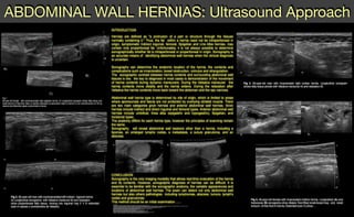
  • 1. ABDOMINAL WALL HERNIAS: Ultrasound Approach REFERENCES 1.B. A. Urban, E. K. Fishman. Tailored Helical CT Evaluation of Acute Abdomen : RadioGraphics, May 1, 2000; 20(3): 725 - 749. 2.T. Rettenbacher, A. Hollerweger, P. Macheiner, N. Gritzmann, T. Gotwald, R. Frass, B. Schneider. Abdominal Wall Hernias: Cross-Sectional Imaging Signs of Incarceration Determined with Sonography. Am. J. Roentgenol;, November 1, 2001; 177(5): 1061 - 1066. 3.Yokoyama T, Munakata Y, Ogiwara M, Kamijima T, Kitamura H, Kawasaki S. Preoperative diagnosis of strangulated obturator hernia using ultrasonography. Am J Surg 1997;174:76 -78 4.Mufid MM, Abu-Yousef MM, Kakish ME, Urdaneta LF, Al-Jurf AS. Spigelian hernia: diagnosis by high-resolution real-time sonography. J Ultrasound Med 1997;16:183 -187 6.Van den Berg JC, Strijk SP. Groin hernia: role of herniography. Radiology 1992;184:191 -194 7.Subramanyam BR, Balthazar EJ, Raghavendra NB, Horii SC, Hilton S. Sonographic diagnosis of scrotal hernia. AJR 1982;139:535 -538 8.Sutphen JH, Hitchcock DA, King DC. Ultrasonic demonstration of Spigelian hernia. AJR 1980;134:174 -175 9.IWantz GE. A 65 year-old man with an inguinal hernia JAMA 1997:277: 663-668 10.Miller PA. Mezwa DG, Feezko PJ, Jafri ZH, Mareazo SL. Imaging of abdominal hernias. Radiographics 1995: 15:333-347. 11.Nehme AE. Groin hernias in elderly patients management and prognosis. Am J Surg. 1983: 146:257 -260. 12.Hodgson T J, Collins Me. Anterior abdominal wall hernias: diagnosis by ultrasound and tangential radiographs. Clin. Radiol.1991 :44185-188. 13.Holder LE, Schneider HJ. Spigelian hernias: anatomy and roentgenography manifestations. Radiology 1974:112309-313. Fig.1 50-year-old female with nonincarcerated right spigelian hernia. A) Longitudinal sonogram shows fatty tissue and small amount of free fluid. B)en un barrido extendido se demuestra mejor la hernia C) con reconstrucción en 3D se observa las diferentes capas compromete la hernia. A) B) C) D)C) Fig 3. 50-year-old man with incarcerated right lumbar hernia. Longitudinal sonogram shows fatty tissue persist with Valsalva maneuver A) and relaxation B) A) B) A) B) C) Fig 4. 46-year-old female with incarcerated midline hernia. Longitudinal (A) and transverse (B) sonograms show dilated, fluid-filled small-bowel loop and small amount of free fluid in hernia. Extended scan C) show ... INTRODUCTION Hernias are defined as "a protrusion of a part or structure through the tissues normally containing it." Thus, the fat within a hernia need not be intraperitoneal in origin, symptomatic Indirect inguinal, femoral, Spigelian and Line Alba hernias, may contain only properitoneal fat. Unfortunately, it is not always possible to determine sonographically whether fat is intraperitoneal or properitoneal in origin. Sonography is an accurate means of identifying abdominal wall hernias when the clinical diagnosis is uncertain. Sonography can determine the anatomic location of the hernia, the contents and complications such as incarceration, bowel obstruction, volvulus and strangulation. The sonographic contrast between hernia contents and surrounding abdominal wall tissues is low, the key to diagnosis in most cases is demonstration of the movement of hernia contents during dynamic maneuvers. During the Valsalva maneuver the hernia contents move distally and the hernia widens. During the relaxation after Valsalva the hernia contents move back toward the abdomen and the sac narrows. Abdominal wall hernia type is determined by site of origin, which is limited to areas where aponeurosis and fascia are not protected by overlying striated muscle. There are two main categories groin hernias and anterior abdominal wall hernias. Groin hernias include indirect and direct inguinal and femoral types. Anterior abdominal wall hernias include umbilical, linea alba (epigastric and hypogastric), Spigelian, and incisional types. The anatomy differs for each hernia type, however the principles of scanning remain the same. Sonography will reveal abdominal wall lessions other than a hernia, including a lipomas, an enlarged lymphs nodes, a metastasis, a suture granuloma, and an abscess. CONCLUSION Sonography is the only imaging modality that allows real-time evaluation of the hernia and its contents. However, sonographic diagnosis of hernias can be difficult, It is essential to be familiar with the sonographic anatomy, the variable appearances and locations of abdominal wall hernias. The exam can detect not only abdominal wall hernias but also others pathologies including lymphomas, abscess, tumors, lymph's nodes and granulomas. This method should be an initial examination ...... A) B) A) B) Fig 2. 65-year-old man with nonincarcerated left indirect inguinal hernia. A) Longitudinal sonograms with Valsalva maneuver B) and relaxation show properitoneal fatty tissue, moving into inguinal ring C Y D extended scan in repose y conmaniobra de Valsalva