SlideShare a Scribd company logo
1 of 57
CIRCULATION
dr.aari
dr.aarif
Circulatory System
Blood Vascular
System
Lymphatic
System
Blood
Heart
Blood Vessels
Lymph
Lymph vessels
Lymph nodes
Functions of circulatory system:
1. Transport of nutrients from the digestive organs to different tissues.
2. Transport of waste products from different tissues to the excretory organs.
3. Transport of respiratory gases namely, O2 from the respiratory organs to the tissues and
CO2 from the tissues to the respiratory organs for its removal.
4. Transport of hormones from the site of origin (i.e. endocrine glands) to the tissues,
which they stimulate (i.e. target-organs).
5. Distribution of water to the tissues to prevent their desiccation (drying).
6. Maintenance of temperature by distributing heat uniformly all over the body.
7. Transport of electrolytes for maintenance of homeostasis in the body.
8. Protection of the body as it contains W.B.C.s, which destroy microorganisms.
9. Besides these, the circulatory fluids help in the clinical diagnosis of variety of diseases
dr.aarif
Blood Vascular
Systems
Open Circulatory System Closed Circulatory System
1. Blood does not remain confined to
blood vessels.
2. Exchange of material takes place
between tissue cells and the blood
directly. There are no veins and
capillaries.
3. Blood of these animals does not contain
respiratory pigment.
4. Hemoglobin is dissolved in plasma and
not in cells. Blood is colourless and is
called haemolymph.
5. Blood pressure is low. Efficiency of
system is less.
6. Open blood vascular system is present
in leech, crab, prawn, lobster, cockroach,
spider, snail, bivalve and herdmania
1. Blood flows through closed blood vessels
and never comes in direct contact with cells.
2. Blood flows through blood vessels like
arteries, veins and capillaries.
3. Exchange of metabolic substances takes
place through the thin wall of capillaries.
4. Blood flows with high blood pressure and
shows high efficiency.
5. Closed blood vascular system is found in
earthworm, sepia and all vertebrates.
dr.aarif
Circulation in Humans
1. The heart (a muscular, contractile organ) pumps
the blood into arteries
2. They branch into smaller vessels called arterioles.
3. The arterioles further divide into many tiny, thin-
walled vessels called capillaries.
4. Nutrients, water, respiratory-gases, electrolytes,
hormones etc. are exchanged between blood and
surrounding tissue cells across the capillary wall.
5. The capillaries unite into small vessels called
venules.
6. The venules join together to form bigger vessels
called, veins which finally returns the blood back
to the heart.
Advantages of closed circulatory system:
1. The blood flows faster in closed blood vessels
than in wide-open spaces and hence a sufficiently
high blood pressure can be maintained (important
for brain, kidney etc.).
2. Due to the rapid flow of blood, the supply and
removal of gases and materials to and fro the
tissues by the blood is quickened.
dr.aarif
BLOOD
1. Blood is a fluid connective tissue derived from the mesoderm.
2. 5-6 liters of blood which is 6% to 8% of body weight .
3. Study of blood is called Haematology
4. It is bright red, slightly alkaline (pH 7.4), salty, viscous fluid heavier than water
PLASMA:
1. 55%of blood, straw coloured, alkaline fluid.
2. 90%- 92% water and 8%-10% suspended and dissolved substances.
3. Plasma proteins (7%)
a) Fibrinogens - clotting or coagulation of blood.
b) Globulins -defence mechanisms of the body
c) Albumin - osmotic balance.
d) Factors for coagulation or clotting of blood are also present in the plasma in an inactive form.
Plasma without the clotting factors is called serum.
4. Other solutes are - nutrients like glucose (0.1%),amino acids, fatty acids and glycerol;
- nitrogenous wastes such as urea, uric acid, ammonia and creatinine;
- gases like oxygen, carbon dioxide nitrogen;
- regulatory substances like enzymes & hormones,
- immune components like antibodies;
- inorganic substances like HCO3 , Cl-, PO4 & SO4 of Na+ , K+, Ca++, Mg++ etc.
BLOOD Plasma (55%) + Cells (45%)=
dr.aarif
Red Blood Corpuscles / Erythrocytes
Structure:
- Circular, biconcave, non-nucleated cells
- Diameter is 7.2 μm and thickness is 2-2.2 μm.
- count in adult males is 5.0 to 5.5 million per mm3
- in adult females is 4.5 to 5.0 million per mm3.
- life span of a single RBC is 100 to 120 days.
Characteristics:
- Formation of RBC's is called erythropoiesis.
- In foetus - yolk sac, kidney, spleen and liver.
- In adults - red bone marrow.
- Destroyed by spleen & liver.
- Iron containing complex protein called haemoglobin
- Hemoglobin has great affinity for oxygen and carbon-di-oxide.
- 13 -18 mg/100 ml of blood in males
- 11.5 -16.5 mg/100 ml of blood.
Function:
- transport of respiratory gases
- maintain blood pH as haemoglobin acts as a buffer.
- maintain the viscosity of blood
dr.aarif
Life Cycle of RBC
White Blood Corpuscles / Leucocytes
Structure:
Colourless, nucleated, amoeboid, phagocytic cells.
They can easily pass through the blood vessels.
This process is called diapedesis.
WBC count is 4000-11000 per cu mm and size is
about 8μ to 15μ.
Life span is3-4 days.
Leukemia is pathological increase in number and is
commonly called blood cancer.
Formation of WBCs is called leucopoiesis.
Leucocytes are produced in red bone marrow,
spleen, lymph nodes, tonsils, thymus, Payer's
patches
WBC - Granulocytes
NEUTROPHILS :
Stained by neutral stain, 60-65% of WBC’s
Nucleus is multi-lobed.
They are the chief phagocytic cells and engulf microorganisms.
BASOPHILS:
Basic stains, about 0.5% of total WBC.
twisted nucleus ,non-phagocytic ,release heparin in the blood stream
along with histamine and serotinin.
EOSINOPHILS (ACIDOPHILS):
stained by acidic stains (like eosin), about 2-3% of total WBC.
Nucleus is bi-lobed.
Non-phagocytic and their number increases during allergic conditions.
They show anti-histaminic property.
Increase of eosinophils is known as eosinophilia
WBC - Agranulocytes LYMPHOCYTES:
large rounded nucleus , about 20-30 % of total WBC's.
produce antibodies and opsonins to neutralize harmful effect of toxins
Two major types – ‘B’ and ‘T’ forms.
Both B and T lymphocytes are responsible for immune responses in body
MONOCYTES :
largest , 6-8 % of the total WBC's.
kidney shaped nucleus.
At the site of infection, enlarge to become macrophages and become
phagocytic and engulf microorganisms.
dr.aarif
THROMBOCYTES:
These are fragments of large cells
called megakaryocytes in the bone
marrow.
They are non-nucleated, round and bi-convex.
These are smallest elements of blood of2.5μm to 5μm.
The average count is about 1.5-3.5 lakhs per cubic millimeter.
Decrease in the number of platelets is called thrombocytopenia
while; increase in number of platelets is called thrombocytosis.
They help in formation of clot over the injury. Thus, loss of
blood is prevented.
A reduction in their number can lead to clotting disorders which
will lead to excessive loss of blood from the body.
BLOOD PLATELETS (THROMBOCYTES)
dr.aarif
FUNCTIONS OF BLOOD
Distribution : supplies digested and absorbed food from alimentary canal to all parts.
Transport: It carries hormones , respiratory gases , nitrogenous wastes
Production of antibodies: Antibodies are produced in response to foreign substances thus
help in defense mechanism.
Thermoregulation: It distributes heat and helps in maintaining body temperature.
Prevention of water loss: It distributes adequate quantity of water to all cells and prevents
dehydration.
Protection: It prevents loss of blood by formation of blood clot.
Phagocytosis by neutrophils and monocytes, which engulf and digest bacteria. Thus, it gives
protection from pathogens.
Osmotic balance: The proteins and minerals in plasma exert osmotic pressure and regulate
blood pressure
dr.aarif
CLOTTING OF BLOOD
BLOOD CLOTTING OR COAGULATION:
- initiated by platelets.
- The injured cells release substances that
attract the platelets.
- They gather at and stick to the injured
surface of the blood vessel.
- The mass of aggregated platelets alone may
physically plug a cut in a very small vessel.
- The contact of platelets with the collagen
fibers exposed by the injury causes them to
disintegrate and release two substances:
serotonin and thromboplastin, which minimize
loss of blood from the injury in two ways.
dr.aarif
CLOTTING OF BLOOD
Vasocontraction:
Serotonin causes the blood vessels at the site of
bleeding to contract.
Clot Formation:
Thromboplastin, a lipoprotein,
helps in clot formation
dr.aarif
CLOTTING OF BLOOD
dr.aarif
BLOOD GROUPS
ABO Blood Groups:
- Karl Landsteiner (1900) recognized four types of blood groups in human beings
(commonly known as ABO blood groups).
- The ABO blood groups are determined by the gene I having three alleles(IA, IB, and IO)
- In human beings, there are present two antigens A and B produced by IA and IB alleles
respectively.
- These antigens are always present on the surface of red blood cells.
- Also are present two antibodies in the plasma a and b
dr.aarif
dr.aarif
Rh (Rhesus) System:
Human blood possesses another factor antigen D i.e. Rh factor.
Accordingly, human blood can be categorized into two groups: Rh-positive and Rh-negative.
Those human beings who had this antigen were termed as Rh-positive (Rh+), (80% of the
population)
The persons, in whom Rh antigen was absent, were designated as Rh-negative (Rh-), (20%
of the population)
It is thought that Rh+ is a dominant character over Rh-which is recessive
ERYTHROBLASTOSIS
FOETALIS
(Haemolytic Disease of
Newborn)
dr.aarif
DOUBLE CIRCULATION
Systemic Circulation:
- The oxygenated blood from the left ventricle of
the heart is sent to the different parts of the
body (except the lungs) via the aorta and its
branches.
- At the same time deoxygenated blood is collected
from all the parts of the body via veins and is
brought back to the right-auricle by the superior
and inferior vena-cavas.
- The blood is carried from the heart to the organs
and back to the heart.
Pulmonary Circulation:
- Deoxygenated blood from the right ventricle is
carried to the lungs via the pulmonary arteries.
- Oxygenation of the blood takes place in the lungs
and oxygenated blood is returned from the lungs
to the left auricle through pulmonary veins.
- Thus blood is carried from the heart to the lungs
and back to the heart.
dr.aarif
HUMAN HEART EXTERNAL STRUCTURE OF THE HEART
Position:
1. Hollow, muscular, conical, pumping-organ
2. situated in the mid-ventral side of the body in
the thoracic cavity between the lungs and
behind the sternum in a space called
mediastinum.
3. Lies slanting a little to the left.
4. The apex (narrow end) is towards the front and
left resting on the diaphragm and base (broad-
end) is directed upwards.
Shape & Size:
1. It is conical in shape and approximately of the
size of one's own-fist.
2. It measures about 12 cm in length, 9 cm in
breadth. It weighs about 300 grams in males
and 250 grams in females
EXTERNAL STRUCTURE OF THE HEARTHUMAN HEART
Bony Protection : sternum ventrally,
vertebral column dorsally
rib cage on the sides
Immediate protection:
By double walled membrane called pericardium.
1. The Fibrous-Pericardium: outer, tough, fibrous
connective tissue covering.
2. The Serous-Pericardium: inner , double-layered,
vascularized covering around the heart.
A. The Parietal layer: outer,
B. The Visceral layer: inner in contact with the
heart and is called the epicardium.
In between the parietal and visceral folds of pericardium,
there is a fluid present called pericardial fluid (about 50
ml).
Functions:
1. Acts as a shock absorber and protects the heart from
mechanical-shocks.
2. Prevents friction between the two-pericardial
membranes.
dr.aarif
dr.aarif
EXTERNAL STRUCTURE OF THE HEARTHUMAN HEART
dr.aarif
EXTERNAL STRUCTURE OF THE HEARTHUMAN HEART
dr.aarif
Chambers:
The human-heart is four chambered, made up of 2 atria and 2 ventricles.
In between the atria and ventricles, there is a transverse groove called the coronary sulcus.
The two ventricles are separated by anterior and posterior inter-ventricular-sulci.
Atria:
They are the upper chambers of the heart.
They are thin-walled and small-chambered.
They form the base of the heart.
Each atrium has a main cavity and an appendage called an auricle
The atria receive blood from the organs to the heart i.e. receiving-chambers.
Right-atrium receives deoxygenated blood from the two major veins:
Superior vena cava, which brings blood from the head region.
Inferior-vena-cava, which brings blood from the body region (except from the lungs and heart
itself)
Left-atrium receives oxygenated blood from the lungs through the four pulmonary veins, two from each lung opening
dorsally in the left atrium.
Ventricles:
They are the lower chambers of the heart.
They arc thick-walled and large-chambered.
The left ventricle is bigger than the right ventricle and forms the apex of the heart.
The ventricles push the blood from the heart to the organs i.e. distributing chambers.
Right-Ventricle pumps the blood (deoxygenated) into the pulmonary-aorta to be carried to the lungs.
Left-Ventricle pumps the blood (oxygenated) into the systemic-aorta to be distributed all over the body.
dr.aarif
Blood-supply of the heart :
Blood vessels are present on the surface of the
ventricles i.e. the coronary arteries and veins.
1. Two coronary arteries arise from the base of the
aorta i.e. the left and right coronary arteries, which
supply oxygenated blood to the muscles of the heart
(seen on ventral-side).
2. The coronary veins collect deoxygenated blood
from the heart muscles, and collect to form the main
coronary vein called coronary sinus and opens into the
right atrium (seen on the dorsal-side)
Embryonic Connection:
The pulmonary trunk and systemic aorta are connected
by ligamentum arteriosum (It is the remnant of ductus
arteriosus a blood vessel, which used to bypass the
blood to the heart in foetus, as the lungs are not
functioning till the child is born.)
After birth once the lungs start functioning, it starts to
close and is completely closed by second month)that
represents remnant of embryonic connection between
the two blood vessels.
dr.aarif
INTERNAL STRUCTURE OF THE HEARTHUMAN HEART
dr.aarif
INTERNAL STRUCTURE OF THE HEARTHUMAN HEART
dr.aarif
INTERNAL STRUCTURE OF THE HEARTHUMAN HEART
THE ATRIA:
upper, thin-walled, small chambers.
separated from each other by inter-atrial septum.
The inter atrial septum shows a shallow saucer shaped depression in the lower part
called as fossa ovalis, which is the remnant of the opening foramen ovale of the foetal
stage when the lungs were not functioning.
Right-Atrium:
openings for the three major-veins:
Superior vena cava bringing deoxygenated blood from the head-region.
Inferior-vena-cava bringing deoxygenated blood from the body-region, guarded by
rudimentary valve of the inferior vena cava (Eustachian valve).
Coronary sinus, the major coronary vein bringing deoxygenated blood from the heart
muscles guarded by the valve of the coronary sinus (Thebesian valve).
Left-Atrium:
This left upper chamber shows 4 openings,
two for the right and two for the left pulmonary veins.
dr.aarif
THE VENTRICLES:
lower, thick-walled and large chambers.
The left ventricle is larger than the right ventricle.
Also, the walls of the left ventricle are thicker than those of the right ventricle. The left
ventricle also forms the apex of the heart.
Both the ventricles are separated from each other by the vertical partition called inter-
ventricular-septum.
The inner surface of both the ventricles is thrown into muscular ridges called trabeculae-
carnae.
Some particular types of these ridges are pillar like and are called papillary-muscles. They
are three and two respectively in the right and left ventricles.
Right-Ventricle:
A pulmonary-trunk arises anteriorly from the right ventricle, and divides into the right and
left pulmonary arteries carrying deoxygenated blood from it to respective lungs.
Left ventricle:
A systemic-aorta arises anteriorly from the left-ventricle carrying oxygenated blood from it
to all the parts of the body.
INTERNAL STRUCTURE OF THE HEARTHUMAN HEART
dr.aarif
INTERNAL STRUCTURE OF THE HEARTHUMAN HEART
Atrio-Ventricular (A-V Valves):
The right atrio-ventricular-aperture is guarded by three
thin leaf -like cusps (flaps) called as the tricuspid valve.
The left atrio-ventricular-aperture is guarded by two
thin leaf- like cusps (flaps) called as the bicuspid
valve(mitral valve).
Each valve is connected by delicate cords (chordae-
tendinae) to one papillary-muscle each of the
ventricles, which prevent the opening of the valves into
the atria.
Semilunar-valves:
The opening of the systemic aorta into the left ventricle
is guarded by three somewhat half-moon shaped cusps
(flaps) which is called as the aortic-semilunar-valve.
Similarly the opening of the pulmonary aorta into the
right ventricle is guarded by the pulmonary-semilunar-
valve. TRICUSPID VALVE
dr.aarif
CARDIAC CYCLE
•The blood is kept in a constant circulation
through blood vessels by the pumping
action or beating of the heart throughout
life.
•The rhythmic contraction of the heart is
called systole followed by its dilatation is
called diastole.
•One complete systole and diastole of the
heart makes one heartbeat or cardiac-
cycle.
•During each heartbeat, the chambers of
the heart contract and relax in a specific
sequence and are repeated in a cyclic
manner.
These movements take place in a definite 3 phases:
Phase I: Atrial-systole (simultaneous contraction of
both the atria) lasting about 0.1 sec.
Phase II: Ventricular systole (simultaneous
contraction of both the ventricles) lasting about0.3
sec.
Phase III:Joint-diastole (simultaneous relaxation of
both, the 2 atria and the 2 ventricles) lasting about
0.4 sec.
dr.aarif
CARDIAC CYCLE
Phase I:
Both the atria simultaneously contract(the
ventricles are still relaxing).
As a result of which the A-V valves completely
open and both the ventricles get filled with
blood(flow increases by 30 %).
The atrial muscles contract and constrict the
openings of the vena-cavas and pulmonary
veins and prevent the backflow of blood into
these vessels.
dr.aarif
CARDIAC CYCLE
Phase II:
It is followed immediately by the ventricular systole
(contraction), while the atria start relaxing.
The bicuspid and tricuspid valves now close sharply
to prevent the backflow of blood into the atria.
Their closure produces the first heart sound (lubb) a
fairly loud sound.
Due to the increased pressure in the ventricles, the
blood tries to push both the A-V valves in the
upward direction.
The papillary muscles contract and there is a pulling
of the cusps by the stretched chordae-tendinae in
the downward direction.
dr.aarif
CARDIAC CYCLE
Due to these opposite actions, the opening of
the valves into the atria is prevented. Thus the
backflow of blood into the atria is prevented.
The semilunar valves now open as the
pressure in the ventricles increases and the
deoxygenated blood from the right ventricle
enters the pulmonary artery and goes to the
lungs while the oxygenated blood from the left
ventricle enters the aorta and goes to all the
parts of the body.
.
dr.aarif
Phase III (joint-diastole):
The ventricles relax now.
As the ventricular pressure decreases the semilunar
valves close to prevent the backflow of blood from
the vessels into the ventricles.
Their closure produces the second heart sound
(dubb).
The atria are already relaxing at this point (hence
joint-diastole) and receiving blood from the vena-
cavas and pulmonary veins. The pressure of blood in
the atria keeps on increasing.
The ventricles are relaxing as 'closed chambers' (i.e.
without any blood in them and all the valves closed)
and hence the pressure in the ventricles decreases
rapidly. Thus the pressure in the atria rises more
than in the ventricles.
Hence the A-V valves slightly open and blood
trickles from the atria into the ventricles again and a
new cycle is repeated.
dr.aarif
dr.aarif
CONDUCTING SYSTEM OF THE HEARTHUMAN HEART
dr.aarif
CONDUCTING SYSTEM OF THE HEARTHUMAN HEART
dr.aarif
NERVOUS CONTROL OF HEARTHUMAN HEART
dr.aarif
ELECTROCARDIOGRAM (ECG)HUMAN HEART
dr.aarif
ELECTROCARDIOGRAM (ECG)HUMAN HEART
The ECG is a composite record of action
potentials produced byall the heart muscle
fibers during each heartbeat
1. In clinical practice, electrodes are positioned
on the arms and legs (limb leads) and at six
positions on the chest (chest leads) to
record the ECG.
2. The electrocardiograph amplifies the
heart’s electrical signals and produces 12
different tracings from different
combinations of limb and chest leads.
3. Each limb and chest electrode records
slightly different electrical activity because
of the difference in its position relative to
the heart.
4. By comparing these records with one
another and with normal records, it is
possible to determine
(a) if the conducting pathway is abnormal,
(b) if the heart is enlarged,
(c) if certain regions of the heart damaged
(d) the cause of chest pain.
dr.aarif
ELECTROCARDIOGRAM (ECG)HUMAN HEART
dr.aarif
ELECTROCARDIOGRAM (ECG)HUMAN HEART
dr.aarif
ELECTROCARDIOGRAM (ECG)HUMAN HEART
In reading an ECG, the size of the waves can provide clues to abnormalities.
1. Larger P waves indicate enlargement of an atrium;
2. an enlarged Q wave may indicate a myocardial infarction;
3. enlarged R wave generally indicates enlarged ventricles.
4. The T wave is flatter than normal when the heart muscle is receiving insufficient oxygen—as, for
example, in coronary artery disease.
5. The T wave may be elevated in hyperkalemia (high blood K level).
Analysis of an ECG also involves measuring the time spans between waves, which are called intervals or
segments.
1. The P-Q interval is the time from the beginning of the P wave to the beginning of the QRS complex. It
represents the conduction time from the beginning of atrial excitation to the beginning of ventricular
excitation. Put another way, the P-Q interval is the time required for the action potential to travel
through the atria, atrio ventricular node, and the remaining fibers of the conduction system. As the
action potential is forced to detour around scar tissue caused by disorders such as coronary artery
disease and rheumatic fever, the P-Q interval lengthens.
2. The S-T segment, which begins at the end of the S wave and ends at the beginning of the T wave,
represents the time when the ventricular contractile fibers are depolarized during the plateau phase of
the action potential. The S-T segment is elevated (above the baseline) in acute myocardial infarction
and depressed (below the baseline) when the heart muscle receives insufficient oxygen.
3. The Q-T interval extends from the start of the QRS complex to the end of the T wave. It is the time from
the beginning of ventricular depolarization to the end of ventricular repolarization. The Q-T interval
may be lengthened by myocardial damage, myocardial ischemia (decreased blood flow), or
conduction abnormalities.
dr.aarif
STRUCTURE OF ARTEY & VEIN
dr.aarif
BLOOD PRESSURE
Definition:
It is defined as ‘the lateral pressure exerted by a column of blood against the wall of artery’.
Measurement:
It is usually measured as the blood pressure in the artery of the elbow region, by an
mercury filled instrument called sphygmomanometer(sphygmos= pulse, manometer =
device to measure pressure).
Hence measurements are in units of mm of Hg.
Systolic pressure : 120 mm Hg
Diastolic pressure : 80 mm Hg
dr.aarif
FACTORS AFFECTING BLOOD PRESSURE
BLOOD
PRESSURE
Total blood volume
Cardiac output
Viscosity of blood
Physiological factors
Neuro – endocrine
Peripheral resistance
Venous return
Elasticity of arteries
HYPERTENSION: (I.E. HIGH BLOOD PRESSURE):
Systolic pressure > 140 mm of Hg & Diastolic pressure > 90 mm of Hg
Causes:
- Hereditary.
- High intake of salt.
- Excessive smoking.
- Physical or emotional stress.
- Obesity (excessively fat) leading to fatty deposits in inner walls of arteries in the old age
(atherosclerosis) causing loss of elasticity of arteries. (i.e. arteriosclerosis).
- Kidney diseases (increases secretion of renin, epinephrine or aldosterone).
Symptoms:
- Throbbing headache.
- Sweating.
- Palpitations of the heart.
- Dizziness.
- Exhaustion.
Complications:
- Heart attack (i.e. myocardial infarction).
- Stroke (cerebro vascular accidents) leading to paralysis.
- Kidney-failure (nephritis).
- Rupturing of blood vessels of the eyes (causing blindness).
dr.aarif
HIGH BLOOD PRESSURE (Hypertension)
dr.aarif
HYPOTENSION: (I.E. LOW BLOOD PRESSURE):
- Systolic pressure < 110 mm of Hg
- Diastolic pressure < 60 mm of Hg
Causes:
- Chronic anemia.
- Disorder of nerves (neuropathy).
- Disorder of hormones (hypothyroidism).
- Disease like tuberculosis and cancer.
Complications:
- Giddiness.
- Syncope (i.e. temporary loss of consciousness)
LOW BLOOD PRESSURE (Hypotension)
dr.aarif
CARDIOVASCULAR DISORDERS
CORONARY ARTERY DISEASE (CAD):
- Coronary Artery Disease, often referred to as
atherosclerosis, causes narrowing of coronary
arteries so that the blood flow to the heart is
reduced.
- This results in coronary heart disease, a
condition in which the heart muscle is
damaged because of an inadequate amount
blood and thus oxygen due to obstruction of
its blood supply. Depending on the degree of
obstruction, symptoms may be mild chest pain
(angina pectoris) or heart attack (myocardial
infarction).
- Atherosclerosis refers to the deposition of
substances in the lining of arteries. Deposits of
calcium, fat, cholesterol and fibrous tissues
cause it, which makes the lumen of arteries
narrower. This results in the formation of an
atherosclerotic plaque that decreases the size
of the arterial lumen.
dr.aarif
ANGINA:
- It is also called ‘angina pectoris’.
- A symptom of acute chest pain appears,
when not enough oxygen is reaching the
heart muscle because of a reduction in
blood supply to cardiac muscle due to
narrowed and hardened coronary arteries
(arteriosclerosis).
- It is characterized by heaviness and severe
pain in the chest. The pain can also be
referred to the neck, lower jaw, left arm
and left shoulder.
- Angina pectoris often occurs during exer-
tion, when the heart demands more oxygen
and narrowed blood vessels cannot supply.
- It disappears with rest.
- Angina can occur in men and women of any
age but it is more common among the
middle-aged and elders
CARDIOVASCULAR DISORDERS
dr.aarif
HEART FAILURE:
- Heart failure is the result of progressive weakening
of heart muscles and hence not able to pump
blood effectively enough to meet the needs of the
body.
Causes :
- Hypertension increases the load on the heart, can
produce significant enlargement of the heart
(cardiomegaly), and can finally result in heart
failure.
- Advanced age, malnutrition, chronic infections,
toxins, severe anaemia or hyperthyroidism can
cause degeneration of heart muscle, resulting in
heart failure.
- It is sometimes called congestive cardiac failure
(c.c.f) because congestion of the lungs is one of the
main symptoms of this disease.
- Heart failure is not the same as cardiac arrest
(when the heart stops beating due to a defect in the
electrical conducting system of the heart) or a
heart attack (when the heart muscle is suddenly
damaged by an inadequate blood supply, due to
blocks in the coronary artery
CARDIOVASCULAR DISORDERS
dr.aarif
LYMPHATIC SYSYTEM
dr.aarif
LYMPHATIC SYSYTEM
Lymph:
- It is a colourless, extracellular, extra-vascular, liquid connective tissue.
- As the blood passes through the capillaries in tissues, some water with many small
water soluble substances move out, in between the cells of tissues leaving the larger
proteins and most of the cells (RBCs) in the blood vessels.
- The tissue fluid that bathes the cells is called lymph. It is collected in lymphatic
capillaries.
- Lymph is blood minus RBCs, platelets and some plasma proteins. It contains carbon
dioxide and metabolic wastes.
dr.aarif
LYMPHATIC SYSYTEM
Lymphatic capillaries :
- These are thin walled vessels present in all the tissue spaces.
- They are interwoven with the blood capillaries, but are not connected with them.
- They are blind at one end and are wider than blood capillaries. Each is lined by
endothelium of thin and flat cells.
dr.aarif
LYMPHATIC SYSYTEM
Lymphatic vessels :
- Lymphatic capillaries unite to form larger tubes called lymphatic vessels. These have
thinner walls and numerous valves to prevent backflow.
- The two main lymph vessels are - thoracic or left lymphatic duct and right lymphatic
duct.
- The thoracic duct is the main collecting duct of the lymphatic system. It receives lymph
from left side of the head, neck, chest, left upper extremity and entire body below the
ribs. The lymphatic vessel coming from the intestinal villi contains absorbed fats. Hence
they are milky in appearance and are called lacteals (L. lactis- milk).
- The right lymphatic duct drains lymph from the upper right side of the body.
dr.aarif
LYMPHATIC SYSYTEM
Lymph nodes :
- These are small, oval or bean shaped bodies placed in the course of lymphatic
vessels.
- Lymph nodes are scattered throughout the body but are maximum in neck. armpit
and groin.
- They act as filters, as macrophages of lymph nodes remove bacteria, foreign material
and cell debris.
- They also produce lymphocytes and antibodies.

More Related Content

What's hot

The Respiratory System
The Respiratory SystemThe Respiratory System
The Respiratory Systemmpattani
 
Circulatory System.ppt
Circulatory System.pptCirculatory System.ppt
Circulatory System.pptShama
 
The circulatory system
The circulatory systemThe circulatory system
The circulatory systemMike Tamas
 
the cardiovascular system and Physiology of heart
the cardiovascular system and Physiology of heartthe cardiovascular system and Physiology of heart
the cardiovascular system and Physiology of heartbhupendra kumar
 
Circulatory system
Circulatory system Circulatory system
Circulatory system X-tian Mike
 
arteries, arterioles, veins, venules, capillary and fluid or gaseous exchange...
arteries, arterioles, veins, venules, capillary and fluid or gaseous exchange...arteries, arterioles, veins, venules, capillary and fluid or gaseous exchange...
arteries, arterioles, veins, venules, capillary and fluid or gaseous exchange...Nikita Sharma
 
Digestive system - anatomy
Digestive system - anatomy   Digestive system - anatomy
Digestive system - anatomy Areej Abu Hanieh
 
Transport system in humans
Transport system in humansTransport system in humans
Transport system in humansFaiz Abdullah
 
Blood vessels: Arteries, Veins and Capillaries
Blood vessels: Arteries, Veins and CapillariesBlood vessels: Arteries, Veins and Capillaries
Blood vessels: Arteries, Veins and CapillariesAmir Rifaat
 
Bloodvessels
BloodvesselsBloodvessels
Bloodvesselsscuffruff
 
Circulatory system
Circulatory systemCirculatory system
Circulatory systemjvcole1
 
The circulatory system complete Info ppt
The circulatory system complete Info pptThe circulatory system complete Info ppt
The circulatory system complete Info pptDr. Armaan Singh
 

What's hot (20)

Powerpoint circulatory system
Powerpoint circulatory systemPowerpoint circulatory system
Powerpoint circulatory system
 
Muscles
MusclesMuscles
Muscles
 
The Respiratory System
The Respiratory SystemThe Respiratory System
The Respiratory System
 
Circulatory System.ppt
Circulatory System.pptCirculatory System.ppt
Circulatory System.ppt
 
The circulatory system
The circulatory systemThe circulatory system
The circulatory system
 
General physiology
General physiologyGeneral physiology
General physiology
 
the cardiovascular system and Physiology of heart
the cardiovascular system and Physiology of heartthe cardiovascular system and Physiology of heart
the cardiovascular system and Physiology of heart
 
Circulatory system
Circulatory system Circulatory system
Circulatory system
 
arteries, arterioles, veins, venules, capillary and fluid or gaseous exchange...
arteries, arterioles, veins, venules, capillary and fluid or gaseous exchange...arteries, arterioles, veins, venules, capillary and fluid or gaseous exchange...
arteries, arterioles, veins, venules, capillary and fluid or gaseous exchange...
 
Circulatory System
Circulatory SystemCirculatory System
Circulatory System
 
Digestive system - anatomy
Digestive system - anatomy   Digestive system - anatomy
Digestive system - anatomy
 
Transport system in humans
Transport system in humansTransport system in humans
Transport system in humans
 
Digestive system
Digestive systemDigestive system
Digestive system
 
Cardiac muscle tissue
Cardiac muscle tissueCardiac muscle tissue
Cardiac muscle tissue
 
Blood vessels: Arteries, Veins and Capillaries
Blood vessels: Arteries, Veins and CapillariesBlood vessels: Arteries, Veins and Capillaries
Blood vessels: Arteries, Veins and Capillaries
 
Bloodvessels
BloodvesselsBloodvessels
Bloodvessels
 
Human heart
Human heartHuman heart
Human heart
 
Circulatory system
Circulatory systemCirculatory system
Circulatory system
 
2.1 blood circulation
2.1 blood circulation2.1 blood circulation
2.1 blood circulation
 
The circulatory system complete Info ppt
The circulatory system complete Info pptThe circulatory system complete Info ppt
The circulatory system complete Info ppt
 

Similar to Circulation

Human circulatory system
Human circulatory systemHuman circulatory system
Human circulatory systemMohdMafuz1
 
04 Blood.ppt
04 Blood.ppt04 Blood.ppt
04 Blood.pptShama
 
1854964 635072095191071250
1854964 6350720951910712501854964 635072095191071250
1854964 635072095191071250Omkar Khandagale
 
Forensic Existence of Blood as a Dynamic Evidence
Forensic Existence of Blood as a Dynamic EvidenceForensic Existence of Blood as a Dynamic Evidence
Forensic Existence of Blood as a Dynamic Evidenceijtsrd
 
Circulatory system
Circulatory systemCirculatory system
Circulatory systemlaxmi3112
 
Chapter 20 Blood Circulatory System
Chapter 20 Blood Circulatory System Chapter 20 Blood Circulatory System
Chapter 20 Blood Circulatory System mae dessa matuguina
 
Anatomy of Circulatory system and lymphatic system
Anatomy of Circulatory system and lymphatic system Anatomy of Circulatory system and lymphatic system
Anatomy of Circulatory system and lymphatic system DR .PALLAVI PATHANIA
 
CLASS 11 TH Body fluids and circulation
 CLASS 11 TH Body fluids and circulation CLASS 11 TH Body fluids and circulation
CLASS 11 TH Body fluids and circulationDHARUN MUGHILAN
 
Chapter 6
Chapter 6Chapter 6
Chapter 6msefah
 
ANATOMY AND PHYSIOLOGY II.ppt
ANATOMY AND PHYSIOLOGY II.pptANATOMY AND PHYSIOLOGY II.ppt
ANATOMY AND PHYSIOLOGY II.pptCentiBlogs
 
6 anatomy circulatory system
6 anatomy circulatory system6 anatomy circulatory system
6 anatomy circulatory systemHome
 

Similar to Circulation (20)

Human circulatory system
Human circulatory systemHuman circulatory system
Human circulatory system
 
04 Blood.ppt
04 Blood.ppt04 Blood.ppt
04 Blood.ppt
 
Q & A. on Physiology for Exam 17november13
Q & A.  on  Physiology for Exam 17november13Q & A.  on  Physiology for Exam 17november13
Q & A. on Physiology for Exam 17november13
 
1854964 635072095191071250
1854964 6350720951910712501854964 635072095191071250
1854964 635072095191071250
 
Forensic Existence of Blood as a Dynamic Evidence
Forensic Existence of Blood as a Dynamic EvidenceForensic Existence of Blood as a Dynamic Evidence
Forensic Existence of Blood as a Dynamic Evidence
 
Circulatory system
Circulatory systemCirculatory system
Circulatory system
 
CIRCULATORY SYSTEM
CIRCULATORY SYSTEMCIRCULATORY SYSTEM
CIRCULATORY SYSTEM
 
Chapter 20 Blood Circulatory System
Chapter 20 Blood Circulatory System Chapter 20 Blood Circulatory System
Chapter 20 Blood Circulatory System
 
Anatomy of Circulatory system and lymphatic system
Anatomy of Circulatory system and lymphatic system Anatomy of Circulatory system and lymphatic system
Anatomy of Circulatory system and lymphatic system
 
blood-conversion
blood-conversionblood-conversion
blood-conversion
 
CLASS 11 TH Body fluids and circulation
 CLASS 11 TH Body fluids and circulation CLASS 11 TH Body fluids and circulation
CLASS 11 TH Body fluids and circulation
 
Chapter 6
Chapter 6Chapter 6
Chapter 6
 
Blood
BloodBlood
Blood
 
BLOOD ppt.pptx
BLOOD ppt.pptxBLOOD ppt.pptx
BLOOD ppt.pptx
 
Blood Anatomy and Physiology
Blood Anatomy and PhysiologyBlood Anatomy and Physiology
Blood Anatomy and Physiology
 
ANATOMY AND PHYSIOLOGY II.ppt
ANATOMY AND PHYSIOLOGY II.pptANATOMY AND PHYSIOLOGY II.ppt
ANATOMY AND PHYSIOLOGY II.ppt
 
hematologi
hematologihematologi
hematologi
 
Blood chapter.ppt
Blood chapter.pptBlood chapter.ppt
Blood chapter.ppt
 
6 anatomy circulatory system
6 anatomy circulatory system6 anatomy circulatory system
6 anatomy circulatory system
 
Blood
BloodBlood
Blood
 

More from Aarif Kanadia

More from Aarif Kanadia (14)

Morphology of flowering plants part2
Morphology of flowering plants  part2Morphology of flowering plants  part2
Morphology of flowering plants part2
 
Reproduction in plants
Reproduction in plantsReproduction in plants
Reproduction in plants
 
Earthworm
EarthwormEarthworm
Earthworm
 
Frog
FrogFrog
Frog
 
Morphology of flowering plants - I (root, stem & leaf)
Morphology of flowering plants - I (root, stem & leaf)Morphology of flowering plants - I (root, stem & leaf)
Morphology of flowering plants - I (root, stem & leaf)
 
Study of animal tissue
Study of animal tissueStudy of animal tissue
Study of animal tissue
 
Excretion and osmoregulation
Excretion and osmoregulationExcretion and osmoregulation
Excretion and osmoregulation
 
Kingdom plantae
Kingdom plantaeKingdom plantae
Kingdom plantae
 
Animal types cockroach
Animal types cockroachAnimal types cockroach
Animal types cockroach
 
Human respiration
Human respirationHuman respiration
Human respiration
 
Biomolecules
BiomoleculesBiomolecules
Biomolecules
 
Joints
JointsJoints
Joints
 
Skeleton
SkeletonSkeleton
Skeleton
 
Human nutrition
Human nutritionHuman nutrition
Human nutrition
 

Recently uploaded

1029 - Danh muc Sach Giao Khoa 10 . pdf
1029 -  Danh muc Sach Giao Khoa 10 . pdf1029 -  Danh muc Sach Giao Khoa 10 . pdf
1029 - Danh muc Sach Giao Khoa 10 . pdfQucHHunhnh
 
How to Make a Pirate ship Primary Education.pptx
How to Make a Pirate ship Primary Education.pptxHow to Make a Pirate ship Primary Education.pptx
How to Make a Pirate ship Primary Education.pptxmanuelaromero2013
 
microwave assisted reaction. General introduction
microwave assisted reaction. General introductionmicrowave assisted reaction. General introduction
microwave assisted reaction. General introductionMaksud Ahmed
 
POINT- BIOCHEMISTRY SEM 2 ENZYMES UNIT 5.pptx
POINT- BIOCHEMISTRY SEM 2 ENZYMES UNIT 5.pptxPOINT- BIOCHEMISTRY SEM 2 ENZYMES UNIT 5.pptx
POINT- BIOCHEMISTRY SEM 2 ENZYMES UNIT 5.pptxSayali Powar
 
Arihant handbook biology for class 11 .pdf
Arihant handbook biology for class 11 .pdfArihant handbook biology for class 11 .pdf
Arihant handbook biology for class 11 .pdfchloefrazer622
 
Employee wellbeing at the workplace.pptx
Employee wellbeing at the workplace.pptxEmployee wellbeing at the workplace.pptx
Employee wellbeing at the workplace.pptxNirmalaLoungPoorunde1
 
Software Engineering Methodologies (overview)
Software Engineering Methodologies (overview)Software Engineering Methodologies (overview)
Software Engineering Methodologies (overview)eniolaolutunde
 
Introduction to AI in Higher Education_draft.pptx
Introduction to AI in Higher Education_draft.pptxIntroduction to AI in Higher Education_draft.pptx
Introduction to AI in Higher Education_draft.pptxpboyjonauth
 
Sanyam Choudhary Chemistry practical.pdf
Sanyam Choudhary Chemistry practical.pdfSanyam Choudhary Chemistry practical.pdf
Sanyam Choudhary Chemistry practical.pdfsanyamsingh5019
 
Accessible design: Minimum effort, maximum impact
Accessible design: Minimum effort, maximum impactAccessible design: Minimum effort, maximum impact
Accessible design: Minimum effort, maximum impactdawncurless
 
18-04-UA_REPORT_MEDIALITERAСY_INDEX-DM_23-1-final-eng.pdf
18-04-UA_REPORT_MEDIALITERAСY_INDEX-DM_23-1-final-eng.pdf18-04-UA_REPORT_MEDIALITERAСY_INDEX-DM_23-1-final-eng.pdf
18-04-UA_REPORT_MEDIALITERAСY_INDEX-DM_23-1-final-eng.pdfssuser54595a
 
Presentation by Andreas Schleicher Tackling the School Absenteeism Crisis 30 ...
Presentation by Andreas Schleicher Tackling the School Absenteeism Crisis 30 ...Presentation by Andreas Schleicher Tackling the School Absenteeism Crisis 30 ...
Presentation by Andreas Schleicher Tackling the School Absenteeism Crisis 30 ...EduSkills OECD
 
Measures of Central Tendency: Mean, Median and Mode
Measures of Central Tendency: Mean, Median and ModeMeasures of Central Tendency: Mean, Median and Mode
Measures of Central Tendency: Mean, Median and ModeThiyagu K
 
Separation of Lanthanides/ Lanthanides and Actinides
Separation of Lanthanides/ Lanthanides and ActinidesSeparation of Lanthanides/ Lanthanides and Actinides
Separation of Lanthanides/ Lanthanides and ActinidesFatimaKhan178732
 
URLs and Routing in the Odoo 17 Website App
URLs and Routing in the Odoo 17 Website AppURLs and Routing in the Odoo 17 Website App
URLs and Routing in the Odoo 17 Website AppCeline George
 

Recently uploaded (20)
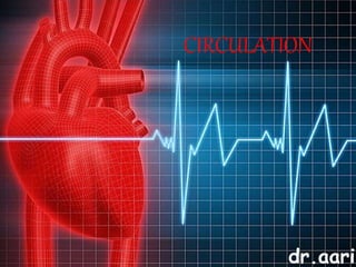
1029 - Danh muc Sach Giao Khoa 10 . pdf
1029 -  Danh muc Sach Giao Khoa 10 . pdf1029 -  Danh muc Sach Giao Khoa 10 . pdf
1029 - Danh muc Sach Giao Khoa 10 . pdf
 
Staff of Color (SOC) Retention Efforts DDSD
Staff of Color (SOC) Retention Efforts DDSDStaff of Color (SOC) Retention Efforts DDSD
Staff of Color (SOC) Retention Efforts DDSD
 
TataKelola dan KamSiber Kecerdasan Buatan v022.pdf
TataKelola dan KamSiber Kecerdasan Buatan v022.pdfTataKelola dan KamSiber Kecerdasan Buatan v022.pdf
TataKelola dan KamSiber Kecerdasan Buatan v022.pdf
 
How to Make a Pirate ship Primary Education.pptx
How to Make a Pirate ship Primary Education.pptxHow to Make a Pirate ship Primary Education.pptx
How to Make a Pirate ship Primary Education.pptx
 
microwave assisted reaction. General introduction
microwave assisted reaction. General introductionmicrowave assisted reaction. General introduction
microwave assisted reaction. General introduction
 
POINT- BIOCHEMISTRY SEM 2 ENZYMES UNIT 5.pptx
POINT- BIOCHEMISTRY SEM 2 ENZYMES UNIT 5.pptxPOINT- BIOCHEMISTRY SEM 2 ENZYMES UNIT 5.pptx
POINT- BIOCHEMISTRY SEM 2 ENZYMES UNIT 5.pptx
 
Arihant handbook biology for class 11 .pdf
Arihant handbook biology for class 11 .pdfArihant handbook biology for class 11 .pdf
Arihant handbook biology for class 11 .pdf
 
Mattingly "AI & Prompt Design: Structured Data, Assistants, & RAG"
Mattingly "AI & Prompt Design: Structured Data, Assistants, & RAG"Mattingly "AI & Prompt Design: Structured Data, Assistants, & RAG"
Mattingly "AI & Prompt Design: Structured Data, Assistants, & RAG"
 
Código Creativo y Arte de Software | Unidad 1
Código Creativo y Arte de Software | Unidad 1Código Creativo y Arte de Software | Unidad 1
Código Creativo y Arte de Software | Unidad 1
 
INDIA QUIZ 2024 RLAC DELHI UNIVERSITY.pptx
INDIA QUIZ 2024 RLAC DELHI UNIVERSITY.pptxINDIA QUIZ 2024 RLAC DELHI UNIVERSITY.pptx
INDIA QUIZ 2024 RLAC DELHI UNIVERSITY.pptx
 
Employee wellbeing at the workplace.pptx
Employee wellbeing at the workplace.pptxEmployee wellbeing at the workplace.pptx
Employee wellbeing at the workplace.pptx
 
Software Engineering Methodologies (overview)
Software Engineering Methodologies (overview)Software Engineering Methodologies (overview)
Software Engineering Methodologies (overview)
 
Introduction to AI in Higher Education_draft.pptx
Introduction to AI in Higher Education_draft.pptxIntroduction to AI in Higher Education_draft.pptx
Introduction to AI in Higher Education_draft.pptx
 
Sanyam Choudhary Chemistry practical.pdf
Sanyam Choudhary Chemistry practical.pdfSanyam Choudhary Chemistry practical.pdf
Sanyam Choudhary Chemistry practical.pdf
 
Accessible design: Minimum effort, maximum impact
Accessible design: Minimum effort, maximum impactAccessible design: Minimum effort, maximum impact
Accessible design: Minimum effort, maximum impact
 
18-04-UA_REPORT_MEDIALITERAСY_INDEX-DM_23-1-final-eng.pdf
18-04-UA_REPORT_MEDIALITERAСY_INDEX-DM_23-1-final-eng.pdf18-04-UA_REPORT_MEDIALITERAСY_INDEX-DM_23-1-final-eng.pdf
18-04-UA_REPORT_MEDIALITERAСY_INDEX-DM_23-1-final-eng.pdf
 
Presentation by Andreas Schleicher Tackling the School Absenteeism Crisis 30 ...
Presentation by Andreas Schleicher Tackling the School Absenteeism Crisis 30 ...Presentation by Andreas Schleicher Tackling the School Absenteeism Crisis 30 ...
Presentation by Andreas Schleicher Tackling the School Absenteeism Crisis 30 ...
 
Measures of Central Tendency: Mean, Median and Mode
Measures of Central Tendency: Mean, Median and ModeMeasures of Central Tendency: Mean, Median and Mode
Measures of Central Tendency: Mean, Median and Mode
 
Separation of Lanthanides/ Lanthanides and Actinides
Separation of Lanthanides/ Lanthanides and ActinidesSeparation of Lanthanides/ Lanthanides and Actinides
Separation of Lanthanides/ Lanthanides and Actinides
 
URLs and Routing in the Odoo 17 Website App
URLs and Routing in the Odoo 17 Website AppURLs and Routing in the Odoo 17 Website App
URLs and Routing in the Odoo 17 Website App
 

Circulation

  • 2. dr.aarif Circulatory System Blood Vascular System Lymphatic System Blood Heart Blood Vessels Lymph Lymph vessels Lymph nodes Functions of circulatory system: 1. Transport of nutrients from the digestive organs to different tissues. 2. Transport of waste products from different tissues to the excretory organs. 3. Transport of respiratory gases namely, O2 from the respiratory organs to the tissues and CO2 from the tissues to the respiratory organs for its removal. 4. Transport of hormones from the site of origin (i.e. endocrine glands) to the tissues, which they stimulate (i.e. target-organs). 5. Distribution of water to the tissues to prevent their desiccation (drying). 6. Maintenance of temperature by distributing heat uniformly all over the body. 7. Transport of electrolytes for maintenance of homeostasis in the body. 8. Protection of the body as it contains W.B.C.s, which destroy microorganisms. 9. Besides these, the circulatory fluids help in the clinical diagnosis of variety of diseases
  • 3. dr.aarif Blood Vascular Systems Open Circulatory System Closed Circulatory System 1. Blood does not remain confined to blood vessels. 2. Exchange of material takes place between tissue cells and the blood directly. There are no veins and capillaries. 3. Blood of these animals does not contain respiratory pigment. 4. Hemoglobin is dissolved in plasma and not in cells. Blood is colourless and is called haemolymph. 5. Blood pressure is low. Efficiency of system is less. 6. Open blood vascular system is present in leech, crab, prawn, lobster, cockroach, spider, snail, bivalve and herdmania 1. Blood flows through closed blood vessels and never comes in direct contact with cells. 2. Blood flows through blood vessels like arteries, veins and capillaries. 3. Exchange of metabolic substances takes place through the thin wall of capillaries. 4. Blood flows with high blood pressure and shows high efficiency. 5. Closed blood vascular system is found in earthworm, sepia and all vertebrates.
  • 4. dr.aarif Circulation in Humans 1. The heart (a muscular, contractile organ) pumps the blood into arteries 2. They branch into smaller vessels called arterioles. 3. The arterioles further divide into many tiny, thin- walled vessels called capillaries. 4. Nutrients, water, respiratory-gases, electrolytes, hormones etc. are exchanged between blood and surrounding tissue cells across the capillary wall. 5. The capillaries unite into small vessels called venules. 6. The venules join together to form bigger vessels called, veins which finally returns the blood back to the heart. Advantages of closed circulatory system: 1. The blood flows faster in closed blood vessels than in wide-open spaces and hence a sufficiently high blood pressure can be maintained (important for brain, kidney etc.). 2. Due to the rapid flow of blood, the supply and removal of gases and materials to and fro the tissues by the blood is quickened.
  • 5. dr.aarif BLOOD 1. Blood is a fluid connective tissue derived from the mesoderm. 2. 5-6 liters of blood which is 6% to 8% of body weight . 3. Study of blood is called Haematology 4. It is bright red, slightly alkaline (pH 7.4), salty, viscous fluid heavier than water PLASMA: 1. 55%of blood, straw coloured, alkaline fluid. 2. 90%- 92% water and 8%-10% suspended and dissolved substances. 3. Plasma proteins (7%) a) Fibrinogens - clotting or coagulation of blood. b) Globulins -defence mechanisms of the body c) Albumin - osmotic balance. d) Factors for coagulation or clotting of blood are also present in the plasma in an inactive form. Plasma without the clotting factors is called serum. 4. Other solutes are - nutrients like glucose (0.1%),amino acids, fatty acids and glycerol; - nitrogenous wastes such as urea, uric acid, ammonia and creatinine; - gases like oxygen, carbon dioxide nitrogen; - regulatory substances like enzymes & hormones, - immune components like antibodies; - inorganic substances like HCO3 , Cl-, PO4 & SO4 of Na+ , K+, Ca++, Mg++ etc. BLOOD Plasma (55%) + Cells (45%)=
  • 6. dr.aarif Red Blood Corpuscles / Erythrocytes Structure: - Circular, biconcave, non-nucleated cells - Diameter is 7.2 μm and thickness is 2-2.2 μm. - count in adult males is 5.0 to 5.5 million per mm3 - in adult females is 4.5 to 5.0 million per mm3. - life span of a single RBC is 100 to 120 days. Characteristics: - Formation of RBC's is called erythropoiesis. - In foetus - yolk sac, kidney, spleen and liver. - In adults - red bone marrow. - Destroyed by spleen & liver. - Iron containing complex protein called haemoglobin - Hemoglobin has great affinity for oxygen and carbon-di-oxide. - 13 -18 mg/100 ml of blood in males - 11.5 -16.5 mg/100 ml of blood. Function: - transport of respiratory gases - maintain blood pH as haemoglobin acts as a buffer. - maintain the viscosity of blood
  • 8. White Blood Corpuscles / Leucocytes Structure: Colourless, nucleated, amoeboid, phagocytic cells. They can easily pass through the blood vessels. This process is called diapedesis. WBC count is 4000-11000 per cu mm and size is about 8μ to 15μ. Life span is3-4 days. Leukemia is pathological increase in number and is commonly called blood cancer. Formation of WBCs is called leucopoiesis. Leucocytes are produced in red bone marrow, spleen, lymph nodes, tonsils, thymus, Payer's patches
  • 9. WBC - Granulocytes NEUTROPHILS : Stained by neutral stain, 60-65% of WBC’s Nucleus is multi-lobed. They are the chief phagocytic cells and engulf microorganisms. BASOPHILS: Basic stains, about 0.5% of total WBC. twisted nucleus ,non-phagocytic ,release heparin in the blood stream along with histamine and serotinin. EOSINOPHILS (ACIDOPHILS): stained by acidic stains (like eosin), about 2-3% of total WBC. Nucleus is bi-lobed. Non-phagocytic and their number increases during allergic conditions. They show anti-histaminic property. Increase of eosinophils is known as eosinophilia WBC - Agranulocytes LYMPHOCYTES: large rounded nucleus , about 20-30 % of total WBC's. produce antibodies and opsonins to neutralize harmful effect of toxins Two major types – ‘B’ and ‘T’ forms. Both B and T lymphocytes are responsible for immune responses in body MONOCYTES : largest , 6-8 % of the total WBC's. kidney shaped nucleus. At the site of infection, enlarge to become macrophages and become phagocytic and engulf microorganisms.
  • 10. dr.aarif THROMBOCYTES: These are fragments of large cells called megakaryocytes in the bone marrow. They are non-nucleated, round and bi-convex. These are smallest elements of blood of2.5μm to 5μm. The average count is about 1.5-3.5 lakhs per cubic millimeter. Decrease in the number of platelets is called thrombocytopenia while; increase in number of platelets is called thrombocytosis. They help in formation of clot over the injury. Thus, loss of blood is prevented. A reduction in their number can lead to clotting disorders which will lead to excessive loss of blood from the body. BLOOD PLATELETS (THROMBOCYTES)
  • 11. dr.aarif FUNCTIONS OF BLOOD Distribution : supplies digested and absorbed food from alimentary canal to all parts. Transport: It carries hormones , respiratory gases , nitrogenous wastes Production of antibodies: Antibodies are produced in response to foreign substances thus help in defense mechanism. Thermoregulation: It distributes heat and helps in maintaining body temperature. Prevention of water loss: It distributes adequate quantity of water to all cells and prevents dehydration. Protection: It prevents loss of blood by formation of blood clot. Phagocytosis by neutrophils and monocytes, which engulf and digest bacteria. Thus, it gives protection from pathogens. Osmotic balance: The proteins and minerals in plasma exert osmotic pressure and regulate blood pressure
  • 12. dr.aarif CLOTTING OF BLOOD BLOOD CLOTTING OR COAGULATION: - initiated by platelets. - The injured cells release substances that attract the platelets. - They gather at and stick to the injured surface of the blood vessel. - The mass of aggregated platelets alone may physically plug a cut in a very small vessel. - The contact of platelets with the collagen fibers exposed by the injury causes them to disintegrate and release two substances: serotonin and thromboplastin, which minimize loss of blood from the injury in two ways.
  • 13. dr.aarif CLOTTING OF BLOOD Vasocontraction: Serotonin causes the blood vessels at the site of bleeding to contract. Clot Formation: Thromboplastin, a lipoprotein, helps in clot formation
  • 15. dr.aarif BLOOD GROUPS ABO Blood Groups: - Karl Landsteiner (1900) recognized four types of blood groups in human beings (commonly known as ABO blood groups). - The ABO blood groups are determined by the gene I having three alleles(IA, IB, and IO) - In human beings, there are present two antigens A and B produced by IA and IB alleles respectively. - These antigens are always present on the surface of red blood cells. - Also are present two antibodies in the plasma a and b
  • 17. dr.aarif Rh (Rhesus) System: Human blood possesses another factor antigen D i.e. Rh factor. Accordingly, human blood can be categorized into two groups: Rh-positive and Rh-negative. Those human beings who had this antigen were termed as Rh-positive (Rh+), (80% of the population) The persons, in whom Rh antigen was absent, were designated as Rh-negative (Rh-), (20% of the population) It is thought that Rh+ is a dominant character over Rh-which is recessive ERYTHROBLASTOSIS FOETALIS (Haemolytic Disease of Newborn)
  • 18. dr.aarif DOUBLE CIRCULATION Systemic Circulation: - The oxygenated blood from the left ventricle of the heart is sent to the different parts of the body (except the lungs) via the aorta and its branches. - At the same time deoxygenated blood is collected from all the parts of the body via veins and is brought back to the right-auricle by the superior and inferior vena-cavas. - The blood is carried from the heart to the organs and back to the heart. Pulmonary Circulation: - Deoxygenated blood from the right ventricle is carried to the lungs via the pulmonary arteries. - Oxygenation of the blood takes place in the lungs and oxygenated blood is returned from the lungs to the left auricle through pulmonary veins. - Thus blood is carried from the heart to the lungs and back to the heart.
  • 19. dr.aarif HUMAN HEART EXTERNAL STRUCTURE OF THE HEART Position: 1. Hollow, muscular, conical, pumping-organ 2. situated in the mid-ventral side of the body in the thoracic cavity between the lungs and behind the sternum in a space called mediastinum. 3. Lies slanting a little to the left. 4. The apex (narrow end) is towards the front and left resting on the diaphragm and base (broad- end) is directed upwards. Shape & Size: 1. It is conical in shape and approximately of the size of one's own-fist. 2. It measures about 12 cm in length, 9 cm in breadth. It weighs about 300 grams in males and 250 grams in females
  • 20. EXTERNAL STRUCTURE OF THE HEARTHUMAN HEART Bony Protection : sternum ventrally, vertebral column dorsally rib cage on the sides Immediate protection: By double walled membrane called pericardium. 1. The Fibrous-Pericardium: outer, tough, fibrous connective tissue covering. 2. The Serous-Pericardium: inner , double-layered, vascularized covering around the heart. A. The Parietal layer: outer, B. The Visceral layer: inner in contact with the heart and is called the epicardium. In between the parietal and visceral folds of pericardium, there is a fluid present called pericardial fluid (about 50 ml). Functions: 1. Acts as a shock absorber and protects the heart from mechanical-shocks. 2. Prevents friction between the two-pericardial membranes. dr.aarif
  • 21. dr.aarif EXTERNAL STRUCTURE OF THE HEARTHUMAN HEART
  • 22. dr.aarif EXTERNAL STRUCTURE OF THE HEARTHUMAN HEART
  • 23. dr.aarif Chambers: The human-heart is four chambered, made up of 2 atria and 2 ventricles. In between the atria and ventricles, there is a transverse groove called the coronary sulcus. The two ventricles are separated by anterior and posterior inter-ventricular-sulci. Atria: They are the upper chambers of the heart. They are thin-walled and small-chambered. They form the base of the heart. Each atrium has a main cavity and an appendage called an auricle The atria receive blood from the organs to the heart i.e. receiving-chambers. Right-atrium receives deoxygenated blood from the two major veins: Superior vena cava, which brings blood from the head region. Inferior-vena-cava, which brings blood from the body region (except from the lungs and heart itself) Left-atrium receives oxygenated blood from the lungs through the four pulmonary veins, two from each lung opening dorsally in the left atrium. Ventricles: They are the lower chambers of the heart. They arc thick-walled and large-chambered. The left ventricle is bigger than the right ventricle and forms the apex of the heart. The ventricles push the blood from the heart to the organs i.e. distributing chambers. Right-Ventricle pumps the blood (deoxygenated) into the pulmonary-aorta to be carried to the lungs. Left-Ventricle pumps the blood (oxygenated) into the systemic-aorta to be distributed all over the body.
  • 24. dr.aarif Blood-supply of the heart : Blood vessels are present on the surface of the ventricles i.e. the coronary arteries and veins. 1. Two coronary arteries arise from the base of the aorta i.e. the left and right coronary arteries, which supply oxygenated blood to the muscles of the heart (seen on ventral-side). 2. The coronary veins collect deoxygenated blood from the heart muscles, and collect to form the main coronary vein called coronary sinus and opens into the right atrium (seen on the dorsal-side) Embryonic Connection: The pulmonary trunk and systemic aorta are connected by ligamentum arteriosum (It is the remnant of ductus arteriosus a blood vessel, which used to bypass the blood to the heart in foetus, as the lungs are not functioning till the child is born.) After birth once the lungs start functioning, it starts to close and is completely closed by second month)that represents remnant of embryonic connection between the two blood vessels.
  • 25. dr.aarif INTERNAL STRUCTURE OF THE HEARTHUMAN HEART
  • 26. dr.aarif INTERNAL STRUCTURE OF THE HEARTHUMAN HEART
  • 27. dr.aarif INTERNAL STRUCTURE OF THE HEARTHUMAN HEART THE ATRIA: upper, thin-walled, small chambers. separated from each other by inter-atrial septum. The inter atrial septum shows a shallow saucer shaped depression in the lower part called as fossa ovalis, which is the remnant of the opening foramen ovale of the foetal stage when the lungs were not functioning. Right-Atrium: openings for the three major-veins: Superior vena cava bringing deoxygenated blood from the head-region. Inferior-vena-cava bringing deoxygenated blood from the body-region, guarded by rudimentary valve of the inferior vena cava (Eustachian valve). Coronary sinus, the major coronary vein bringing deoxygenated blood from the heart muscles guarded by the valve of the coronary sinus (Thebesian valve). Left-Atrium: This left upper chamber shows 4 openings, two for the right and two for the left pulmonary veins.
  • 28. dr.aarif THE VENTRICLES: lower, thick-walled and large chambers. The left ventricle is larger than the right ventricle. Also, the walls of the left ventricle are thicker than those of the right ventricle. The left ventricle also forms the apex of the heart. Both the ventricles are separated from each other by the vertical partition called inter- ventricular-septum. The inner surface of both the ventricles is thrown into muscular ridges called trabeculae- carnae. Some particular types of these ridges are pillar like and are called papillary-muscles. They are three and two respectively in the right and left ventricles. Right-Ventricle: A pulmonary-trunk arises anteriorly from the right ventricle, and divides into the right and left pulmonary arteries carrying deoxygenated blood from it to respective lungs. Left ventricle: A systemic-aorta arises anteriorly from the left-ventricle carrying oxygenated blood from it to all the parts of the body. INTERNAL STRUCTURE OF THE HEARTHUMAN HEART
  • 29. dr.aarif INTERNAL STRUCTURE OF THE HEARTHUMAN HEART Atrio-Ventricular (A-V Valves): The right atrio-ventricular-aperture is guarded by three thin leaf -like cusps (flaps) called as the tricuspid valve. The left atrio-ventricular-aperture is guarded by two thin leaf- like cusps (flaps) called as the bicuspid valve(mitral valve). Each valve is connected by delicate cords (chordae- tendinae) to one papillary-muscle each of the ventricles, which prevent the opening of the valves into the atria. Semilunar-valves: The opening of the systemic aorta into the left ventricle is guarded by three somewhat half-moon shaped cusps (flaps) which is called as the aortic-semilunar-valve. Similarly the opening of the pulmonary aorta into the right ventricle is guarded by the pulmonary-semilunar- valve. TRICUSPID VALVE
  • 30. dr.aarif CARDIAC CYCLE •The blood is kept in a constant circulation through blood vessels by the pumping action or beating of the heart throughout life. •The rhythmic contraction of the heart is called systole followed by its dilatation is called diastole. •One complete systole and diastole of the heart makes one heartbeat or cardiac- cycle. •During each heartbeat, the chambers of the heart contract and relax in a specific sequence and are repeated in a cyclic manner. These movements take place in a definite 3 phases: Phase I: Atrial-systole (simultaneous contraction of both the atria) lasting about 0.1 sec. Phase II: Ventricular systole (simultaneous contraction of both the ventricles) lasting about0.3 sec. Phase III:Joint-diastole (simultaneous relaxation of both, the 2 atria and the 2 ventricles) lasting about 0.4 sec.
  • 31. dr.aarif CARDIAC CYCLE Phase I: Both the atria simultaneously contract(the ventricles are still relaxing). As a result of which the A-V valves completely open and both the ventricles get filled with blood(flow increases by 30 %). The atrial muscles contract and constrict the openings of the vena-cavas and pulmonary veins and prevent the backflow of blood into these vessels.
  • 32. dr.aarif CARDIAC CYCLE Phase II: It is followed immediately by the ventricular systole (contraction), while the atria start relaxing. The bicuspid and tricuspid valves now close sharply to prevent the backflow of blood into the atria. Their closure produces the first heart sound (lubb) a fairly loud sound. Due to the increased pressure in the ventricles, the blood tries to push both the A-V valves in the upward direction. The papillary muscles contract and there is a pulling of the cusps by the stretched chordae-tendinae in the downward direction.
  • 33. dr.aarif CARDIAC CYCLE Due to these opposite actions, the opening of the valves into the atria is prevented. Thus the backflow of blood into the atria is prevented. The semilunar valves now open as the pressure in the ventricles increases and the deoxygenated blood from the right ventricle enters the pulmonary artery and goes to the lungs while the oxygenated blood from the left ventricle enters the aorta and goes to all the parts of the body. .
  • 34. dr.aarif Phase III (joint-diastole): The ventricles relax now. As the ventricular pressure decreases the semilunar valves close to prevent the backflow of blood from the vessels into the ventricles. Their closure produces the second heart sound (dubb). The atria are already relaxing at this point (hence joint-diastole) and receiving blood from the vena- cavas and pulmonary veins. The pressure of blood in the atria keeps on increasing. The ventricles are relaxing as 'closed chambers' (i.e. without any blood in them and all the valves closed) and hence the pressure in the ventricles decreases rapidly. Thus the pressure in the atria rises more than in the ventricles. Hence the A-V valves slightly open and blood trickles from the atria into the ventricles again and a new cycle is repeated.
  • 36. dr.aarif CONDUCTING SYSTEM OF THE HEARTHUMAN HEART
  • 37. dr.aarif CONDUCTING SYSTEM OF THE HEARTHUMAN HEART
  • 38. dr.aarif NERVOUS CONTROL OF HEARTHUMAN HEART
  • 40. dr.aarif ELECTROCARDIOGRAM (ECG)HUMAN HEART The ECG is a composite record of action potentials produced byall the heart muscle fibers during each heartbeat 1. In clinical practice, electrodes are positioned on the arms and legs (limb leads) and at six positions on the chest (chest leads) to record the ECG. 2. The electrocardiograph amplifies the heart’s electrical signals and produces 12 different tracings from different combinations of limb and chest leads. 3. Each limb and chest electrode records slightly different electrical activity because of the difference in its position relative to the heart. 4. By comparing these records with one another and with normal records, it is possible to determine (a) if the conducting pathway is abnormal, (b) if the heart is enlarged, (c) if certain regions of the heart damaged (d) the cause of chest pain.
  • 43.
  • 44. dr.aarif ELECTROCARDIOGRAM (ECG)HUMAN HEART In reading an ECG, the size of the waves can provide clues to abnormalities. 1. Larger P waves indicate enlargement of an atrium; 2. an enlarged Q wave may indicate a myocardial infarction; 3. enlarged R wave generally indicates enlarged ventricles. 4. The T wave is flatter than normal when the heart muscle is receiving insufficient oxygen—as, for example, in coronary artery disease. 5. The T wave may be elevated in hyperkalemia (high blood K level). Analysis of an ECG also involves measuring the time spans between waves, which are called intervals or segments. 1. The P-Q interval is the time from the beginning of the P wave to the beginning of the QRS complex. It represents the conduction time from the beginning of atrial excitation to the beginning of ventricular excitation. Put another way, the P-Q interval is the time required for the action potential to travel through the atria, atrio ventricular node, and the remaining fibers of the conduction system. As the action potential is forced to detour around scar tissue caused by disorders such as coronary artery disease and rheumatic fever, the P-Q interval lengthens. 2. The S-T segment, which begins at the end of the S wave and ends at the beginning of the T wave, represents the time when the ventricular contractile fibers are depolarized during the plateau phase of the action potential. The S-T segment is elevated (above the baseline) in acute myocardial infarction and depressed (below the baseline) when the heart muscle receives insufficient oxygen. 3. The Q-T interval extends from the start of the QRS complex to the end of the T wave. It is the time from the beginning of ventricular depolarization to the end of ventricular repolarization. The Q-T interval may be lengthened by myocardial damage, myocardial ischemia (decreased blood flow), or conduction abnormalities.
  • 46. dr.aarif BLOOD PRESSURE Definition: It is defined as ‘the lateral pressure exerted by a column of blood against the wall of artery’. Measurement: It is usually measured as the blood pressure in the artery of the elbow region, by an mercury filled instrument called sphygmomanometer(sphygmos= pulse, manometer = device to measure pressure). Hence measurements are in units of mm of Hg. Systolic pressure : 120 mm Hg Diastolic pressure : 80 mm Hg
  • 47. dr.aarif FACTORS AFFECTING BLOOD PRESSURE BLOOD PRESSURE Total blood volume Cardiac output Viscosity of blood Physiological factors Neuro – endocrine Peripheral resistance Venous return Elasticity of arteries
  • 48. HYPERTENSION: (I.E. HIGH BLOOD PRESSURE): Systolic pressure > 140 mm of Hg & Diastolic pressure > 90 mm of Hg Causes: - Hereditary. - High intake of salt. - Excessive smoking. - Physical or emotional stress. - Obesity (excessively fat) leading to fatty deposits in inner walls of arteries in the old age (atherosclerosis) causing loss of elasticity of arteries. (i.e. arteriosclerosis). - Kidney diseases (increases secretion of renin, epinephrine or aldosterone). Symptoms: - Throbbing headache. - Sweating. - Palpitations of the heart. - Dizziness. - Exhaustion. Complications: - Heart attack (i.e. myocardial infarction). - Stroke (cerebro vascular accidents) leading to paralysis. - Kidney-failure (nephritis). - Rupturing of blood vessels of the eyes (causing blindness). dr.aarif HIGH BLOOD PRESSURE (Hypertension)
  • 49. dr.aarif HYPOTENSION: (I.E. LOW BLOOD PRESSURE): - Systolic pressure < 110 mm of Hg - Diastolic pressure < 60 mm of Hg Causes: - Chronic anemia. - Disorder of nerves (neuropathy). - Disorder of hormones (hypothyroidism). - Disease like tuberculosis and cancer. Complications: - Giddiness. - Syncope (i.e. temporary loss of consciousness) LOW BLOOD PRESSURE (Hypotension)
  • 50. dr.aarif CARDIOVASCULAR DISORDERS CORONARY ARTERY DISEASE (CAD): - Coronary Artery Disease, often referred to as atherosclerosis, causes narrowing of coronary arteries so that the blood flow to the heart is reduced. - This results in coronary heart disease, a condition in which the heart muscle is damaged because of an inadequate amount blood and thus oxygen due to obstruction of its blood supply. Depending on the degree of obstruction, symptoms may be mild chest pain (angina pectoris) or heart attack (myocardial infarction). - Atherosclerosis refers to the deposition of substances in the lining of arteries. Deposits of calcium, fat, cholesterol and fibrous tissues cause it, which makes the lumen of arteries narrower. This results in the formation of an atherosclerotic plaque that decreases the size of the arterial lumen.
  • 51. dr.aarif ANGINA: - It is also called ‘angina pectoris’. - A symptom of acute chest pain appears, when not enough oxygen is reaching the heart muscle because of a reduction in blood supply to cardiac muscle due to narrowed and hardened coronary arteries (arteriosclerosis). - It is characterized by heaviness and severe pain in the chest. The pain can also be referred to the neck, lower jaw, left arm and left shoulder. - Angina pectoris often occurs during exer- tion, when the heart demands more oxygen and narrowed blood vessels cannot supply. - It disappears with rest. - Angina can occur in men and women of any age but it is more common among the middle-aged and elders CARDIOVASCULAR DISORDERS
  • 52. dr.aarif HEART FAILURE: - Heart failure is the result of progressive weakening of heart muscles and hence not able to pump blood effectively enough to meet the needs of the body. Causes : - Hypertension increases the load on the heart, can produce significant enlargement of the heart (cardiomegaly), and can finally result in heart failure. - Advanced age, malnutrition, chronic infections, toxins, severe anaemia or hyperthyroidism can cause degeneration of heart muscle, resulting in heart failure. - It is sometimes called congestive cardiac failure (c.c.f) because congestion of the lungs is one of the main symptoms of this disease. - Heart failure is not the same as cardiac arrest (when the heart stops beating due to a defect in the electrical conducting system of the heart) or a heart attack (when the heart muscle is suddenly damaged by an inadequate blood supply, due to blocks in the coronary artery CARDIOVASCULAR DISORDERS
  • 54. dr.aarif LYMPHATIC SYSYTEM Lymph: - It is a colourless, extracellular, extra-vascular, liquid connective tissue. - As the blood passes through the capillaries in tissues, some water with many small water soluble substances move out, in between the cells of tissues leaving the larger proteins and most of the cells (RBCs) in the blood vessels. - The tissue fluid that bathes the cells is called lymph. It is collected in lymphatic capillaries. - Lymph is blood minus RBCs, platelets and some plasma proteins. It contains carbon dioxide and metabolic wastes.
  • 55. dr.aarif LYMPHATIC SYSYTEM Lymphatic capillaries : - These are thin walled vessels present in all the tissue spaces. - They are interwoven with the blood capillaries, but are not connected with them. - They are blind at one end and are wider than blood capillaries. Each is lined by endothelium of thin and flat cells.
  • 56. dr.aarif LYMPHATIC SYSYTEM Lymphatic vessels : - Lymphatic capillaries unite to form larger tubes called lymphatic vessels. These have thinner walls and numerous valves to prevent backflow. - The two main lymph vessels are - thoracic or left lymphatic duct and right lymphatic duct. - The thoracic duct is the main collecting duct of the lymphatic system. It receives lymph from left side of the head, neck, chest, left upper extremity and entire body below the ribs. The lymphatic vessel coming from the intestinal villi contains absorbed fats. Hence they are milky in appearance and are called lacteals (L. lactis- milk). - The right lymphatic duct drains lymph from the upper right side of the body.
  • 57. dr.aarif LYMPHATIC SYSYTEM Lymph nodes : - These are small, oval or bean shaped bodies placed in the course of lymphatic vessels. - Lymph nodes are scattered throughout the body but are maximum in neck. armpit and groin. - They act as filters, as macrophages of lymph nodes remove bacteria, foreign material and cell debris. - They also produce lymphocytes and antibodies.