Dr Patrick Treacy treating Cutaneous Malignant Melanoma
•
0 likes•529 views

Dr Patrick Treacy shares some of his most challenging cases. This month he talks about treating Cutaneous Malignant Melanoma. Melanoma, also known as malignant melanoma, is a type of cancer that develops from the pigment-containing cells known as melanocytes. They typically occur in the skin but may rarely occur in the mouth, intestines, or eye. In women they most commonly occur on the legs, while in men they are most common on the back. Sometimes they develop from a mole with concerning changes including an increase in size, irregular edges, change in color, itchiness, or skin breakdown

Recommended
Recommended
More Related Content
What's hot
What's hot (20)
Viewers also liked
Similar to Dr Patrick Treacy treating Cutaneous Malignant Melanoma
Similar to Dr Patrick Treacy treating Cutaneous Malignant Melanoma (20)
More from Dr. Patrick J. Treacy
More from Dr. Patrick J. Treacy (20)
Recently uploaded
Recently uploaded (20)
Dr Patrick Treacy treating Cutaneous Malignant Melanoma
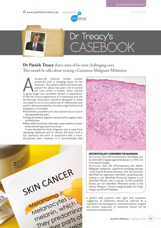
- 1. S K I N / D E R M AT O L O G Y 45Aesthetic Medicine • July/August 2015 SPONSORED BY CASE FILESwww.aestheticmed.co.uk Dr Patrick Treacy shares some of his most challenging cases. This month he talks about treating s Cutaneous Malignant Melanoma Dr Treacy’s CASEBOOK A 23-year-old Siberian female patient presented with a changing lesion on her abdomen. The patient stated the lesion was present for about two years and it started off from within a freckle, which started to grow larger and somewhat darken in appearance. It had the clinical appearance of a melanoma and the dermoscopy three-point checklist (designed to allow non-experts not to miss detection of melanomas) was used to determine whether this had a high likelihood of malignancy. It included: Asymmetry: asymmetry of colour and structure in one or two perpendicular axes Atypical network: pigment network with irregular holes and thick lines Blue-white structures: there was some evidence of blue- white veil and regression structures It was decided the likely diagnosis was a superficial spreading melanoma and to remove the lesion with a 1cm clearance and work in association with a multi- disciplinary team. However, it is recommended that a patient who presents with signs and symptoms suggestive of melanoma should be referred to a consultant dermatologist or consultant plastic surgeon and lesions suspicious of melanoma should not be removed in primary care. HISTOPATHOLOGY CONFIRMED THE DIAGNOSIS Macroscopy: Skin, left inframammary: Skin ellipse, 15 x 9 x 3mm with irregular pigmented lesion, 5 x 7mm, 1cm from nearest margin. Microscopy: Skin, left infra-mammary, skin ellipse: Malignant melanoma, superficial spreading subtype - Clark level III. Breslow thickness: 1mm. No ulceration identified No regression identified. Lymphovascular invasion is not identified. Perineural invasion is not identified. Mitotic rate is two per 10. Microsatellite lesions are not identified. Melanoma arises in the naevus. Margins: - Closest margin (radial): 1cm. Deep margin: 4mm Prof K Sheahan
- 2. 46 Aesthetic Medicine • July/August 2015 SPONSORED BYCASE FILES www.aestheticmed.co.uk S K I N / D E R M AT O L O G Y Please advise on captions HISTOPATHOLOGY EXPLAINED The pathologist’s report above includes a macroscopic description (the naked eye view), of the specimen and a microscopic description. The following features suggest an invasive melanoma. Diagnosis of primary melanoma Breslow thickness to the nearest 0.1 mm Clark level of invasion Margins of excision i.e. the normal tissue around the tumour Mitotic rate – a measure of how fast the cells are proliferating Whether or not there is ulceration The report includes comments about cell type and its growth pattern, invasionofbloodvesselsornerves,inflammatoryresponse, regression and whether there is associated in-situ disease Breslow thickness is reported for invasive melanomas. It is measured vertically in millimetres from the top of the granular layer (or base of superficial ulceration) to the deepest point of tumour involvement. It is a strong predictor of outcome; the thicker the melanoma, the more likely it is to metastasize. CLARK LEVEL Clark level indicates the anatomic plane of invasion. The deeper the Clark level, the greater the risk of metastasis. It is useful in predicting outcome in thin tumours, and less useful for thicker ones TYPES OF MELANOMA Melanomas are described according to their appearance and behaviour. Those that start off as flat patches (i.e. have Level 1 In situ melanoma Level 2 Melanoma has invaded papillary dermis Level 3 Melanoma has filled papillary dermis Level 4 Melanoma has invaded reticular dermis Level 5 Melanoma has invaded subcutaneous tissue a horizontal growth phase) include: (1)Superficialspreadingmelanoma(SSM)-70%ofall melanomas (2)Lentigomalignamelanoma(sundamaged skinofface,scalpandneck) (3) Acral lentiginous melanoma (on soles of feet, palms of hands or under the nails) (*subungual melanoma) These superficial forms of melanoma tend to grow slowly, but at any time, they may begin to thicken up or develop a nodule (i.e. progress to a vertical growth phase). Melanomas that quickly invade deeper tissues include: (1) Nodular melanoma (presenting as a rapidly enlarging lump) 15–30% of all melanomas (2) Spitzoidmelanoma(anodulethatresemblesaSpitznaevus) (3) Mucosalmelanoma(arisingonlips,eyelids,vulva,penis,anus) (4) Neurotropic and desmoplastic melanoma (fibrous tumour with a tendency to nerves) Histologic factors that affect metastatic potential include ulceration of the tumour, mitotic rate, presence of lymphovascular invasion, microsatellites, regression, perineural invasion, and the presence of lymphocytes infiltrating the tumour. WHO IS AT RISK OF MELANOMA? The main risk factors for developing superficial spreading melanoma include: Affluence, Increasing age, female Fair skin that burns easily. Red or light coloured hair, Light coloured eyes and light coloured skin (Anglo-Celtic) (Gandini S et al 2005). Multiple (5) atypical nevi (moles that are histologically dysplastic) (Garbe C et al 1994). Many benign melanocytic naevi person (Ferrone CR et al 2005). Giant congenital melanotic naevi ≥20cm in diameter Previousinvasivemelanomaormelanomainsitu(Goggins WB et al 2003). Family history of melanoma. Two first-degree relatives affected (Florrell Sr et al 2005). UVB (280- 320nm) solar radiation (causing sunburn) is the The primary mode of treatment for localised cutaneous melanoma is surgery. Surgical margins of 5mm are currently recommended for melanoma in situ, and margins of 1cm are recommended for melanomas ≤1 mm in depth
- 3. S K I N / D E R M AT O L O G Y 47Aesthetic Medicine • July/August 2015 SPONSORED BY CASE FILESwww.aestheticmed.co.uk Please advise on captions principle cause of melanoma. UVA may be involved in the pathogenesis of melanoma (Wang SQ et al 2001). CHECKING FOR A MELANOMA Glasgow 7-point checklist The ABCDs of Melanoma STAGING OF MELANOMA Melanoma staging means finding out if the melanoma has spread from its original site in the skin. The stages are: TREATMENT OF MELANOMA The primary mode of treatment for localized cutaneous melanoma is surgery. Surgical margins of 5mm are currently recommended for melanoma in situ, and margins of 1cm are recommended for melanomas ≤1 mm in depth1. For tumors of intermediate thickness (1–4 mm Breslow depth), randomized prospective studies show that 2cm margins are appropriate, although 1cm margins have been proven effective for tumors of 1-2mm thickness.2,3 Margins of 2cm are recommended for cutaneous melanomas greater than 4mm in thickness (high-riskprimaries)topreventpotentiallocalrecurrence in or around the scar site. Numerous adjuvant therapies have been investigated for the treatment of localized cutaneous melanoma following complete surgical removal. Adjuvant interferon (IFN) alfa-2b is the only adjuvant therapy approved by the US Food and Drug Administration for high-risk melanoma.4 While early-stage melanomas can often be cured with surgery, more advanced melanomas can be much harder to treat. But in recent years, newer types of immunotherapy and targeted therapies have shown a great deal of promise and have changed the treatment of this disease. Major features Minor features • Change in size • Diameter 7mm • Irregular shape • Inflammation • Irregular colour • Oozing • Change in sensation A Asymmetry B Border irregularity C Colour variation D Diameter over 6 mm E Evolving (enlarging, changing) Stage Characteristics Level 1 In situ melanoma Level 2 Melanoma has invaded papillary dermis Level 3 Melanoma has filled papillary dermis Level 4 Melanoma has invaded reticular dermis Level 5 Melanoma has invaded subcutaneous tissue
- 4. 48 CASE FILES www.aestheticmed.co.uk Aesthetic Medicine • July/August 2015 S K I N / D E R M AT O L O G Y REFERENCES (1) NIH Consensus conference. Diagnosis and treatment of early melanoma. JAMA. 268(10):1314–9. 9. (2) Balch CM, Urist MM, Karakousis CP, Smith TJ, Temple WJ, Drzewiecki K, et al. Efficacy of 2-cm surgical margins for intermediate-thickness melanomas (1 to 4 mm). Results of a multi-institutional randomized surgical trial. Ann Surg. 1993;218(3):262–7. (3) Veronesi U, Cascinelli N, Adamus J, Balch C, Bandiera D, Barchuk A, et al. Thin stage I primary cutaneous malignant melanoma. Comparison of excision with margins of 1 or 3 cm. N Engl J Med.1988;318(18):1159–62. (4) Veronesi U, Adamus J, Aubert C, Bajetta E, Beretta G, Bonadonna G, et al. A randomized trial of adjuvant chemotherapy and immunotherapy in cutaneous melanoma. N Engl J Med. 1982;307(15):913–6. (5) Rager EL, Bridgeford EP, Ollila DW. Cutaneous melanoma: update on prevention, screening, diagnosis, and treatment. Am Fam Physician. 2005;72(2):269–76. (6) Whiteman DC, Whiteman CA, Green AC. Childhood sun exposure as a risk factor for melanoma: a systematic review of epidemiologic studies. Cancer Causes Control. 2001;12(1):69–82. (7) Anderson WF, Pfeiffer RM, Tucker MA, Rosenberg PS. Divergent cancer pathways for early-onset and late-onset cutaneous malignant melanoma. Cancer. 2009 Sep 15;115(18):4176-85. doi: 10.1002/cncr.24481. PubMed PMID: 19536874; PubMed Central PMCID: PMC2741537. (8) Cohn-Cedarmark G, Rutqvist LE, Anderson R et al. Long-term results of a randomised study by the Swedish Melanoma Study Group on 2-cm versus 5-cm resection margins for patients with cutaneous melanoma with a tumour thickness of 0.8-2.0mm.Cancer 2000; 89: 1495-1501. (9) Revised U.K. guidelines for the management of cutaneous melanoma 2010. JR Marsden, JA Newton-Bishop, L Burrows, M Cook, PG Corrie, NH Cox, ME Gore, P Lorigan, R MacKie, P Nathan, H Peach, B Powell, C Walker, BJD, Vol. 163, No. 2, August 2010 (p238-256) (10) Wang SQ, Setlow R, Berwick M, Polsky D,Marghoob AA, Kopf AW, Bart RS. Ultraviolet A and melanoma: a review. J Am Acad Dermatol 2001; 44: 837-846. (11) The prevention, diagnosis, referral and management of melanoma of the skin: concise guidelines (Newton Bishop J, Bataille V, Gavin A, Lens M, Marsden J, Mathews T, Ormerod A, Wheelhouse C). Royal College of Physicians and British Association of Dermatologists. Concise guidance to good practice series, No 7. London : RCP, September 2007 (12) Cutaneous melanoma: update on prevention, screening, diagnosis, and treatment. Rager EL, Bridgeford EP, Ollila DW Am Fam Physician. 2005 Jul 15; 72(2):269-76. (13) Abrahamsen HN, Hamilton-Dutoit SJ, Larsen J, Steiniche T. Sentinel lymph nodes in malignant melanoma: extended histopathological evaluation improves diagnostic precision. Cancer 2004; 100: 1683-1691. (14) Balch CM, Buzaid AC, Soong SJ et al. Final version of the American Joint Committee on Cancer staging system for cutaneous melanoma. J Clin Oncol 2001; 19: 3635-3648. (15) Annual report to the nation on the status of cancer, 1973-1999, featuring implications of age and aging on U.S. cancer burden. Edwards BK, Howe HL, Ries LA, Thun MJ, Rosenberg HM, Yancik R, Wingo PA, Jemal A, Feigal EG Cancer. 2002 May 15; 94(10):2766-92. (16) Gandini S, Sera F, Cattaruzza MS, Pasquini P, Picconi O, Boyle P, Melchi CF. Meta-analysis of risk factors for cutaneous melanoma: I. Common and atypical naevi. Eur J Cancer 2005; 41: 28-44. (17) Wang SQ, Setlow R, Berwick M, Polsky D,Marghoob AA, Kopf AW, Bart RS. Ultraviolet A and melanoma: a review. J Am Acad Dermatol 2001; 44: 837-846. DrPatrickTreacy is CEO Ailesbury Clinics, chairman of the Irish Association of Cosmetic Doctors and Irish regional representative of the British College of Aesthetic Medicine (BCAM). He is also president of the World Trichology Association. Dr Treacy has won a number of awards for his contributions to facial aesthetics and hair transplants including the AMEC Award in Paris in 2014. Dr Treacy also sits on the editorial boards of three international journals and features regularly on international television and radio programmes. He is scientific committee for AMWC Monaco 2015, AMWC Eastern Europe 2015, AMWC Latin America 2015, RSM ICG7 (London) and Faculty IMCAS Paris 2015 and IMCAS China 2015. IMMUNOTHERAPY Drugs that block CTLA-4: Ipilimumab targets CTLA-4, a protein that normally suppresses the T-cell immune response, which helps melanoma cells survive. Ipilimumab has been shown to help people with advanced melanomas live longer. Combining ipilimumab with GM-CSF is better than using ipilimumab alone. The combination has fewer serious side effects. Drugs that block PD-1 or PD-L1: Melanoma cells also use pathways in the body to avoid being detected, and a protein called PD-L1 on their surface helps them evade the immune system. Two drugs that block PD-1, pembrolizumab (Keytruda) and nivolumab (Opdivo), are now approved to treat advanced melanoma. Melanoma vaccines: These are experimental therapies that have not yet been proven to be helpful. CONCLUSION Skin cancer is the most common malignancy in the United Kingdom and Ireland.5 Malignant melanoma is the most deadly cutaneous neoplasm. Numerous risk factors for development of melanoma have been identified, including white skin, fair hair, light eyes, sun sensitivity and a tendency to freckle. Other factors, include family history of melanoma, dysplastic nevi, increased numbers of typical nevi, large congenital nevi and immunosuppression. Although sun exposure is a risk factor for melanoma, cutaneous melanomas can also arise in areas of the body not exposed to the sun. Sun exposure in childhood and having more than one blistering sunburn in childhood are associated with an increased risk of melanoma.6 Most melanomas arise as superficial tumors confined to the epidermis. The prognosis for melanoma is closely related to the thickness of the tumour. In order to effectively treat melanomas, drugs that target proteins that normally suppresses the T-cell immune response or block ones that help them evade the immune system provide the best chance for treating patients with advanced melanoma. In early studies, combination drugs have shrunk tumors in about one half of pateints with melanoma. AM