Acute Coronary Syndrome - Overview
•Download as PPTX, PDF•
221 likes•66,154 views

Definition, Diagnosis, Types and therapy in Acute coronary syndrome. NSTEMI/UA and STEMI Causes of chest pain Thrombolysis/ fibrinolysis PCI Treatment Algorithms

Recommended
More Related Content
What's hot
What's hot (20)
Similar to Acute Coronary Syndrome - Overview
Similar to Acute Coronary Syndrome - Overview (20)
More from Rahul Varshney
More from Rahul Varshney (6)
Recently uploaded
Recently uploaded (20)
Acute Coronary Syndrome - Overview
- 1. ACUTE CORONARY SYNDROME Dr RAHUL VARSHNEY
- 3. Hospitalizations in the U.S. Due to Acute Coronary Syndromes (ACS) Acute Coronary Syndromes* 1.57 Million Hospital Admissions - ACS UA/NSTEMI† STEMI 1.24 million Admissions per year .33 million Admissions per year Heart Disease and Stroke Statistics – 2007 Update. Circulation 2007; 115:69-171. *Primary and secondary diagnoses. †About 0.57 million NSTEMI and 0.67 million UA.
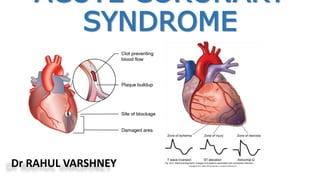
- 4. Acute Coronary Syndrome Definition: The spectrum of acute ischemia related syndromes ranging from UA to MI with or without ST elevation that are secondary to acute plaque rupture or plaque erosion. [----UA---------NSTEMI----------STEMI----] A constellation of symptoms related to obstruction of coronary arteries with chest pain being the most common symptom in addition to nausea, vomiting, diaphoresis etc.
- 5. Signs and symptoms of ACS presentation Symptoms may include: • chest discomfort (tightness, pressure, heaviness) at rest or for a prolonged period (> 10 minutes, not relieved by sublingual nitrates) • recurrent chest discomfort • discomfort associated with syncope/acute heart failure. The pain may spread to other parts of the upper body, including: • Back, neck, jaw, arm(s), shoulder(s) or epigastric pain. The person may also experience: • Dyspnoea (shortness of breath), diaphoresis (profuse perspiration), dizziness, nausea or vomiting • Recent research shows that women, the elderly and people with diabetes are less likely to experience chest pain as a symptom.
- 7. Anginal equivalent syndrome • Patients who are experiencing ACS, yet do not complain of typical chest pain; rather, these patients note atypical pain, dyspnea, weakness, diaphoresis, or emesis—these complaints, in fact, are the manifestation of the ACS event. • Patients with altered cardiac pain perception e.g., the elderly or patients with longstanding diabetes mellitus, are potentially at risk to present with anginal equivalent syndromes. • The most frequently encountered anginal equivalent chief complaint is dyspnea, which is found in 10% to 30% of patients with AMI, often due to pulmonary edema.
- 8. CHARACTERISTICS OF TYPICAL ANGINAL CHEST PAIN CHARACTERISTIC SUGGESTIVE OF ANGINA LESS SUGGESTIVE OF ANGINA TYPE OF PAIN DULL PRESSURE/CRUSHING PAIN SHARP/STABBING DURATION 2-5 MIN, <20 MIN SECONDSTO HOURS/CONTINUOUS ONSET GRADUAL RAPID LOCATION/CHEST WALL TENDERNESS SUBSTERNAL, NOT TENDER TO PALP. LATERAL CHEST WALL/TENDER TO PALP. REPRODUCIBALITY WITH EXERTION/ACTIVITY WITH BREATHING/MOVING AUTONOMIC SYMPTOMS PRESENT USUALLY ABSENT
- 9. • STEMI: ST elevation, elevated cardiac enzymes • NSTEMI: ST depression, T-wave inversion, elevated cardiac enzymes • Unstable Angina: Non specific EKG changes, normal cardiac enzymes Based on ECG and cardiac enzymes, ACS is classified into:
- 12. Pathophysiology of ACS Evolution of Coronary Thrombosis
- 13. Pathophysiology ACS Decreased O2 Supply •Flow- limiting stenosis •Anemia •Plaque rupture/clot Increased O2 Demand Myocardial ischemia → Necrosis O2 supply/demand mismatch → Ischemia Asymptomatic Angina Pathophysiology of Stable Angina and ACS
- 14. Unstable Angina STEMINSTEMI Non occlusive thrombus Non specific ECG Normal cardiac enzymes Non-occlusive thrombus sufficient to cause tissue damage & mild myocardial necrosis ST depression +/- T wave inversion on ECG Elevated cardiac enzymes Complete thrombus occlusion ST elevations on ECG or new LBBB Elevated cardiac enzymes More severe symptoms
- 15. Risk Factors
- 16. Gender Differences in MI Females, when compared to males: -present with MI later in life -have poorer prognosis and high morbidity -are 2x as likely to die in the first weeks -are more likely to die from the first MI -have higher rates of unrecognized MI
- 17. At least 2 of the following • History ( angina or angina equivalent) • Acute ischemic ECG changes • Typical rise and fall of cardiac markers • Absence of another identifiable etiology Diagnosis of ACS
- 18. Diagnosis Diagnosis requires a rise and/or fall in serum levels of cardiac markers (preferably troponin) together with: Defined clinically by patient history ECG (new ST-T wave changes, new left bundle branch block or evolving pathological Q waves) Imaging evidence of new regional wall motion abnormality.
- 19. STEMI • Compromised left ventricular (LV) function may result in pulmonary edema with development of pulmonary bibasilar crackles and jugular venous distention; a fourth heart sound can be present with small infarcts or even mild ischemia, but a third heart sound is usually indicative of more extensive damage. Pericardial rub may be heard.
- 20. • An ST-segment elevation myocardial infarction (STEMI) can be confirmed by an ECG. • A 12-lead ECG should be performed and interpreted expeditiously. • STEMI is defined as presentation with clinical symptoms consistent with an ACS with ECG features including any of: Persistent ST-segment elevation ≥ 1 mm in two contiguous limb leads ST-segment elevation ≥ 2 mm in two contiguous chest leads New left bundle branch block (LBBB) pattern. STEMI – what is it?
- 21. Acute ST Elevation MI
- 22. Normal ECG
- 23. Serum cardiac markers IDEAL MARKER: • High concentration in myocardium • Myocardium specific • Released early in injury • Proportionate to injury • Non expensive testing
- 24. Advantages Myoglobin 1. High sensitivity 2. Useful in early detection of MI 3. Detection of reperfusion 4. Most useful in ruling out MI CK-MB 1. Rapid, cost-efficient, accurate assays 2. Ability to detect early reinfarction Troponins 1. Powerful for stratification 2. Greater sensitivity and specificity than CK-MB 3. Detection of recent MI up to 2 weeks after onset 4. Useful for selection of therapy 5. Detection of reperfusion
- 25. Diadvantages CK-MB 1. Lack of specificity with skeletal muscle disease/injury 2. Low sensitivity during early MI (<6 h) or late (>36 h) after symptom onset and for minor myocardial damage Myoglobin 1. Very low specificity with skeletal muscle injury or disease 2. Rapid return to normal Troponins 1. Low sensitivity in early phase of MI (<6 h after symptom onset) 2. Limited ability to detect late minor re-infarction
- 27. • Cardiac Imaging 2D ECHO may reveal wall motion abnormality and LV dysfunction. It may also detect RV infarction, Ventricular aneurysms, pericardial effusion and LV Thrombus. Myocardial perfusion imaging with TI201 and Tc99m - sestamibi reveals defect in most patient.
- 28. Time from symptom onset and likely outcome < 1 hour Aborted heart attack or only little heart muscle damage 1–2 hours Minor heart muscle damage only 2–4 hours Some heart muscle damage with moderate heart muscle salvage 4–6 hours Significant heart muscle damage with only minor heart muscle salvage 6–12 hours No heart muscle salvage (permanent loss) with potential infarct healing benefit > 12 hours Reperfusion is not routinely recommended if the patient is asymptomatic and haemodynamically stable
- 29. Therapy STEMI • Once STEMI is suspected or diagnosed, the immediate concerns are to ensure the patients stability and to intervene to limit infarct size by restoring the blood flow to the infarct artery as soon as possible. • Abolute and relative contraindication to therapies must be considered and relative risk assessed. Choices may be limited for the availability of specialized procedures, need to transport to another facility, significant co- morbidities, bleeding risk,etc.
- 30. Early Therapy • Patients presenting with suspected myocardial ischemia should undergo a rapid evaluation, that is a 12 lead ECG, cardiac markers and related laboratory tests. • Should be treated with oxygen • Sublingual Nitroglycerin (unless systolic pressure is <90 mm Hg), and Aspirin, 160 to 325 mg orally. • Opiates relieve pain and also reduce anxiety, the salutary effects of which have been known for decades and should not be underestimated.
- 31. Fibrinolytic therapy • Early reperfusion of an occluded coronary artery is indicated for all eligible candidates. • Fibrinolytic agents administered early in the course of an acute MI reduce infarct size, preserve LV function, and reduce short-term and long-term mortality. • Multiple studies conclude that greatest mortality benefit is seen if fibrinolytics are administered within the first 12 hours of symptom onset, but it is reasonable to administer fibrinolytics to patients whose onset of symptoms exceeds 12 hours but who have continued clinical or ECG evidence of ischemia.
- 32. INDICATIONS FOR AND CONTRAINDICATIONS TO FIBRINOLYTIC THERAPY IN ACUTE MYOCARDIAL INFARCTION
- 33. • After administration of fibrinolytics for STEMI, the patient should be monitored for signs and symptoms of adequate reperfusion within 90 minutes, as indicated by relief of symptoms and/or hemodynamic/ electrical instability coupled with at least a 50% resolution of the highest initial ST elevation. • Patients who receive fibrinolytics for STEMI and have at least one high-risk feature should have cardiac catheterization for risk stratification and potential percutaneous revascularization, even if this involves immediate transfer from the presenting hospital to a PCI- capable facility. High-risk features include • Extensive ST-segment elevation (>2 mm ST elevation in two anterior leads), • New-onset LBBB, • Previous MI, • Killip class 2 or 3 or Left ventricular ejection fraction (LVEF) ≤ 35%, • Systolic blood pressure ≤ 100 mm Hg, • Heart rate ≥ 100 bpm, or • Right ventricular involvement.
- 34. Fibrinolytic agents Used in ST elevation MI Streptokinase 1.5 million intravenous over 30-60 mins Alteplase (tPA) a 15-mg bolus, then 0.75 mg/kg (up to 50 mg) IV over the initial 30 minutes, and 0.5 mg/kg (up to 35 mg) over the next 60 minutes Reteplase (rPA) two 10-U boluses 30 minutes apart Tenecteplase (TNK-tPA) IV Bolus adjusted for weight (30mg if < 60 kg; 35mg if 60-70 kg; 40 mg if 70-80 kg, 45mg if 80-90 kg; 50mg if > 90 kg)
- 35. Reperfusion Therapy • It includes angioplasty, with or without deployment of an intracoronary Bare- metal or Drug-eluting stent, with support of pharmacological measures to prevent thrombosis. i. Percutaneous coronary intervention • Recent meta-analyses comparing direct PTCA with fibrinolytic therapy have suggested lower rates of mortality and re-infarction among those receiving direct PTCA. Thus direct angioplasty, if performed in a timely manner (ideally within 60 minutes) by highly experienced personnel, may be the preferred method of revascularization, since it offers more complete revascularization with improved restoration of normal coronary blood flow and detailed information about coronary anatomy.
- 36. There are certain subpopulations in which primary PCI is clearly preferred, and other populations in which the data are suggestive of benefit.
- 37. Coronary Stents • The use of coronary stents has been shown to reduce restenosis and adverse cardiac outcomes in both routine and high-risk PCI. • A large, randomized, multicenter trial involving 900 patients did not show a difference in mortality at 6 months but did show improvement in ischemia driven target vessel revascularization and less angina in the stented patients compared to balloon angioplasty alone. • Whether to use a bare metal stent or a drug-eluting stent in acute MI is a question that has not yet been addressed definitively by clinical trials; selection is currently based on both patient and angiographic characteristics.
- 38. • Early recognition and diagnosis of STEMI are key to achieving the desired door-to-needle (or medical contact–to-needle) time for initiation of fibrinolytic therapy of 30 minutes or door-to-balloon (or medical contact–to balloon) time for PCI under 90 minutes. • Achieving reperfusion in timely matter correlates with improvement in ultimate infarct size, LV function, and survival. • The ultimate goal is to restore adequate blood flow through the infarct-related artery to the infarct zone, as well as to limit microvascular damage and reperfusion injury. • The latter is accomplished with adjunctive and ancillary treatments.
- 40. Adjunctive therapy 1. Aspirin: • Reduce mortality in acute infarction to the same degree as fibrinolytic therapy, and its effects are additive to fibrinolytics. In addition, aspirin reduces the risk of reinfarction. • Unless contraindicated, all patients with a suspected ACS (STEMI, NSTEMI, UA) should be given aspirin as soon as possible. 2. Thienopyridines: • Clopidagrel • Prasugrel Patients presenting with MI should be considered for a thienopyridine regardless of whether or not they underwent reperfusion therapy. The duration of thienopyridine use in this population has yet to be defined.
- 41. 3. Anticoagulants: Administration of full-dose heparin after fibrinolytic therapy with tPA is essential to diminish re-occlusion after successful reperfusion. 4. Glycoprotein llb/IIIa Receptor antagonist: Role in STEMI uncertain. Not routinely recommended.
- 43. 5. Nitrates: • They reduce myocardial oxygen demand by decreasing preload and afterload, and they may also improve myocardial oxygen supply by increasing sub-endocardial perfusion and collateral blood flow to the ischemic region. • Also reduce platelet aggregation. • Nitrates are first-line agents for the symptomatic relief of angina pectoris and when MI is complicated by congestive heart failure.
- 44. 6. β Blockers: • Routine use of intravenous beta-blockers in the absence of systemic hypertension is no longer recommended. • Relative contraindications to oral beta-blockers include, heart rate less than 60 bpm, systolic arterial pressure less than 100 mm Hg, moderate or severe LV failure, signs of peripheral hypoperfusion, shock, PR interval greater than 0.24 second, second- or third-degree AV block, active asthma, or reactive airway disease.
- 45. 7. ACE inhibitors • Improve hemodynamic, functional capacity and symptoms, and survival in patients with chronic congestive heart failure. Moreover, ACE inhibitors prevent the development of congestive heart failure in patients with asymptomatic LV dysfunction 8. Lipid Lowering agents • All patients with ACS should be started on a statin prior to discharge unless there is a contraindication 9. Calcium channel blockers • May be useful for patients whose postinfarction course is complicated by recurrent angina, because these agents not only reduce myocardial oxygen demand but also inhibit coronary vasoconstriction.
- 46. Treatment algorithm for ST-segment elevation myocardial infarction (STEMI)
- 47. NTSEMI and Unstable angina • Non-ST-elevation MI (NSTEMI) applies to patients with suspected MI in the absence of other plausible causes of troponin elevation (e.g. sepsis, pulmonary embolus). • On physical examination, patients with NSTEMI may have a ‘normal’ ECG reading, or show minor changes (occurs in up to 50% of patients). • All patients with NSTEMI should have their risk stratified to direct management decisions. • The management of patients with NSTEACS requires evolving risk stratification: Clinical assessment, assessment of Cardiac biomarkers and Time.
- 48. ECG Changes NSTEMI: • ST depressions (0.5 mm at least) or T wave inversions ( 1.0 mm at least) without Q waves in 2 contiguous leads with prominent R wave or R/S ratio >1. • Isolated T wave inversions: • can correlate with increased risk for MI • may represent Wellen’s syndrome: • critical LAD stenosis • >2mm inversions in anterior precordial leads Unstable Angina: • May present with nonspecific or transient ST segment depressions or elevations
- 49. Diagnosis
- 50. Evolving Risk Stratification • Provides prognostic information • Determines treatment and level of intervention Low risk patients ---> early discharge, High risk ---> admission to high care • Helps decongest the casualty and make available medical resources to more needy patients. • Risk stratification should be ongoing – at admission, 6-8 hrs, 24hrs, discharge
- 51. Evolving risk stratification… Admit to coronary care unit or high dependency unit: • Estimate ischemic risk, estimate bleeding risk, choose augmented antithrombotic therapy → refer for angiography to determine surgery/PCI, or medical therapy.
- 53. Evolving risk stratification… Intermediate-risk NSTEACS Recurrent ischemia or elevated troponin? YES • admit to CCU or high dependency unit: estimate ischemic risk, estimate bleeding risk, choose augmented antithrombotic therapy →refer for angiography to determine surgery/PCI, or medical therapy. NO • undertake stress test (e.g. exercise ECG): →positive – refer for angiography to determine surgery/PCI, or medical therapy →negative – proceed to discharge patient with urgent cardiac follow-up (on upgraded medical therapy) according to long-term management after control of myocardial ischemia.
- 54. Evolving risk stratification… Appropriate period of observation. Consider if stress test (e.g. exercise ECG) needed? Stress test (e.g. exercise ECG) using treadmill. YES Proceed to discharge patient with urgent cardiac follow-up (on upgraded medical therapy) according to long-term management after control of myocardial ischemia. NO
- 55. • Age ≥ 65 years =1 point • At least 3 risk factors for CAD =1 point • Prior coronary stenosis of ≥ 50% =1 point • ST-segment deviation on ECG presentation =1 point • At least 2 anginal events in prior 24 hours =1 point • Use of aspirin in prior 7 days =1 point • Elevated serum cardiac biomarkers =1 point Variables Used in the TIMI Risk Score
- 56. TIMI Risk Score
- 57. GRACE Risk Score Variable Odds ratio Older age 1.7 per 10 y Killip class 2.0 per class Systolic BP 1.4 per 20 mm Hg ↑ ST-segment deviation 2.4 Cardiac arrest during presentation 4.3 Serum creatinine level 1.2 per 1-mg/dL ↑ Positive initial cardiac biomarkers 1.6 Heart rate 1.3 per 30-beat/min ↑
- 58. Management • It Includes increasing myocardial oxygen demand delivery by improving perfusion and decreasing myocardial oxygen demand. • Reversing myocardial ischemia and confirming the diagnosis of ACS is essential priority. • Oxygen should be administered to patients with dyspnea, hypoxemia or evidence of heart failure or shock. • If precipitating reversible causes such as fever, anemia, hypoxemia, infection, hypertension, anxiety, hyperthyroidism, arrhythmias or any drug ingestion (eg cocaine, ephedrine) can be identified, they should be treated aggressively. • Further management includes relief of pain and anti- ischemic therepy, therepy for platelet aggregation/thrombosis, ongoing risk stratification and consideration of invasive reperfusion procedures.
- 60. 2. Antiplatelet therapy • The current guidelines recommend a loading dose of 300 to 600mg of clopidogrel in patients of UA/NSTEMI, followed by 75 mg daily. The duration of clopidogrel may depend upon whether or not the patient has received stent. 3. Anti thrombin therapy Recommendation Low risk : Aspirin + Anticoagulant + either a glycoprotein IIb/IIIa inhibitor or a thienopyridine High Risk: Aspirin + Anticoagulant + a glycoprotein IIb/IIIa inhibitor + a thienopyridine
- 61. Interventional Management In patients with High Risk, following treatment with anti-ischemic and antithrombotic agents, coronary arteriography is carried out within ~ 48 h of admission, followed by coronary revascularization (PCI or coronary artery bypass grafting), depending on the coronary anatomy. In low-risk patients, the outcomes from an invasive strategy are similar to those obtained from a conservative strategy, which consists of anti-ischemic and antithrombotic therapy followed by “watchful waiting,” and in which coronary arteriography is carried out only if rest pain or ST-segment changes recur or there is evidence of ischemia on a stress test.
- 63. Complications • Ventricular dysfunction • Cardiogenic shock • Right Ventricular Infarction • Pericarditis • Thromboembolism • Left ventricular aneurysm • Sinus bradycardia • AV Block • Ventricular tachycardia and fibrillation
- 64. Take all these pills daily until a new clinical trial is published THANK YOU