SlideShare a Scribd company logo
1 of 58
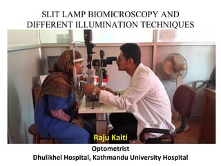
SLIT LAMP BIOMICROSCOPY AND
DIFFERENT ILLUMINATION TECHNIQUES
Raju Kaiti
Optometrist
Dhulikhel Hospital, Kathmandu University Hospital
INTRODUCTION
• Slit lamp biomicroscope was invented in 1911 AD by Gullstrand.
• It is named so because it enables the practitioner to observe the living
tissues of the eye under magnification in slit and wide view.
Introduction
• An instrument consisting of a high-intensity light source that can be focused
to shine as a slit.
• Used in conjunction with a microscope.
• The lamp facilitates an examination which looks at anterior segment, or
frontal structures, of the human eye, which includes the
– Eyelid
– Cornea
– Sclera
– Conjunctiva
– Iris
– Anterior chamber
– Natural crystalline lens and
– Anterior vitreous.
• Three parts:
– Illumination system
– Observation System
– Physical support
• Optics
– Works on the same principle as a Compound Microscope
» Objective lens(+22.00D) & eye piece(+10.00-
+14.00D)
» Adjustable illumination system
• The illumination system is in the form of a bright focal source of light with a
slit mechanism and circular apertures of various sizes, prism, lamp housing
unit, slit width and height control, neutral density filter, cobalt blue light, red-
free (green) filter, field size control, diffuser.
• The observation system is a binocular microscope capable of a wide range of
magnification, camera/video adaptor, observation tube, magnification changer.
• The mechanical support is provided by joystick, headrest and chinrest, screw
arrangement for moving instrument up and down, control for slit, width height
,magnification control ,on/off switch and fixation target, locking controls.
Observation system Illumination system Mechanical support
MAGNIFICATIONS
• Low magnification:
– 7X - 10X : General eye
– Lids.
– Bulbar conjunctiva/sclera.
– Cornea/limbus.
– Tears.
– Anterior chamber/iris/crystalline lens.
• Medium magnification:
– 20X - 25X : Structure of individual
layers
– Epithelium/epithelial breakdown.
– Stroma.
– Endothelium.
– Contact lens fit/lens condition.
• High magnification:
– 30X - 40X : Details
• Epithelium
– vacuoles
– microcysts
– dystrophies.
• Stroma
– striae
– folds.
• Endothelium
– Polymegathism
– guttata
– blebs
– cell density.
ptmetrist
Chinrest
Optometris
t
PRINCIPLE
• A narrow "slit" beam of very bright light produced by lamp.
This beam is focused on to the eye which is then viewed
under magnification with a microscope
TYPES
• There are 2 types of slit lamp biomicroscope
1)Zeiss slit lamp biomicroscope
2)Haag streit slit lamp biomicroscope
• In Zeiss type light source is at the base of the instrument while in Haag
streit type it is at the top of the instrument.
How to start?
• Focus the eye piece
• Adjust the headrest
• Position the fixation target
• Decrease the room illumination
• Start with diffuse illumination
• Use appropriate magnification
ILLUMINATION TECHNIQUES
• Diffuse Illumination
• Direct Illumination
• Indirect Illumination
• Retro illumination
• Specular Reflection
• Sclerotic Scatter
• Tangential Illumination
• Oscillation Technique
DIFFUSE ILLUMINATION
• A diffuse broad beam of light is used and general view of
anterior segment of eye is observed
• 45 degrees angle between light and microscope
• Open the slit fully
• Diffusing filter
• Variable magnification(Low to High)
DIFFUSE ILLUMINATION
• Overall View of:
– Lids and lashes
– Conjunctiva
– Cornea
– Sclera
– Iris
– Pupil
DIFFUSE ILLUMINATION
Mature cataract
Hyphema
DIFFUSE ILLUMINATION
Giant papillae in a case of VKC Iris tear with lens subluxation
secondary to trauma
DIRECT ILLUMINATION
• Observation and Illumination system are focused at the same point
• Angular separation 40 to 50 degrees between light source and microscope
• Vary angle of Illumination
• Low to high magnification
• Vary width and height of light source
• Optic section
– Tall but narrow slit of focused light
– Less than 0.25 mm wide
• Parallelepiped section
– Wider beam of focused light
– 1 to 2 mm in width
• Conical Beam
– Small circular or square spot of light
– Formed by reducing the height of parallelepiped beam
DIRECT ILLUMINATION
• Optic Section
• Used primarily to evaluate structural layers of the cornea and lens
• Estimating the depth of an abnormality such as corneal foreign body
or position of cataract
• Anterior chamber angle- The depth of AC can be observed by Van
Herrick's test in which optic section of light is used at corneal limbal
area.
• Localize
– nerve fibers, blood vessels, infiltrates, and cataracts.
Optic Section
OPTIC SECTION
Corneal FB
Corneal opacityDifferent forms of
cataracts
Patches of iris atrophy
Disc Edema Optic disc coloboma
Enlarged Cupping Macular region
DIRECT ILLUMINATION
• Parallepiped
– Provides a layered view of the cornea and lens
– Probably the most commonly used
– Broader view with extensive examination
– Depth and extent of corneal abrasions, scarring, or foreign
bodies
Parallelepiped
DIRECT ILLUMINATION
• Conical Beam
– Most useful technique when examining the transparency of the anterior
chamber for the evidence of floating cells and flares, as seen in anterior
uveitis
– Best in darkened room
Anterior Chamber cells
INDIRECT ILLUMINATION
• Beam is narrowed to 1 to 2 mm in width
• The beam is then focused on an area adjacent to the ocular
tissue observed and this area is observed in shadow by virtue
of scattered light.
• The foci of the light source and microscope are not coincident
INDIRECT ILLUMINATION
• Angle of illumination can be varied
• Beam width can be varied
• Low to high magnification
• Purpose is to provide somewhat “softer” illumination to give
better definition of the structural components of the iris,
epithelial corneal edema, pigment spots and corneal foreign
bodies
• Also valuable for observing
– Epithelial vesicles
– Epithelial erosions
– Iris sphincter
RETRO ILLUMINATION
• Formed by reflecting light of the slit beam from a structure more
posterior than the structure under observation
• Purpose is to place the object of regard against a bright background,
allowing the object to appear dark or black
• Search for Keratic precipitates and other debris on corneal
endothelium
• The crystalline lens can be retro illuminated for viewing water clefts
and vacuoles of the anterior lens and of posterior subcapsular cataract
• Corneal edema, neovascularization, microcyst and infiltrates
• Contact lens deposits
RETROILLUMINATION
SPECULAR REFLECTION
• Formed by separating the microscope and slit beam by equal angles
from the normal to the cornea i.e. angle of incidence equals angle of
reflection
• Separation for best specular reflection is about 50 degrees
• Under specular reflection, the anterior corneal surface appears as a
white, uniform surface, and the corneal endothelium takes on a golden
mosaic pattern
• Valuable for observing
– Irregularities, deposits or excavations in the smooth corneal surfaces (will fail to
reflect light and thus will appear darker than the surroundings)
– Changes in endothelium
– Tear film debris
– Tear film lipid layer thickness
SCLEROTIC SCATTER
• Formed by focusing a bright but narrow (1mm), slit beam on the limbus and
using the microscope on low magnification. This causes the cornea to take on
total internal reflection
• Slit beam should be approximately 40-60 degrees from microscope
• Halo glow of light around limbus
• As the light is internally reflected, no light will emerge toward the examiner
and so the cornea will appear black
• An area of reduced light transmission within the cornea however will appear
gray
• Valuable for observing
– Central corneal epithelial edema
– Corneal abrasions
– Corneal nebulae and maculae
– Foreign bodies in the cornea
SCLEROTIC SCATTER
TANGENTIAL ILLUMINATION
• Large angle of 70-80 degrees between illumination and
observation system
• Valuable for observing
– Iris freckles
– Tumors
– General integrity of cornea and iris
OSCILLATORY ILLUMINATION
– the microscope is kept in focus on the structure to be
observed and beam is oscillated back and forth alternately
resulting in direct and indirect illumination
– Quick to and fro movement
– Minutes objects in AC
– the microscope is kept in focus on the structure to be
observed and beam is oscillated back and forth alternately
resulting in direct and indirect illumination
FILTERED ILLUMINATION
• Cobalt Blue
• Red Free filter (green)
• Neutral Density
• Valuable for observing
– Tear layer
– Ocular staining
– RGP lens fitting pattern
FILTERS
• Cobalt blue filter is used to locate any lesion in the cornea with the help of
fluorescein staining.
• Also during fitting and trial of RGP lenses.
• Red free filter i.e. green filter is used to see the vascular pattern of choroid
and see any abnormalities in the disc of fundus.
• Yellow filter is used for general view of anterior segment.
Fluorescein and cobalt blue filter
Mires in applanation tonometry
Fluorescein stain after corneal FB removal
Fluorescein pattern in RGP trial
Fluorescein pattern in RGP
trial
Fluorescein and cobalt blue filter
CLINICAL USES
• Diagnostic
– Anterior segment Evaluation
– Goldmann Applanation Tonometry
– TBUT test
– Staining (Fluorescein, Rose Bengal etc.)
– Visiometry
– Gonioscopy
– FFA and Clinical Photography
• Therapeutic
– Epilation
– Foreign Body Removal
– Contact lens trial
– Contact lens fitting characters- static and dynamic
– Corneal epithelial debridement (herpetic keratitis)
– Insertion of punctal plugs
Corneal Foreign Body Removal
Slit lamp techniques in Contact Lens fitting
• Using the slit lamp, set with a broad beam and low magnification, the
practitioner should carry out sweep of the anterior segment and adnexa. Start
by examining the lids and lid margins, looking for signs of cysts, styes,
meibomian gland dysfunction and other.
• The cornea is examined next, initially with a medium magnification (x20 to
x25), again using a sweep to assess the entire cornea, using a beam width of
approximately 2mm (Parallelepiped), initially.
• Should any anomaly be detected, the magnification can be increased and the
beam width also adjusted, accordingly.
• The cornea should also be assessed after fluorescein installation, prior to any
contact lens fitting. Fluorescein stain in conjunction with the cobalt blue light
on the slit lamp is used to assess the integrity of the cornea epithelium, which
highlights damaged epithelial tissue, in a very reliable and effective manner.
• Tear film evaluation either tear prism height or TBUT should be assessed
using diffuse light.
• For TBUT cobalt blue filter in conjunction with fluorescein staining is
preferred technique.
• For soft lens trial and RGP lens trial also diffuse illumination is preferred.
• The static (how the back surface of the lens aligns to the cornea) is well
assessed by diffuse illumination but after staining and with cobalt blue
filter.
• Dynamic (how the lens centers and moves on the eye) fitting is also
assessed by diffuse illumination
• Parallelepiped and high magnification- for observation of striae and folds
• Microcysts seen under high magnification with marginal retro-
illumination.
• Corneal edema, neovascularization, microcyst and infiltrates and Contact
lens deposits all are seen through retro- illumination.
Slit lamp techniques.pptx

More Related Content

What's hot

Special purpose frames
Special purpose framesSpecial purpose frames
Special purpose framesEyenirvaan
 
Accommodation/ Accommodation of Eye, Measurement of Accommodation of Eye (hea...
Accommodation/ Accommodation of Eye, Measurement of Accommodation of Eye (hea...Accommodation/ Accommodation of Eye, Measurement of Accommodation of Eye (hea...
Accommodation/ Accommodation of Eye, Measurement of Accommodation of Eye (hea...Bikash Sapkota
 
Hirschberg and krimsky test.pptx
Hirschberg and krimsky test.pptxHirschberg and krimsky test.pptx
Hirschberg and krimsky test.pptxjyotishah48
 
WORTH FOUR DOT TEST.pptx
WORTH FOUR DOT TEST.pptxWORTH FOUR DOT TEST.pptx
WORTH FOUR DOT TEST.pptxKAUSTAV GOGOI
 
Introduction to binocular single vision (BSV)
Introduction to binocular single vision (BSV)Introduction to binocular single vision (BSV)
Introduction to binocular single vision (BSV)Anis Suzanna Mohamad
 
Corneal topography
Corneal topographyCorneal topography
Corneal topographySatish Jeria
 
Confrontation test
Confrontation testConfrontation test
Confrontation testSheim Elteb
 
Non Surgical Treatment of Strabismus
 Non Surgical Treatment of Strabismus Non Surgical Treatment of Strabismus
Non Surgical Treatment of StrabismusMeghna Verma
 
Assesssment of strabismus
Assesssment of strabismusAssesssment of strabismus
Assesssment of strabismusanwesha manna
 

What's hot (20)

Binocular balancing
Binocular balancing Binocular balancing
Binocular balancing
 
Special purpose frames
Special purpose framesSpecial purpose frames
Special purpose frames
 
Aphakia
AphakiaAphakia
Aphakia
 
Accommodation/ Accommodation of Eye, Measurement of Accommodation of Eye (hea...
Accommodation/ Accommodation of Eye, Measurement of Accommodation of Eye (hea...Accommodation/ Accommodation of Eye, Measurement of Accommodation of Eye (hea...
Accommodation/ Accommodation of Eye, Measurement of Accommodation of Eye (hea...
 
Tinted lenses
Tinted lensesTinted lenses
Tinted lenses
 
Jackson cross cylinder
Jackson cross cylinderJackson cross cylinder
Jackson cross cylinder
 
VISUALACUITY CHARTS
VISUALACUITY CHARTSVISUALACUITY CHARTS
VISUALACUITY CHARTS
 
Hirschberg and krimsky test.pptx
Hirschberg and krimsky test.pptxHirschberg and krimsky test.pptx
Hirschberg and krimsky test.pptx
 
Colour vision test
Colour vision testColour vision test
Colour vision test
 
WORTH FOUR DOT TEST.pptx
WORTH FOUR DOT TEST.pptxWORTH FOUR DOT TEST.pptx
WORTH FOUR DOT TEST.pptx
 
Macular function tests
Macular function testsMacular function tests
Macular function tests
 
Introduction to binocular single vision (BSV)
Introduction to binocular single vision (BSV)Introduction to binocular single vision (BSV)
Introduction to binocular single vision (BSV)
 
Corneal topography
Corneal topographyCorneal topography
Corneal topography
 
keratometry
 keratometry keratometry
keratometry
 
Confrontation test
Confrontation testConfrontation test
Confrontation test
 
Non Surgical Treatment of Strabismus
 Non Surgical Treatment of Strabismus Non Surgical Treatment of Strabismus
Non Surgical Treatment of Strabismus
 
Synoptophore
SynoptophoreSynoptophore
Synoptophore
 
Maddox rod n wing
Maddox rod n wingMaddox rod n wing
Maddox rod n wing
 
A scan biometry
A scan biometryA scan biometry
A scan biometry
 
Assesssment of strabismus
Assesssment of strabismusAssesssment of strabismus
Assesssment of strabismus
 

Similar to Slit lamp techniques.pptx

Similar to Slit lamp techniques.pptx (20)

Slit lamp biomicroscopy
Slit lamp biomicroscopySlit lamp biomicroscopy
Slit lamp biomicroscopy
 
Slit lamp biomicroscopy
Slit lamp biomicroscopySlit lamp biomicroscopy
Slit lamp biomicroscopy
 
Slitlamp bimicroscopy
Slitlamp bimicroscopy Slitlamp bimicroscopy
Slitlamp bimicroscopy
 
Anterior segment examination
Anterior segment examinationAnterior segment examination
Anterior segment examination
 
SLIT LAMP AND ITS DIFFERENT ILLUMINATION TECHNIQUES.pptx
SLIT LAMP AND ITS DIFFERENT ILLUMINATION TECHNIQUES.pptxSLIT LAMP AND ITS DIFFERENT ILLUMINATION TECHNIQUES.pptx
SLIT LAMP AND ITS DIFFERENT ILLUMINATION TECHNIQUES.pptx
 
slit-lamp-160212142229.pdf
slit-lamp-160212142229.pdfslit-lamp-160212142229.pdf
slit-lamp-160212142229.pdf
 
Slit lamp ..
Slit lamp ..Slit lamp ..
Slit lamp ..
 
Slit Lamp Biomicroscopy.
Slit Lamp Biomicroscopy.Slit Lamp Biomicroscopy.
Slit Lamp Biomicroscopy.
 
Slit lamp
Slit lampSlit lamp
Slit lamp
 
Visual acuity, lens and fundus oculi
Visual acuity, lens and fundus oculiVisual acuity, lens and fundus oculi
Visual acuity, lens and fundus oculi
 
Ophthalmoscopy
OphthalmoscopyOphthalmoscopy
Ophthalmoscopy
 
SLIT LAMP BIOMICROSCOPE MOPB 021.pptx
SLIT LAMP BIOMICROSCOPE MOPB 021.pptxSLIT LAMP BIOMICROSCOPE MOPB 021.pptx
SLIT LAMP BIOMICROSCOPE MOPB 021.pptx
 
Slit lamp – biomicroscopy of eye
Slit lamp – biomicroscopy of eyeSlit lamp – biomicroscopy of eye
Slit lamp – biomicroscopy of eye
 
Slit lamp biomicroscope
Slit lamp biomicroscopeSlit lamp biomicroscope
Slit lamp biomicroscope
 
Dark room tests in ophthalmology
Dark room tests in ophthalmologyDark room tests in ophthalmology
Dark room tests in ophthalmology
 
Vision1
Vision1Vision1
Vision1
 
Slit lamp biomicroscope
Slit lamp biomicroscopeSlit lamp biomicroscope
Slit lamp biomicroscope
 
Slit lamp examination lecture
Slit lamp examination lectureSlit lamp examination lecture
Slit lamp examination lecture
 
Oct in post seg disorders
Oct in post seg disordersOct in post seg disorders
Oct in post seg disorders
 
Sl exam pt ii
Sl exam pt iiSl exam pt ii
Sl exam pt ii
 

More from Raju Kaiti

Frames:types, materials and designs
Frames:types, materials and designsFrames:types, materials and designs
Frames:types, materials and designsRaju Kaiti
 
Exophthalmometry
ExophthalmometryExophthalmometry
ExophthalmometryRaju Kaiti
 
Inconcomitant strabismus types and different tests
Inconcomitant strabismus types and different testsInconcomitant strabismus types and different tests
Inconcomitant strabismus types and different testsRaju Kaiti
 
Principle of visual acuity charts class
Principle of visual acuity charts classPrinciple of visual acuity charts class
Principle of visual acuity charts classRaju Kaiti
 
Spherical, cylindrical and toric lenses
Spherical, cylindrical and toric lensesSpherical, cylindrical and toric lenses
Spherical, cylindrical and toric lensesRaju Kaiti
 
Preliminary examination
Preliminary examinationPreliminary examination
Preliminary examinationRaju Kaiti
 
History taking
History takingHistory taking
History takingRaju Kaiti
 
Prism & it's uses
Prism & it's uses Prism & it's uses
Prism & it's uses Raju Kaiti
 
Magnification and it's clinical uses
Magnification and it's clinical usesMagnification and it's clinical uses
Magnification and it's clinical usesRaju Kaiti
 
Polarization and it's application in Ophthalmology
Polarization and it's application in OphthalmologyPolarization and it's application in Ophthalmology
Polarization and it's application in OphthalmologyRaju Kaiti
 
Low vision introduction
Low vision introductionLow vision introduction
Low vision introductionRaju Kaiti
 
Laser and it's clinical application
Laser and it's clinical applicationLaser and it's clinical application
Laser and it's clinical applicationRaju Kaiti
 
Coneal topography instrumentation, techniques, procedures, limitations, advan...
Coneal topography instrumentation, techniques, procedures, limitations, advan...Coneal topography instrumentation, techniques, procedures, limitations, advan...
Coneal topography instrumentation, techniques, procedures, limitations, advan...Raju Kaiti
 
Color vision physiology, defects and different testing Procedures
Color vision physiology, defects and different testing ProceduresColor vision physiology, defects and different testing Procedures
Color vision physiology, defects and different testing ProceduresRaju Kaiti
 
Low vision non optical devices
Low vision non optical devicesLow vision non optical devices
Low vision non optical devicesRaju Kaiti
 
Color vision and physiological processes
Color vision and physiological processesColor vision and physiological processes
Color vision and physiological processesRaju Kaiti
 
Congenital nasolacrimal duct obstruction
Congenital nasolacrimal duct obstructionCongenital nasolacrimal duct obstruction
Congenital nasolacrimal duct obstructionRaju Kaiti
 
Nightblindness and xerophthalmia
Nightblindness and xerophthalmiaNightblindness and xerophthalmia
Nightblindness and xerophthalmiaRaju Kaiti
 
Pediatric Ophthalmic dispensing in different visual problems
Pediatric Ophthalmic dispensing in different visual problemsPediatric Ophthalmic dispensing in different visual problems
Pediatric Ophthalmic dispensing in different visual problemsRaju Kaiti
 
RGP lens care and maintenance
RGP  lens care and maintenanceRGP  lens care and maintenance
RGP lens care and maintenanceRaju Kaiti
 

More from Raju Kaiti (20)

Frames:types, materials and designs
Frames:types, materials and designsFrames:types, materials and designs
Frames:types, materials and designs
 
Exophthalmometry
ExophthalmometryExophthalmometry
Exophthalmometry
 
Inconcomitant strabismus types and different tests
Inconcomitant strabismus types and different testsInconcomitant strabismus types and different tests
Inconcomitant strabismus types and different tests
 
Principle of visual acuity charts class
Principle of visual acuity charts classPrinciple of visual acuity charts class
Principle of visual acuity charts class
 
Spherical, cylindrical and toric lenses
Spherical, cylindrical and toric lensesSpherical, cylindrical and toric lenses
Spherical, cylindrical and toric lenses
 
Preliminary examination
Preliminary examinationPreliminary examination
Preliminary examination
 
History taking
History takingHistory taking
History taking
 
Prism & it's uses
Prism & it's uses Prism & it's uses
Prism & it's uses
 
Magnification and it's clinical uses
Magnification and it's clinical usesMagnification and it's clinical uses
Magnification and it's clinical uses
 
Polarization and it's application in Ophthalmology
Polarization and it's application in OphthalmologyPolarization and it's application in Ophthalmology
Polarization and it's application in Ophthalmology
 
Low vision introduction
Low vision introductionLow vision introduction
Low vision introduction
 
Laser and it's clinical application
Laser and it's clinical applicationLaser and it's clinical application
Laser and it's clinical application
 
Coneal topography instrumentation, techniques, procedures, limitations, advan...
Coneal topography instrumentation, techniques, procedures, limitations, advan...Coneal topography instrumentation, techniques, procedures, limitations, advan...
Coneal topography instrumentation, techniques, procedures, limitations, advan...
 
Color vision physiology, defects and different testing Procedures
Color vision physiology, defects and different testing ProceduresColor vision physiology, defects and different testing Procedures
Color vision physiology, defects and different testing Procedures
 
Low vision non optical devices
Low vision non optical devicesLow vision non optical devices
Low vision non optical devices
 
Color vision and physiological processes
Color vision and physiological processesColor vision and physiological processes
Color vision and physiological processes
 
Congenital nasolacrimal duct obstruction
Congenital nasolacrimal duct obstructionCongenital nasolacrimal duct obstruction
Congenital nasolacrimal duct obstruction
 
Nightblindness and xerophthalmia
Nightblindness and xerophthalmiaNightblindness and xerophthalmia
Nightblindness and xerophthalmia
 
Pediatric Ophthalmic dispensing in different visual problems
Pediatric Ophthalmic dispensing in different visual problemsPediatric Ophthalmic dispensing in different visual problems
Pediatric Ophthalmic dispensing in different visual problems
 
RGP lens care and maintenance
RGP  lens care and maintenanceRGP  lens care and maintenance
RGP lens care and maintenance
 

Recently uploaded

SURGICAL ANATOMY OF ORAL IMPLANTOLOGY.pptx
SURGICAL ANATOMY OF ORAL IMPLANTOLOGY.pptxSURGICAL ANATOMY OF ORAL IMPLANTOLOGY.pptx
SURGICAL ANATOMY OF ORAL IMPLANTOLOGY.pptxSuresh Kumar K
 
Effects of vaping e-cigarettes on arterial health
Effects of vaping e-cigarettes on arterial healthEffects of vaping e-cigarettes on arterial health
Effects of vaping e-cigarettes on arterial healthCatherine Liao
 
linearity concept of significance, standard deviation, chi square test, stude...
linearity concept of significance, standard deviation, chi square test, stude...linearity concept of significance, standard deviation, chi square test, stude...
linearity concept of significance, standard deviation, chi square test, stude...KavyasriPuttamreddy
 
CURRENT HEALTH PROBLEMS AND ITS SOLUTION BY AYURVEDA.pptx
CURRENT HEALTH PROBLEMS AND ITS SOLUTION BY AYURVEDA.pptxCURRENT HEALTH PROBLEMS AND ITS SOLUTION BY AYURVEDA.pptx
CURRENT HEALTH PROBLEMS AND ITS SOLUTION BY AYURVEDA.pptxDr KHALID B.M
 
Cas 28578-16-7 PMK ethyl glycidate ( new PMK powder) best suppler
Cas 28578-16-7 PMK ethyl glycidate ( new PMK powder) best supplerCas 28578-16-7 PMK ethyl glycidate ( new PMK powder) best suppler
Cas 28578-16-7 PMK ethyl glycidate ( new PMK powder) best supplerSherrylee83
 
Integrated Neuromuscular Inhibition Technique (INIT)
Integrated Neuromuscular Inhibition Technique (INIT)Integrated Neuromuscular Inhibition Technique (INIT)
Integrated Neuromuscular Inhibition Technique (INIT)Anjali Parmar
 
Impact of cancers therapies on the loss in cardiac function, myocardial fffic...
Impact of cancers therapies on the loss in cardiac function, myocardial fffic...Impact of cancers therapies on the loss in cardiac function, myocardial fffic...
Impact of cancers therapies on the loss in cardiac function, myocardial fffic...Catherine Liao
 
Video capsule endoscopy (VCE ) in children
Video capsule endoscopy (VCE ) in childrenVideo capsule endoscopy (VCE ) in children
Video capsule endoscopy (VCE ) in childrenRaju678948
 
CT scan of penetrating abdominopelvic trauma
CT scan of penetrating abdominopelvic traumaCT scan of penetrating abdominopelvic trauma
CT scan of penetrating abdominopelvic traumassuser144901
 
MRI Artifacts and Their Remedies/Corrections.pptx
MRI Artifacts and Their Remedies/Corrections.pptxMRI Artifacts and Their Remedies/Corrections.pptx
MRI Artifacts and Their Remedies/Corrections.pptxDr. Dheeraj Kumar
 
A thorough review of supernormal conduction.pptx
A thorough review of supernormal conduction.pptxA thorough review of supernormal conduction.pptx
A thorough review of supernormal conduction.pptxSergio Pinski
 
Introducing VarSeq Dx as a Medical Device in the European Union
Introducing VarSeq Dx as a Medical Device in the European UnionIntroducing VarSeq Dx as a Medical Device in the European Union
Introducing VarSeq Dx as a Medical Device in the European UnionGolden Helix
 
รายการตํารับยาแผนไทยแห่งชาติ ฉบับ พ.ศ. 2564.pdf
รายการตํารับยาแผนไทยแห่งชาติ ฉบับ พ.ศ. 2564.pdfรายการตํารับยาแผนไทยแห่งชาติ ฉบับ พ.ศ. 2564.pdf
รายการตํารับยาแผนไทยแห่งชาติ ฉบับ พ.ศ. 2564.pdfVorawut Wongumpornpinit
 
Is Rheumatoid Arthritis a Metabolic Disorder.pptx
Is Rheumatoid Arthritis a Metabolic Disorder.pptxIs Rheumatoid Arthritis a Metabolic Disorder.pptx
Is Rheumatoid Arthritis a Metabolic Disorder.pptxSamar Tharwat
 
DIGITAL RADIOGRAPHY-SABBU KHATOON .pptx
DIGITAL RADIOGRAPHY-SABBU KHATOON  .pptxDIGITAL RADIOGRAPHY-SABBU KHATOON  .pptx
DIGITAL RADIOGRAPHY-SABBU KHATOON .pptxSabbu Khatoon
 
Why invest into infodemic management in health emergencies
Why invest into infodemic management in health emergenciesWhy invest into infodemic management in health emergencies
Why invest into infodemic management in health emergenciesTina Purnat
 
NCLEX RN REVIEW EXAM CONTENT BLUE BOOK PDF
NCLEX RN REVIEW EXAM CONTENT BLUE BOOK PDFNCLEX RN REVIEW EXAM CONTENT BLUE BOOK PDF
NCLEX RN REVIEW EXAM CONTENT BLUE BOOK PDFShahid Hussain
 
Relationship between vascular system disfunction, neurofluid flow and Alzheim...
Relationship between vascular system disfunction, neurofluid flow and Alzheim...Relationship between vascular system disfunction, neurofluid flow and Alzheim...
Relationship between vascular system disfunction, neurofluid flow and Alzheim...Catherine Liao
 
Multiple sclerosis diet.230524.ppt3.pptx
Multiple sclerosis diet.230524.ppt3.pptxMultiple sclerosis diet.230524.ppt3.pptx
Multiple sclerosis diet.230524.ppt3.pptxMeenakshiGursamy
 
Antiplatelets in IHD, Dose Duration, DAPT vs SAPT
Antiplatelets in IHD, Dose Duration, DAPT vs SAPTAntiplatelets in IHD, Dose Duration, DAPT vs SAPT
Antiplatelets in IHD, Dose Duration, DAPT vs SAPTAkashGanganePatil1
 

Recently uploaded (20)

SURGICAL ANATOMY OF ORAL IMPLANTOLOGY.pptx
SURGICAL ANATOMY OF ORAL IMPLANTOLOGY.pptxSURGICAL ANATOMY OF ORAL IMPLANTOLOGY.pptx
SURGICAL ANATOMY OF ORAL IMPLANTOLOGY.pptx
 
Effects of vaping e-cigarettes on arterial health
Effects of vaping e-cigarettes on arterial healthEffects of vaping e-cigarettes on arterial health
Effects of vaping e-cigarettes on arterial health
 
linearity concept of significance, standard deviation, chi square test, stude...
linearity concept of significance, standard deviation, chi square test, stude...linearity concept of significance, standard deviation, chi square test, stude...
linearity concept of significance, standard deviation, chi square test, stude...
 
CURRENT HEALTH PROBLEMS AND ITS SOLUTION BY AYURVEDA.pptx
CURRENT HEALTH PROBLEMS AND ITS SOLUTION BY AYURVEDA.pptxCURRENT HEALTH PROBLEMS AND ITS SOLUTION BY AYURVEDA.pptx
CURRENT HEALTH PROBLEMS AND ITS SOLUTION BY AYURVEDA.pptx
 
Cas 28578-16-7 PMK ethyl glycidate ( new PMK powder) best suppler
Cas 28578-16-7 PMK ethyl glycidate ( new PMK powder) best supplerCas 28578-16-7 PMK ethyl glycidate ( new PMK powder) best suppler
Cas 28578-16-7 PMK ethyl glycidate ( new PMK powder) best suppler
 
Integrated Neuromuscular Inhibition Technique (INIT)
Integrated Neuromuscular Inhibition Technique (INIT)Integrated Neuromuscular Inhibition Technique (INIT)
Integrated Neuromuscular Inhibition Technique (INIT)
 
Impact of cancers therapies on the loss in cardiac function, myocardial fffic...
Impact of cancers therapies on the loss in cardiac function, myocardial fffic...Impact of cancers therapies on the loss in cardiac function, myocardial fffic...
Impact of cancers therapies on the loss in cardiac function, myocardial fffic...
 
Video capsule endoscopy (VCE ) in children
Video capsule endoscopy (VCE ) in childrenVideo capsule endoscopy (VCE ) in children
Video capsule endoscopy (VCE ) in children
 
CT scan of penetrating abdominopelvic trauma
CT scan of penetrating abdominopelvic traumaCT scan of penetrating abdominopelvic trauma
CT scan of penetrating abdominopelvic trauma
 
MRI Artifacts and Their Remedies/Corrections.pptx
MRI Artifacts and Their Remedies/Corrections.pptxMRI Artifacts and Their Remedies/Corrections.pptx
MRI Artifacts and Their Remedies/Corrections.pptx
 
A thorough review of supernormal conduction.pptx
A thorough review of supernormal conduction.pptxA thorough review of supernormal conduction.pptx
A thorough review of supernormal conduction.pptx
 
Introducing VarSeq Dx as a Medical Device in the European Union
Introducing VarSeq Dx as a Medical Device in the European UnionIntroducing VarSeq Dx as a Medical Device in the European Union
Introducing VarSeq Dx as a Medical Device in the European Union
 
รายการตํารับยาแผนไทยแห่งชาติ ฉบับ พ.ศ. 2564.pdf
รายการตํารับยาแผนไทยแห่งชาติ ฉบับ พ.ศ. 2564.pdfรายการตํารับยาแผนไทยแห่งชาติ ฉบับ พ.ศ. 2564.pdf
รายการตํารับยาแผนไทยแห่งชาติ ฉบับ พ.ศ. 2564.pdf
 
Is Rheumatoid Arthritis a Metabolic Disorder.pptx
Is Rheumatoid Arthritis a Metabolic Disorder.pptxIs Rheumatoid Arthritis a Metabolic Disorder.pptx
Is Rheumatoid Arthritis a Metabolic Disorder.pptx
 
DIGITAL RADIOGRAPHY-SABBU KHATOON .pptx
DIGITAL RADIOGRAPHY-SABBU KHATOON  .pptxDIGITAL RADIOGRAPHY-SABBU KHATOON  .pptx
DIGITAL RADIOGRAPHY-SABBU KHATOON .pptx
 
Why invest into infodemic management in health emergencies
Why invest into infodemic management in health emergenciesWhy invest into infodemic management in health emergencies
Why invest into infodemic management in health emergencies
 
NCLEX RN REVIEW EXAM CONTENT BLUE BOOK PDF
NCLEX RN REVIEW EXAM CONTENT BLUE BOOK PDFNCLEX RN REVIEW EXAM CONTENT BLUE BOOK PDF
NCLEX RN REVIEW EXAM CONTENT BLUE BOOK PDF
 
Relationship between vascular system disfunction, neurofluid flow and Alzheim...
Relationship between vascular system disfunction, neurofluid flow and Alzheim...Relationship between vascular system disfunction, neurofluid flow and Alzheim...
Relationship between vascular system disfunction, neurofluid flow and Alzheim...
 
Multiple sclerosis diet.230524.ppt3.pptx
Multiple sclerosis diet.230524.ppt3.pptxMultiple sclerosis diet.230524.ppt3.pptx
Multiple sclerosis diet.230524.ppt3.pptx
 
Antiplatelets in IHD, Dose Duration, DAPT vs SAPT
Antiplatelets in IHD, Dose Duration, DAPT vs SAPTAntiplatelets in IHD, Dose Duration, DAPT vs SAPT
Antiplatelets in IHD, Dose Duration, DAPT vs SAPT
 

Slit lamp techniques.pptx

  • 1. SLIT LAMP BIOMICROSCOPY AND DIFFERENT ILLUMINATION TECHNIQUES Raju Kaiti Optometrist Dhulikhel Hospital, Kathmandu University Hospital
  • 2. INTRODUCTION • Slit lamp biomicroscope was invented in 1911 AD by Gullstrand. • It is named so because it enables the practitioner to observe the living tissues of the eye under magnification in slit and wide view.
  • 3.
  • 4. Introduction • An instrument consisting of a high-intensity light source that can be focused to shine as a slit. • Used in conjunction with a microscope. • The lamp facilitates an examination which looks at anterior segment, or frontal structures, of the human eye, which includes the – Eyelid – Cornea – Sclera – Conjunctiva – Iris – Anterior chamber – Natural crystalline lens and – Anterior vitreous.
  • 5. • Three parts: – Illumination system – Observation System – Physical support • Optics – Works on the same principle as a Compound Microscope » Objective lens(+22.00D) & eye piece(+10.00- +14.00D) » Adjustable illumination system
  • 6. • The illumination system is in the form of a bright focal source of light with a slit mechanism and circular apertures of various sizes, prism, lamp housing unit, slit width and height control, neutral density filter, cobalt blue light, red- free (green) filter, field size control, diffuser. • The observation system is a binocular microscope capable of a wide range of magnification, camera/video adaptor, observation tube, magnification changer. • The mechanical support is provided by joystick, headrest and chinrest, screw arrangement for moving instrument up and down, control for slit, width height ,magnification control ,on/off switch and fixation target, locking controls.
  • 7. Observation system Illumination system Mechanical support
  • 8. MAGNIFICATIONS • Low magnification: – 7X - 10X : General eye – Lids. – Bulbar conjunctiva/sclera. – Cornea/limbus. – Tears. – Anterior chamber/iris/crystalline lens. • Medium magnification: – 20X - 25X : Structure of individual layers – Epithelium/epithelial breakdown. – Stroma. – Endothelium. – Contact lens fit/lens condition. • High magnification: – 30X - 40X : Details • Epithelium – vacuoles – microcysts – dystrophies. • Stroma – striae – folds. • Endothelium – Polymegathism – guttata – blebs – cell density.
  • 10. PRINCIPLE • A narrow "slit" beam of very bright light produced by lamp. This beam is focused on to the eye which is then viewed under magnification with a microscope
  • 11. TYPES • There are 2 types of slit lamp biomicroscope 1)Zeiss slit lamp biomicroscope 2)Haag streit slit lamp biomicroscope • In Zeiss type light source is at the base of the instrument while in Haag streit type it is at the top of the instrument.
  • 12. How to start? • Focus the eye piece • Adjust the headrest • Position the fixation target • Decrease the room illumination • Start with diffuse illumination • Use appropriate magnification
  • 13. ILLUMINATION TECHNIQUES • Diffuse Illumination • Direct Illumination • Indirect Illumination • Retro illumination • Specular Reflection • Sclerotic Scatter • Tangential Illumination • Oscillation Technique
  • 14. DIFFUSE ILLUMINATION • A diffuse broad beam of light is used and general view of anterior segment of eye is observed • 45 degrees angle between light and microscope • Open the slit fully • Diffusing filter • Variable magnification(Low to High)
  • 15. DIFFUSE ILLUMINATION • Overall View of: – Lids and lashes – Conjunctiva – Cornea – Sclera – Iris – Pupil
  • 16.
  • 18. DIFFUSE ILLUMINATION Giant papillae in a case of VKC Iris tear with lens subluxation secondary to trauma
  • 19. DIRECT ILLUMINATION • Observation and Illumination system are focused at the same point • Angular separation 40 to 50 degrees between light source and microscope • Vary angle of Illumination • Low to high magnification • Vary width and height of light source • Optic section – Tall but narrow slit of focused light – Less than 0.25 mm wide • Parallelepiped section – Wider beam of focused light – 1 to 2 mm in width • Conical Beam – Small circular or square spot of light – Formed by reducing the height of parallelepiped beam
  • 20.
  • 21. DIRECT ILLUMINATION • Optic Section • Used primarily to evaluate structural layers of the cornea and lens • Estimating the depth of an abnormality such as corneal foreign body or position of cataract • Anterior chamber angle- The depth of AC can be observed by Van Herrick's test in which optic section of light is used at corneal limbal area. • Localize – nerve fibers, blood vessels, infiltrates, and cataracts.
  • 22.
  • 24. OPTIC SECTION Corneal FB Corneal opacityDifferent forms of cataracts Patches of iris atrophy
  • 25. Disc Edema Optic disc coloboma Enlarged Cupping Macular region
  • 26. DIRECT ILLUMINATION • Parallepiped – Provides a layered view of the cornea and lens – Probably the most commonly used – Broader view with extensive examination – Depth and extent of corneal abrasions, scarring, or foreign bodies
  • 27.
  • 28.
  • 30.
  • 31. DIRECT ILLUMINATION • Conical Beam – Most useful technique when examining the transparency of the anterior chamber for the evidence of floating cells and flares, as seen in anterior uveitis – Best in darkened room
  • 33. INDIRECT ILLUMINATION • Beam is narrowed to 1 to 2 mm in width • The beam is then focused on an area adjacent to the ocular tissue observed and this area is observed in shadow by virtue of scattered light. • The foci of the light source and microscope are not coincident
  • 34.
  • 35. INDIRECT ILLUMINATION • Angle of illumination can be varied • Beam width can be varied • Low to high magnification • Purpose is to provide somewhat “softer” illumination to give better definition of the structural components of the iris, epithelial corneal edema, pigment spots and corneal foreign bodies • Also valuable for observing – Epithelial vesicles – Epithelial erosions – Iris sphincter
  • 36. RETRO ILLUMINATION • Formed by reflecting light of the slit beam from a structure more posterior than the structure under observation • Purpose is to place the object of regard against a bright background, allowing the object to appear dark or black • Search for Keratic precipitates and other debris on corneal endothelium • The crystalline lens can be retro illuminated for viewing water clefts and vacuoles of the anterior lens and of posterior subcapsular cataract • Corneal edema, neovascularization, microcyst and infiltrates • Contact lens deposits
  • 37.
  • 39. SPECULAR REFLECTION • Formed by separating the microscope and slit beam by equal angles from the normal to the cornea i.e. angle of incidence equals angle of reflection • Separation for best specular reflection is about 50 degrees • Under specular reflection, the anterior corneal surface appears as a white, uniform surface, and the corneal endothelium takes on a golden mosaic pattern • Valuable for observing – Irregularities, deposits or excavations in the smooth corneal surfaces (will fail to reflect light and thus will appear darker than the surroundings) – Changes in endothelium – Tear film debris – Tear film lipid layer thickness
  • 40.
  • 41. SCLEROTIC SCATTER • Formed by focusing a bright but narrow (1mm), slit beam on the limbus and using the microscope on low magnification. This causes the cornea to take on total internal reflection • Slit beam should be approximately 40-60 degrees from microscope • Halo glow of light around limbus • As the light is internally reflected, no light will emerge toward the examiner and so the cornea will appear black • An area of reduced light transmission within the cornea however will appear gray • Valuable for observing – Central corneal epithelial edema – Corneal abrasions – Corneal nebulae and maculae – Foreign bodies in the cornea
  • 42.
  • 44. TANGENTIAL ILLUMINATION • Large angle of 70-80 degrees between illumination and observation system • Valuable for observing – Iris freckles – Tumors – General integrity of cornea and iris
  • 45.
  • 46. OSCILLATORY ILLUMINATION – the microscope is kept in focus on the structure to be observed and beam is oscillated back and forth alternately resulting in direct and indirect illumination – Quick to and fro movement – Minutes objects in AC – the microscope is kept in focus on the structure to be observed and beam is oscillated back and forth alternately resulting in direct and indirect illumination
  • 47. FILTERED ILLUMINATION • Cobalt Blue • Red Free filter (green) • Neutral Density • Valuable for observing – Tear layer – Ocular staining – RGP lens fitting pattern
  • 48. FILTERS • Cobalt blue filter is used to locate any lesion in the cornea with the help of fluorescein staining. • Also during fitting and trial of RGP lenses. • Red free filter i.e. green filter is used to see the vascular pattern of choroid and see any abnormalities in the disc of fundus. • Yellow filter is used for general view of anterior segment.
  • 49. Fluorescein and cobalt blue filter
  • 50. Mires in applanation tonometry Fluorescein stain after corneal FB removal Fluorescein pattern in RGP trial Fluorescein pattern in RGP trial
  • 51. Fluorescein and cobalt blue filter
  • 52. CLINICAL USES • Diagnostic – Anterior segment Evaluation – Goldmann Applanation Tonometry – TBUT test – Staining (Fluorescein, Rose Bengal etc.) – Visiometry – Gonioscopy – FFA and Clinical Photography
  • 53. • Therapeutic – Epilation – Foreign Body Removal – Contact lens trial – Contact lens fitting characters- static and dynamic – Corneal epithelial debridement (herpetic keratitis) – Insertion of punctal plugs
  • 55.
  • 56. Slit lamp techniques in Contact Lens fitting • Using the slit lamp, set with a broad beam and low magnification, the practitioner should carry out sweep of the anterior segment and adnexa. Start by examining the lids and lid margins, looking for signs of cysts, styes, meibomian gland dysfunction and other. • The cornea is examined next, initially with a medium magnification (x20 to x25), again using a sweep to assess the entire cornea, using a beam width of approximately 2mm (Parallelepiped), initially. • Should any anomaly be detected, the magnification can be increased and the beam width also adjusted, accordingly. • The cornea should also be assessed after fluorescein installation, prior to any contact lens fitting. Fluorescein stain in conjunction with the cobalt blue light on the slit lamp is used to assess the integrity of the cornea epithelium, which highlights damaged epithelial tissue, in a very reliable and effective manner.
  • 57. • Tear film evaluation either tear prism height or TBUT should be assessed using diffuse light. • For TBUT cobalt blue filter in conjunction with fluorescein staining is preferred technique. • For soft lens trial and RGP lens trial also diffuse illumination is preferred. • The static (how the back surface of the lens aligns to the cornea) is well assessed by diffuse illumination but after staining and with cobalt blue filter. • Dynamic (how the lens centers and moves on the eye) fitting is also assessed by diffuse illumination • Parallelepiped and high magnification- for observation of striae and folds • Microcysts seen under high magnification with marginal retro- illumination. • Corneal edema, neovascularization, microcyst and infiltrates and Contact lens deposits all are seen through retro- illumination.