SlideShare a Scribd company logo
1 of 83
PHARYNGEAL POUCH
Dr.Ramesh Parajuli
MS (ENT-Head,Neck Surgery)
Chitwan Medical College Teaching Hospital,
Bharatpur-10, Chitwan, Nepal
CONTENTS:
īą Embryology and Anatomy
ī‚¨ Introduction
ī‚¨ Classification
ī‚¨ Aetiology
ī‚¨ Mechanism
ī‚¨ Clinical features
ī‚¨ Treatment
ī‚¨ Future and controversies
Embryology and Anatomy:
īą Pharyngeal Apparatus:
Cleft(Groove)(1st,
2nd
, 3rd
and 4th)
īƒ Ectoderm
Arch(1st
, 2nd
,3rd
, 4th
and 6th)
īƒ Mesoderm
Pouch(1st
, 2nd
, 3rd
and 4th
)īƒ Endoderm
īą Structures of head, neck and mediastinum
Three pharyngeal constrictor muscles:
ī‚¨ Develops from splanchnic mesoderm
ī‚¨ Migrates around the pharynx
ī‚¨ Partially deficient anterolaterally-
neurovascular bundle to each branchial
arch enters the pharynx
ī‚¨ Overlap each other
Superior-innermost
Inferior-outermost
ī‚¨ All inserts into posterior midline raphe
Pharyngeal Constrictor muscles:
Weakareas :
LATERAL :
1. Above the superior
constrictor
2. Between the superior &
middle constrictors
3. Between the middle &
inferior constrictors
4. Below cricopharyngeus-
Killian-jamieson’s area
(betn oblique & transverse
fibers of cricopharyngeus
muscle)
POSTERIOR:
1. Laimer-Hackermann’s area
(betn cricopharyngeus &
superior most oesophageal
circular muscle)
2. Killian’s dehiscence
(betn thyropharyngeus &
cricopharyngeus )
A. Killian's triangle:
Region between the
thyropharyngeus &
cricopharyngeus
B. Laimer's triangle:
Region between the
cricopharyngeal and most
superior esophageal circular
muscle
C. Killian-Jamieson's triangle:
Region between the oblique
and transverse fibers of the
cricopharyngeal muscle
Introduction:
ī‚¨ Oesophageal diverticula – classifications
ī‚¨ 1.Anatomic location :
-Pharyngo-oesophageal
-Middle, thoracic or mid-oesophageal
-Lower or Epiphrenic
ī‚¨ 2.Mechanism of origin :
-Traction diverticula
-Pulsion diverticula
Traction diverticula:
īą Pulling forces external to the oesophagus
-Inflammatory process
-Neoplastic process
īą Usually anterior wall near the tracheal bifurcation
īą Adhesions following surgery to fuse anterior cervical spine after trauma
Pulsion diverticula:
ī‚¨ Herniation of oesophageal
mucosa & submucosa
ī‚¨ Pseudodiverticulum
ī‚¨ Area of weakened musculature
Pharyngo-oesophageal diverticula:
ī‚¨ Pharyngeal:
-Majority arise above the
cricopharyngeus muscle eg. posterior
pharyngeal pulsion diverticulum
(Zenker’s diverticulum)
-Most frequent
īą Oesophageal:
-Arise below cricopharyngeus muscle
-Uncommon
Pharyngo-oesophageal diverticula:
ī‚¨ Congenital or acquired:
ī‚¨ Multiple or single:
ī‚¨ Lateral or posterior:
ī‚¨ Size of the sac may vary from 1cm -12cm or more
ī‚¨ May present at any age
ī‚¨ Most present in later lifeīƒ acquired origin
ī‚¨ Normally curable unless complicated by
carcinoma
Classification of pharyngeal diverticula:
ī‚¨ lateral:
1. Congenital
2. Acquired
(a) Normal bulges
(b) Traumatic
(c) Raised intrapharyngeal pressure
(pharyngocoeles)
ī‚¨ Posterior:
1.Congenital
2.Acquired
(a) Traumatic
(b) Raised intrapharyngo-oesophageal pressure
(c) Posterior pharyngeal pulsion diverticulum
(Zenker’s diverticulum)
Lateral pouches:
ī‚¨ Congenital
ī‚¨ Acquired
1. Normal bulge
2. Traumatic
3. Raised intrapharyngeal
pressure(pharyngocoele)
Lateral pouches:
ī‚¨ Uncommon
ī‚¨ Arise from the posterior faucial pillar or the pyriform
fossa
īą Contrast enhanced cineradiography
īą Clinically –modified valsalva manoeuvre
ī‚¨ Divided into Congenital and Acquired- controversial
Congenital lateral pharyngeal diverticula:
ī‚¨ Extremely rare
ī‚¨ Few cases reported
ī‚¨ First two decades of life
ī‚¨ Recurrent infected neck swelling
with previous treatment
ī‚¨ Developmental defect in Branchial
apparatus
ī‚¨ Branchial pouch derivates
ī‚¨ Diagnosis: barrium swallow
ī‚¨ Treatment:excision of diverticulum
Acquired lateral pharyngeal diverticula:
ī‚¨ Aetiology-argument still continues
ī‚¨ Basic defect – congenital weaknessīƒ Congenital
ī‚¨ Precipitating factor -Raised intrapharyngeal pressure
-Muscular laxity
-Ageing
īą Usually in adultīƒ Acquired
1.Normal bulges:
ī‚¨ Frequent & incidental findings on routine barium
swallow
ī‚¨ Small lateral pharyngeal bulge- Asymptomatic
ī‚¨ Early stage in the evolution of larger diverticula
ī‚¨ Usually bilateral & asymptomaticīƒ thought as normal
variants
ī‚¨ Arise from-pyriform sinus or tonsillar fossa
ī‚¨ Modified valsalva manoeuvre
ī‚¨ More common in elderly - reduced muscular tone
- loss of elasticity of tissues
ī‚¨ Radiological Contrast studies:
smooth,hemispherical prominences arising from the
pyriform sinus or tonsillar fossaīƒ Pharyngeal ‘ears’
ī‚¨
2.Traumatic:
ī‚¨ Self inflicted diverticula: Habitual criminals from India
Repetitive introduction of piece of lead into the tonsillar fossa
ī‚¨ Probably lies between the middle & superior constrictors
ī‚¨ If not maintained, disappears rapidly
3. Pharyngoceles (Raised intra-pharyngeal pressure)
ī‚¨ Large, occasionally symptomatic diverticula
ī‚¨ Usually unilateral but occasionally bilateral
ī‚¨ Predominantly in male (M/F=8:1)
ī‚¨ First described by Wheeler (1886)
ī‚¨ Arise from precursor pharyngeal ‘ears’
ī‚¨ Development
1. Frequent repetitive increase in intrapharyngeal
pressure
2. Loss of muscle resilience
3. Both
Lateral pharyngocele: variable
location above and lateral to the
cricopharyngeus
īą Predisposing factors:
Younger patients- playing wind instruments, violent
sneezing, or coughing
Older patients- laxity of musculature
ī‚¨ Both group: intrinsic weakness in the lateral wall
ī‚¨ Symptoms:
Dysphagia, food regurgitation, halitosis, foul taste,
nocturnal coughing, choking- food entrapment in the diverticulum
Dyshphonia
-Spillage into larynx or
-Compression of recurrent laryngeal nerve
ī‚¨ Signs:
just anterior to SCM palpable lump, soft & compressible
ī‚¨ Indirect laryngoscopy: slit like ostium in the region of the posterior faucial
pillar or the pyriform sinus
ī‚¨ Plain radiograph: translucency-lateral to PFS
ī‚¨ Ultrasonography:
ī‚¨ Cine or videofluoroscopic technique with barium: rounded, contrast lined
opacity communicating with the PFS or tonsillar fossa with neck
ī‚¨ Direct pharyngoscopy:search for opening in those areas
ī‚¨ Treatment : Asymptomatic- no treatment, but follow up
Symptomatic: excision of the diverticulum
Posterior Pouches:
ī‚¨ More common
ī‚¨ Posterior pharyngeal pulsion diverticulum
(zenker’s diverticulum)- most common
ī‚¨ Congenital
ī‚¨ Acquired
1. Traumatic pharyngeal pseudodiverticulum
2. Diverticulum resulting from raised
intrapharyngo-oesophageal pressure
3. Posterior pharyngeal pulsion
diverticulum(zenker’s diverticulum)
Congenital posterior pharyngeal pouch:
ī‚¨ Very rare
ī‚¨ First described in infants –symtoms similar to
oesophageal atresia
ī‚¨ Radiological evidence of air in stomach in the
absence of tracheo-oesophageal
fistulaīƒ oesophageal patency
ī‚¨ Whole diverticulum covered with muscle-
distinguished from acquired pulsion diverticulum
ī‚¨ Treatment: excision of diverticulum
Acquired posterior pharyngeal pouch:
1.traumatic pharyngeal pseudodiverticulum:
ī‚¨ Very rare condition
ī‚¨ Usually presents in newborn infants but reported in adults
too
ī‚¨ Aetiological factor: hypopharyngeal trauma
either from damage caused by the obsterician’s finger
during breech delivery or blind passage of the suction
tubes
īą Spontaneous rupture of a retropharyngeal abscess in
immunocompromised adult patient- reported case
ī‚¨ Abdominal radiograph- air in
stomachīƒ  oesophageal atresia
īą Radiological appearance: irregular
elongated tract originating in the
pharynx & passing behind the
oesophagus into the posterior
mediastinum
ī‚¨ Treatment : not clearly defined
Conservative treatment:
Deterioration of the general condition:
surgical drainage of the
pseudodiverticulum
2. Diverticulum resulting from raised intrapharyngo
-oesophageal pressure:
ī‚¨ Rare
ī‚¨ The laimer-Hackerman area
ī‚¨ Elderly people
ī‚¨ Weakness of the musculature
ī‚¨ Always asymptomatic
ī‚¨ No treatment required
ī‚¨ Vary in size depending on the peristaltic wave
ī‚¨ Some discount their existence altogether
3. Posterior pharyngeal pulsion diverticulum:
(Zenker’s diverticulum)
ī‚¨ Most common
ī‚¨ Many names:
-Pharyngo-oesophageal pouch or
diverticulum
-Retropharyngeal pouch or
diverticulum
-Posterior pharyngeal pouch or
diverticulum
-Zenker’s diverticulum
-Cricopharyngeal achalasia
-Hypopharyngeal diverticulum
Friedrich Albert von Zenker, Professor of
Pathology at Erlangen University (1825–
1898), German pathologist whose name is
associated with Zenker's diverticulum
ī‚¨ Acquired, pulsion diverticulum
between the thyropharyngeus and
the cricopharyngeus muscle in an
area of weakness called Killian’s
dehiscence /triangle/hiatus
ī‚¨ Described by Killian in 1907
ī‚¨ Found almost exclusively in humans
ī‚¨ Hypothesized to be secondary to the large size &
relatively caudal location of the larynxīƒ oblique
orientation of the pharyngeal constrictor muscles
īƒ regions of weakness
ī‚¨ Some animals- pig, camel, monkey & elephant
ī‚¨ Theoretically- diverticulum herniates to the side
of least resistance
ī‚¨ ZD more prone to herniate to the left:
īƒ˜ Left carotid artery located more laterallyīƒ less
adherent to the adjacent prevertebral fascia
īƒ˜ Cervical oesophagus slight convexity to the left
Incidence:
ī‚¨ Difficult to quantify the incidence in
general population
ī‚¨ Incidence of presentation to ENT
specialist- 0.47 cases per 100,000 per
year
ī‚¨ In 1999, Incidence in Oxford region UK-
1/100,000
ī‚¨ Men affected 2-3 times more often than
women
ī‚¨ Usually above 50 yrs, 7th
-8th
decade of life
ī‚¨ Affects caucasians
ī‚¤ Extremely rare in Asian and African
ī‚¨ First case described by Abraham Ludlow,
surgeon from Bristol in 1764
ī‚¨ Ineffectual swallowing attempts leading to
pharyngeal distension
(Sir Charles Bell, 1817)
ī‚¨ Early classifications of oesophageal diverticula
by Zenker called this type ‘pulsion diverticula’
ī‚¨ Zenker and Von Ziemssen (1878) reviewed 22
cases between 1764-1876īƒ  symptoms &
possible pathogenesis
ī‚¨ Spasmodic contraction of the circular fibers at
the upper end of the oesophagus (Killian, 1907)
Aetiology:
īą Unknown
ī‚¨ Conflicting evidence based on anatomical, radiographic, manometric
and electromyographic studies
ī‚¨ Many theories:
1. Spasm of the cricopharyngeus muscle (Negus, 1950)
2. Lack of inhibitory stimuli to the cricopharyngeus (Dohlman and
Mattsson, 1959)
3. The second swallow(due to pharyngeal laxity) (Wilson, 1962)
4. Neuromuscular incoordination and congenital weakness(Korkis, 1958)
1. Spasm of the cricopharyngeus muscle:
ī‚¨ Human evolution to an erect position with
larynx & cricopharyngeus moving lower
down the neck, causing other constrictors
to lie obliquelyīƒ Killian’s dehiscence
(Negus, 1950)
ī‚¨ Persistent, tonic spasm of
cricopharyngeus (inflammation, stenosis,
or neurological deficit)īƒ  high
pressureīƒ herniation of mucosa through
Killian’s dehiscence
(Sutherland, 1962 and Belsey, 1996)
3. The second swallow (due to pharyngeal laxity):
ī‚¨ Due to pharyngeal muscular
laxityīƒ weak pharyngeal
stripping(peristaltic) wave īƒ unable to
clear the whole bolus before the
cricopharyngeal sphincter
contractedīƒ residue left in the pharynx
ī‚¨ Second swallow needed to clear the
residueīƒ against a closed
sphincterīƒ high pressure īƒ  mucosal
bulging,
If long standingīƒ  diverticulum
(Wilson, 1962)
2. Lack of inhibitory stimuli to the cricopharyngeus:
ī‚¨ During deglutition- the larynx elevated
pulling the cricopharyngeus upwards
rather than stretching the muscle which
normally trigger off a reflex arc resulting
sphincter relaxationīƒ readiness for bolus
ī‚¨ The cricopharyngeal sphincter failed to
relaxīƒ increased intrapharyngeal
pressure īƒ mucosal bulging posteriorly
ī‚¨ Prevertebral fascia weakening with age
4. Neuromuscular incoordination and congenital weakness:
ī‚¨ Neurological disorder in presence of
congenital weaknessīƒ  diverticulum
(Korkis, 1958)
ī‚¨ If diverticula were acquired, they should
occur more frequently in women as
dysphagia is more common in women
ī‚¨ But diverticula are more common in men
ī‚¨ Gastro-oesophageal reflux may lead to
cricopharyngeus spasm or incoordination
( Resouly, 1994)
Risk factors:
ī‚¨ Older age
ī‚¨ Male gender
ī‚¨ Hiatal hernia
ī‚¨ Gastro-oesophageal
reflux(GORD)
īą Pulmonary complications: Aspirationīƒ 
Recurrent respiratory infections, pneumonia,
bronchiectasis & lung abscess
īą Hoarseness:
-Laryngitis(aspiration or gastric reflux)
-Compression of recurrent laryngeal nerve
-Carcinoma in the diverticulumīƒ vocal cord paralysis
īą Belching, choking, coughing
Symptoms:
ī‚¨ Symptoms of variable severity not necessarily related
to the size of the pouch
ī‚¨ Longstanding and slowly progressive symptoms
ī‚¨ Dysphagia- most common symptom, virtually in all pts
initially for solids, then semisolid and finally liquid
īą Regurgitation of undigested food- 80% patients
īą Noisy deglutition (borborygmi)
īą Hoarseness
īą Foul taste and halitosis
īą Weight loss & malnutrition
īą Blood in regurgitated food contents- carcinoma
ī‚¨ Pain – carcinoma
īą Patients fail to respond to medication for another
condition (tablets lodging in the sac)
Signs:
īą Usually without any specific findings,
Minimal physical findings
īą Emaciation or dehydration
īą Soft, compressible swelling usually in the
left side in anterior triangle
ī‚¨ Laryingitis or Pooling of saliva in
hypopharynx in I/L examination
ī‚¨ Boyce’s sign (swelling in the neck that
gurgles on palpation)
ī‚¨ Spasm of coughing on palpation(spillage of
contents into larynx)
ī‚¨ Blood in regurgitated contents-carcinoma
Differential diagnosis of dysphagia:
Investigations:
ī‚¨ History and examination:
virtually pathognomic
ī‚¨ Confirm the diagnosis with radiological
evidence
1. Barium swallow
2. Contrast videofluoroscopy
3. Plain radiography
4. Ultrasonography
5. Oesophagoscopy
Barium swallow:
ī‚¨ Internal contour examined
īƒ˜ Irregular or filling defect within diverticulum:
solid food remnants or carcinoma
īƒ˜ Constant filling defect in lower two-third of
sac-carcinoma
īƒ˜ Filling defect in the neck of pouch- food and
air bubbles
ī‚¨ Long term radiographic follow up failed to
show transient diverticulum īƒ  into full
blown diverticulum
īą Contrast video-fluoroscopy:
īƒ˜ Constant monitoring of the swallowing
(single shot barium swallow may miss small
pouch)
īƒ˜ Able to see pouch from different angles
īƒ˜ Size, location, and character of the mucosal
lining
īƒ˜ Function of the pharyngeal muscles
īƒ˜ Presence or absence of gastric reflux
ī‚¨ Contrast study should include lower oesophagus
& stomach – lower oesophageal carcinoma &
hiatal hernia can coexist with pharyngeal pouch
Images obtained during barium swallow videofluoroscopy
demonstrating an intermediate-sized Zenker diverticulum
ī‚¨ Plain radiograph of neck
Triangular lucency in the prevertebral tissues with apex at the level of cricoid(due
to air in the upper part of pouch),base has meniscus(due to fluid in the fundus)
Chest x-ray of a 75-year-old patient with a 6-cm Zenker's diverticulum. A,
Before barium swallow. Note the hazy soft tissue mass in the right upper
lung field (arrowheads) representing the Zenker's diverticulum. B, After
barium swallow in the same patient. Air-fluid level can be seen within the
Zenker's diverticulum.
ī‚¨ Rigid or flexible oesophagoscopy:
-to assess the nature of the mucosa of the
diverticulum
-to exclude the presence of SCC or
carcinoma in situ
Care must be taken with rigid
esophagoscopy to avoid perforating the
Zenker diverticulum
ī‚¨ Esophageal or hypopharyngeal
manometry: does not add to the clinical
workup
ī‚¨ Ultrasonography:
īą CT scan:
There are classifications based on contrast radiography, vertebral body
measurements, and simple radiologic appearance, but categories are becoming
increasingly complex and incorporate elements from several of the classic
classification schemes. Clinical utility is not particularly high
There are classifications based on contrast radiography, vertebral body
measurements, and simple radiologic appearance, but categories are becoming
increasingly complex and incorporate elements from several of the classic
classification schemes. Clinical utility is not particularly high
Pathology:
ī‚¨ Lined with stratified squamous epithelium
ī‚¨ No muscular layer exists
ī‚¨ Fibrosis surrounding the diverticulum is common
The fibrotic tissue limit the spread of any
extravasated material from the diverticulum
during endoscopic procedures īƒ  reduce
likelihood of local abscess
Complications of Zenker’s Diverticulum:
1. Oesophageal obstruction
2. Aspirarion pneumonia, bronchiectasis, lung abscess
Recurrent infection
3. Compression of trachea
4. Ulceration
5. Squamous cell carcinoma: 0.4%
Chronic inflammation of lining of diverticulumīƒ Ca
(Sood and Newbegin, 2000)
6. Diverticulo-tracheal fistula
Additional risk factor in the overall health of the elderly patient.
īƒ deterioration of pulmonary function
īƒ cachexia/dehydration/malnutrition secondary to “fear of eating”
Treatment:
1.Conservative treatment:
- Asymptomatic patients: No treatment but follow
up
-If general condition is poor and medically unfit or
with minimal symptoms: No treatment
2.Pharyngeal pouch surgery:
Symptomatic patients: surgery is the
mainstay of treatment
1.Endoscopic surgery:
2.External approach surgery:
Algorithm to approach a patient with pharyngeal pouch
Surgical treatment methods:
ī‚¨ External:
1. Cricopharyngeal myotomy alone
2. Diverticulectomy (Excision)
3. Diverticulopexy(Suspension)
4. Inversion
īą Endoscopic:
1. Dilatation
2. Diathermy/Electrocoagulation (Dohlman’s
operation)
3. Laser :Co2, KTP
4. Stapling(Endoscopic Staple Diverticulostomy)
External approaches:
1.Diverticulectomy:
ī‚¨ In 1886, Wheeler reported the first
successful excision of pharyngeal pouch
ī‚¨ Oesophagoscopy- openings identified
ribbon gauze soaked with BIPP or
proflavin packed, NG tube inserted
ī‚¨ Transverse incision at the upper border of
the cricoid, extending laterally to the SCM
muscle(usu. left side)
ī‚¨ Retract the SCM muscle and carotid
sheath contents laterally, thyroid glands &
cartilage retracted medially
ī‚¨ Anterior belly of Omohyoid, Middle thyroid
veins identified and divided
ī‚¤ The recurrent laryngeal nerve identified,
Inferior thyroid artery divided
īą Diverticulum fundus grapsed with babcock
forceps & the sac neck dissected free of
oesophagus
ī‚¤ CP sphincter & upper circular fibers of
oesophagus divided posteriorly
ī‚¤ Connell suturing
ī‚¤ Drain
ī‚¨ Especially useful:
-Carcinoma in pouch: diverticulectomy + post-
operative radiotherapy
-Large perforation if happens during attempted
endoscopic stapling
2. Cricopharyngeal Myotomy:
ī‚¨ Richardson in 1899 perfomed first cricopharyngeal
myotomy
ī‚¨ Can be performed alone for small diverticulum
(<2cm) or in combination to other procedure
ī‚¨ Other surgical procedure combined with
itīƒ decreased recurrence
ī‚¨ Creating a tunnel betn circular muscle fibers &
submucosa with curved artery forcepsīƒ dividing
muscle betn opened forceps
ī‚¨ 3-4 cm length divided, as posteriorly as possible to
avoid damage to recurrent laryngeal nerve
3. Diveritculopexy:
ī‚¨ Schmid in 1912 described the
method of diverticulopexy
ī‚¨ For high risk surgical candidates, CP
myotomy and diverticulopexy is
preferred
Diverticulopexy technique: After a cricopharyngeal myotomy is performed
and diverticulum freed, the sac is tacked with 2-0 silk sutures superiorly to
the prevertebral fascia
4. Inversion:
ī‚¨ First described by Girard (1896)
ī‚¨ Bevan (1917) modified by placing series of
purse string suture along the length of sac
to obliterate it
ī‚¨ Carried out in same way as for excision but
After mobilisationn of pouch & CP myotomy,
the pouch is invaginated into the
oesophagus & its neck oversewn with
interrupted catgut sutures instead of being
excised
Endoscopic treatment methods
ī‚¨ In 1917 Mosher first described
endoscopic approach but abandoned
due to complications
ī‚¨ In 1960,Dohlman and Mattsson
popularised the procedure(cautery)
ī‚¨ In 1984, Van Overbeek introduced use
of operating microscope & CO2
LASER(15-20 W power)
ī‚¨ Bent and Kuhn in 1992, used
potassium titanyl phosphate
laser(KTP)
ī‚¨ In 1993, Collard et al introduced
endoscopic stapling technique
Dohlman Portrait: Gosta Dohlman,
Professor of Oto-rhino-laryngology
at Lund University who introduced
endoscopic diathermic
diverticulostomy
Endoscopic treatment methods:
1.Dilatation:
ī‚¨ Lahey in 1946 recommended
cricopharyngeal dilatation
ī‚¨ Early treatment method for dilating
Cricopharyngeal sphincter- using
bouginage or hydrostatic bag
ī‚¨ Temporary relief from symptoms
ī‚¨ Risk of perforation
ī‚¨ Rarely used nowadays except to dilate
post-operative stenosis
2.Endoscopic diathermy (Dohlman’s operation):
ī‚¨ First described by Mosher in 1917
using scissorsīƒ abandoned due to
complications
ī‚¨ Dohlman and Mattsson modified and
popularized in 1960
ī‚¨ Short operation lasting 30 mins
ī‚¨ Can be performed under LA, if GA
contraindicated (useful in elderly &
GA unfit pts)
ī‚¨ Doesn’t remove the pouch
ī‚¨ Relieves the symptoms & restores
swallowing - dividing the
cricopharyngeus & widening the
mouth of the diverticulum
Endoscopic electrocautery technique
3.Endoscopic laser technique:
4. Endoscopic staple diverticulostomy(ESD):
ī‚¨ Modified laryngoscope (Weerda bivalved
laryngoscope) īƒ Visualization of the
diverticulum īƒ expose common wall betn
oesophageal & diverticular lumen
ī‚¨ Magnified view of field-rigid 0 or 30
degree telescope with video camera
ī‚¨ 2-0 silk retraction sutures through lateral
edges of common wall
ī‚¨ Upper blade (long beak) into oesophagus
& lower blade (short beak) into neck of
pouch
ī‚¨ Suspension apparatus connected
ī‚¨ Common wall(cricopharyngeal bar)
divided using staplerīƒ internal
cricopharyngeal myotomy
ī‚¨ Stapler simultaneously cuts &
staples the divided mucosal edges
of common wall
ī‚¨ Single lumen created without
removal of pouch
ī‚¨ Endoscopic staple diverticulostomy is
superior to external as well as other,
endoscopic approaches
(Chang et al, 2003 )
ī‚¨ Endoscopic techniques-performed
faster, short in patient stay,shorter
anaesthetic time (important in elderly &
medically infirm), recover more quickly
ī‚¨ Simultaneously divide & “suture” with
staplesīƒ reduced risk of perforation
ī‚¨ No thermal damage to recurrent
laryngeal nerve
ī‚¨ Not only for ZD, but for all other hypopharyngeal and pharyngeal
diverticula
ī‚¨ ESD can be performed in pts with recurrence of diverticulum after
external or endoscopic approaches
ī‚¨ Endoscopic and external approaches are equally effective
treatments
(Overbeek 1994, Liang et al,1995, and Bonafede 1997)
ī‚¨ The endoscopic stapling technique appears to have an improved
efficacy and safety when compared with the CO2 laser technique
(Miller et al,2006)
Limitations of endoscopic techniques:
ī‚¨ Exposure of the diverticulum may be difficult
or impossible – kyphosis, large cervical
osteophytes or small oropharyngeal
opening
ī‚¨ In smaller pouches(<2cm) insufficient
cricopharyngeal myotomy performed
ī‚¨ Pouch can be inspected throughly & biopsy
taken, but complete specimen for
pathological exam not obtained
ī‚¨ Malignancy –endoscopic method
contraindicated
Advantages/disadvantages of Endoscopic vs External surgery
Current management in pharyngeal pouch surgery by UK
Otorhinolaryngologists (Siddiq Mand Sood S, 2004)
Procedures performed by consultants
Treatment of choice
Audit:
Complications of pharyngeal pouch surgery:
ī‚¨ Immediate:
1. Haemorrhage: Slippage of ligature
2. Pneumothorax: Mobilisation of large pouch with
adhesions
3. Surgical emphysema: Mucosal tear
ī‚¨ Early:
1. Secondary haemorrhage: Usually due to infection
2. Hoarseness: Risk of damage to RLN in external
approach(3-5%)
3. Wound infection or abscess: Spillage of contents
during surgery or through suture line(1.5-5%)
4. Fistula: Secondary to infection(1-8%)
5. Mediastinitis: Leakage
6. Aerocele: Sup.mediastinum
ī‚¨ Late:
1. Persistent hoarseness: Division of recurrent laryngeal nerve
2. Stricture: Excising too much mucosa
3. Recurrence:
-Symptomatic relief after surgery (external or endoscopic
approach)-90% in short term
-All methods have recurrence
-If cricopharyngeal myotomy not done- higher recurrence
-Higher for endoscopic diathermy(6-7%) than
diverticulectomy(0.5-4%)
-Recurrences can easily be treated endoscopically than
externally
-If patient has recurrent symptoms after endoscopic procedure,
contrast studies are rarely helpful
(Jaramilo et al, 2001)
Future and Controversies:
ī‚¨ A complete understanding of the aetiology of pharyngeal pouch
formation is not available
ī‚¨ Further studies focused on the function of the CP muscle are likely to
be fruitful
ī‚¨ The final role for endoscopic procedures (with the laser or stapler)
awaits further analysis and longer-term follow-up studies.
ī‚¨ Flexible endoscopic cricopharyngeal myotomy
(Recipi et al, 2010 )
ī‚¨ Harmonic scalpel in the treatment of Zenker's diverticulum
(Fama et al, 2009)
Pokhara,
Nepal

More Related Content

What's hot

Clinical anatomy of Thyroid gland
Clinical anatomy of Thyroid gland Clinical anatomy of Thyroid gland
Clinical anatomy of Thyroid gland Dr.Bhavin Vadodariya
 
Thyroglossal duct cyst
Thyroglossal duct cystThyroglossal duct cyst
Thyroglossal duct cystJohny Wilbert
 
SURGICAL ANATOMY OF DEEP NECK SPACES
SURGICAL ANATOMY OF DEEP NECK SPACESSURGICAL ANATOMY OF DEEP NECK SPACES
SURGICAL ANATOMY OF DEEP NECK SPACESAjay Manickam
 
Carotid body tumors
Carotid body tumorsCarotid body tumors
Carotid body tumorsJason Lepse
 
Thyroid surgical anatomy
Thyroid surgical anatomyThyroid surgical anatomy
Thyroid surgical anatomyikramdr01
 
Submandibular gland excision
Submandibular gland excisionSubmandibular gland excision
Submandibular gland excisionMamoon Ameen
 
Carcinoma tongue
Carcinoma tongueCarcinoma tongue
Carcinoma tongueViswa Kumar
 
Surgery thyroid
Surgery  thyroidSurgery  thyroid
Surgery thyroidDHANPAL SINGH
 
Zenkers DIverticulum
Zenkers DIverticulumZenkers DIverticulum
Zenkers DIverticulumShravan Nadkarni
 
Mucoceles of the Paranasal Sinuses
Mucoceles of the Paranasal SinusesMucoceles of the Paranasal Sinuses
Mucoceles of the Paranasal SinusesDr. Shilpa M J
 
Anatomy of recurrent laryngeal nerveAnatomy of recurrent laryngeal nerveAnato...
Anatomy of recurrent laryngeal nerveAnatomy of recurrent laryngeal nerveAnato...Anatomy of recurrent laryngeal nerveAnatomy of recurrent laryngeal nerveAnato...
Anatomy of recurrent laryngeal nerveAnatomy of recurrent laryngeal nerveAnato...Ebtisam ~
 

What's hot (20)

Clinical anatomy of Thyroid gland
Clinical anatomy of Thyroid gland Clinical anatomy of Thyroid gland
Clinical anatomy of Thyroid gland
 
Parotidectomy
ParotidectomyParotidectomy
Parotidectomy
 
Fess
FessFess
Fess
 
Functional endoscopic sinus surgery
Functional endoscopic sinus surgeryFunctional endoscopic sinus surgery
Functional endoscopic sinus surgery
 
Thyroidectomy
Thyroidectomy Thyroidectomy
Thyroidectomy
 
Thyroglossal duct cyst
Thyroglossal duct cystThyroglossal duct cyst
Thyroglossal duct cyst
 
SURGICAL ANATOMY OF DEEP NECK SPACES
SURGICAL ANATOMY OF DEEP NECK SPACESSURGICAL ANATOMY OF DEEP NECK SPACES
SURGICAL ANATOMY OF DEEP NECK SPACES
 
Carotid body tumors
Carotid body tumorsCarotid body tumors
Carotid body tumors
 
Hypopharynx anatomy
Hypopharynx anatomyHypopharynx anatomy
Hypopharynx anatomy
 
Thyroid surgical anatomy
Thyroid surgical anatomyThyroid surgical anatomy
Thyroid surgical anatomy
 
Diseases of external ear
Diseases of external earDiseases of external ear
Diseases of external ear
 
Neck swelling
Neck swellingNeck swelling
Neck swelling
 
Submandibular gland excision
Submandibular gland excisionSubmandibular gland excision
Submandibular gland excision
 
Carcinoma tongue
Carcinoma tongueCarcinoma tongue
Carcinoma tongue
 
Surgery thyroid
Surgery  thyroidSurgery  thyroid
Surgery thyroid
 
Zenkers DIverticulum
Zenkers DIverticulumZenkers DIverticulum
Zenkers DIverticulum
 
Embryology nose and paranasal sinuses
Embryology nose and paranasal sinusesEmbryology nose and paranasal sinuses
Embryology nose and paranasal sinuses
 
Mucoceles of the Paranasal Sinuses
Mucoceles of the Paranasal SinusesMucoceles of the Paranasal Sinuses
Mucoceles of the Paranasal Sinuses
 
Neck dissection
Neck dissectionNeck dissection
Neck dissection
 
Anatomy of recurrent laryngeal nerveAnatomy of recurrent laryngeal nerveAnato...
Anatomy of recurrent laryngeal nerveAnatomy of recurrent laryngeal nerveAnato...Anatomy of recurrent laryngeal nerveAnatomy of recurrent laryngeal nerveAnato...
Anatomy of recurrent laryngeal nerveAnatomy of recurrent laryngeal nerveAnato...
 

Viewers also liked

Classifications in ent
Classifications in entClassifications in ent
Classifications in entMTD Lakshan
 
Hoarseness
HoarsenessHoarseness
HoarsenessJack Nain
 
Otolaryngology(ENT) history part 1
Otolaryngology(ENT) history part 1Otolaryngology(ENT) history part 1
Otolaryngology(ENT) history part 1Kartik Deshmukh
 
Zenkers presentation IRAPS
Zenkers presentation IRAPSZenkers presentation IRAPS
Zenkers presentation IRAPSWilliam Boro
 
Development of pharyngeal apparatus
Development of pharyngeal apparatusDevelopment of pharyngeal apparatus
Development of pharyngeal apparatusMohamed Autifi
 
Ludwig’s angina
Ludwig’s anginaLudwig’s angina
Ludwig’s anginaHardik Vora
 
GEMC: Selected E.N.T. Emergencies Related to Sepsis
GEMC: Selected E.N.T. Emergencies Related to SepsisGEMC: Selected E.N.T. Emergencies Related to Sepsis
GEMC: Selected E.N.T. Emergencies Related to SepsisOpen.Michigan
 
Disorders of pharynx, dr.sithanandhakumar,25.07.2016
Disorders of pharynx, dr.sithanandhakumar,25.07.2016Disorders of pharynx, dr.sithanandhakumar,25.07.2016
Disorders of pharynx, dr.sithanandhakumar,25.07.2016ophthalmgmcri
 
Pharyngeal apparatus ii tongue, thyroid
Pharyngeal apparatus ii  tongue, thyroidPharyngeal apparatus ii  tongue, thyroid
Pharyngeal apparatus ii tongue, thyroidmgmcri1234
 
Carotid body and nodal disease
Carotid body and nodal diseaseCarotid body and nodal disease
Carotid body and nodal diseaseClinicas Quirurgicas
 
EPIPHRENIC ESOPHAGEAL DIVERTICULUM
EPIPHRENIC ESOPHAGEAL DIVERTICULUMEPIPHRENIC ESOPHAGEAL DIVERTICULUM
EPIPHRENIC ESOPHAGEAL DIVERTICULUMKETAN VAGHOLKAR
 
Deep neck space infections
Deep neck space infectionsDeep neck space infections
Deep neck space infectionsKhem Chalise
 
History taking in throat disorders
History taking in throat disordersHistory taking in throat disorders
History taking in throat disordersManpreet Nanda
 
Development of pharyngeal apparatus and palate
Development of pharyngeal apparatus and palateDevelopment of pharyngeal apparatus and palate
Development of pharyngeal apparatus and palateAbdul Ansari
 
Benign lesions of larynx
Benign lesions of larynxBenign lesions of larynx
Benign lesions of larynxManpreet Nanda
 
ENT Board Review 2013 Gabriella Minera
ENT Board Review 2013 Gabriella MineraENT Board Review 2013 Gabriella Minera
ENT Board Review 2013 Gabriella MineraTroy Pennington
 
Quiz powerpoint
Quiz powerpointQuiz powerpoint
Quiz powerpointcvanmieghem
 

Viewers also liked (20)

Classifications in ent
Classifications in entClassifications in ent
Classifications in ent
 
Clinical otology
Clinical otologyClinical otology
Clinical otology
 
Hoarseness
HoarsenessHoarseness
Hoarseness
 
Otolaryngology(ENT) history part 1
Otolaryngology(ENT) history part 1Otolaryngology(ENT) history part 1
Otolaryngology(ENT) history part 1
 
Zenkers presentation IRAPS
Zenkers presentation IRAPSZenkers presentation IRAPS
Zenkers presentation IRAPS
 
Development of pharyngeal apparatus
Development of pharyngeal apparatusDevelopment of pharyngeal apparatus
Development of pharyngeal apparatus
 
Ludwigs angina
Ludwigs anginaLudwigs angina
Ludwigs angina
 
Ludwig’s angina
Ludwig’s anginaLudwig’s angina
Ludwig’s angina
 
GEMC: Selected E.N.T. Emergencies Related to Sepsis
GEMC: Selected E.N.T. Emergencies Related to SepsisGEMC: Selected E.N.T. Emergencies Related to Sepsis
GEMC: Selected E.N.T. Emergencies Related to Sepsis
 
Disorders of pharynx, dr.sithanandhakumar,25.07.2016
Disorders of pharynx, dr.sithanandhakumar,25.07.2016Disorders of pharynx, dr.sithanandhakumar,25.07.2016
Disorders of pharynx, dr.sithanandhakumar,25.07.2016
 
Pharyngeal apparatus ii tongue, thyroid
Pharyngeal apparatus ii  tongue, thyroidPharyngeal apparatus ii  tongue, thyroid
Pharyngeal apparatus ii tongue, thyroid
 
Carotid body and nodal disease
Carotid body and nodal diseaseCarotid body and nodal disease
Carotid body and nodal disease
 
EPIPHRENIC ESOPHAGEAL DIVERTICULUM
EPIPHRENIC ESOPHAGEAL DIVERTICULUMEPIPHRENIC ESOPHAGEAL DIVERTICULUM
EPIPHRENIC ESOPHAGEAL DIVERTICULUM
 
7 esophageal diverticula
7 esophageal diverticula7 esophageal diverticula
7 esophageal diverticula
 
Deep neck space infections
Deep neck space infectionsDeep neck space infections
Deep neck space infections
 
History taking in throat disorders
History taking in throat disordersHistory taking in throat disorders
History taking in throat disorders
 
Development of pharyngeal apparatus and palate
Development of pharyngeal apparatus and palateDevelopment of pharyngeal apparatus and palate
Development of pharyngeal apparatus and palate
 
Benign lesions of larynx
Benign lesions of larynxBenign lesions of larynx
Benign lesions of larynx
 
ENT Board Review 2013 Gabriella Minera
ENT Board Review 2013 Gabriella MineraENT Board Review 2013 Gabriella Minera
ENT Board Review 2013 Gabriella Minera
 
Quiz powerpoint
Quiz powerpointQuiz powerpoint
Quiz powerpoint
 

Similar to Pharyngeal pouches

ENT Airway Problems and Emergencies; teaching to Foundation Year (FY) Doctors
ENT Airway Problems and Emergencies; teaching to Foundation Year (FY) DoctorsENT Airway Problems and Emergencies; teaching to Foundation Year (FY) Doctors
ENT Airway Problems and Emergencies; teaching to Foundation Year (FY) DoctorsSanjay Verma
 
(1) gastro
(1) gastro(1) gastro
(1) gastroFatma Ahmed
 
Pelvic organ prolapse
Pelvic organ prolapsePelvic organ prolapse
Pelvic organ prolapseSourav Chowdhury
 
Appendicitis
AppendicitisAppendicitis
AppendicitisMonitoshPaul
 
Nasopharyngeal cancer
Nasopharyngeal cancerNasopharyngeal cancer
Nasopharyngeal cancerDeepika Malik
 
Stridor and management of obstructed airway
Stridor and management of obstructed airwayStridor and management of obstructed airway
Stridor and management of obstructed airwayRamesh Parajuli
 
Deep facial spaces of head and neck
Deep facial spaces of head and neckDeep facial spaces of head and neck
Deep facial spaces of head and neckTejal Ragji
 
Upper Airway Obstruction Dr Juhina Clinical Serise
Upper Airway Obstruction  Dr Juhina Clinical Serise Upper Airway Obstruction  Dr Juhina Clinical Serise
Upper Airway Obstruction Dr Juhina Clinical Serise EM OMSB
 
stridor-170103103611.pdf
stridor-170103103611.pdfstridor-170103103611.pdf
stridor-170103103611.pdfMubasharullahjan
 
stridor-170103103611.pdf
stridor-170103103611.pdfstridor-170103103611.pdf
stridor-170103103611.pdfMubasharullahjan
 
Neurological conditions of larynx ashly
Neurological conditions of larynx ashlyNeurological conditions of larynx ashly
Neurological conditions of larynx ashlyashlyalexanderkiran
 
Deep neck infection
Deep neck infection Deep neck infection
Deep neck infection Ramesh Parajuli
 
E.N.T.Dysphagia.(dr.hewa)
E.N.T.Dysphagia.(dr.hewa)E.N.T.Dysphagia.(dr.hewa)
E.N.T.Dysphagia.(dr.hewa)student
 

Similar to Pharyngeal pouches (20)

ENT Airway Problems and Emergencies; teaching to Foundation Year (FY) Doctors
ENT Airway Problems and Emergencies; teaching to Foundation Year (FY) DoctorsENT Airway Problems and Emergencies; teaching to Foundation Year (FY) Doctors
ENT Airway Problems and Emergencies; teaching to Foundation Year (FY) Doctors
 
Nasopharynx
NasopharynxNasopharynx
Nasopharynx
 
(1) gastro
(1) gastro(1) gastro
(1) gastro
 
Hernias.pptx
Hernias.pptxHernias.pptx
Hernias.pptx
 
Pelvic organ prolapse
Pelvic organ prolapsePelvic organ prolapse
Pelvic organ prolapse
 
Appendicitis
AppendicitisAppendicitis
Appendicitis
 
Nasopharyngeal cancer
Nasopharyngeal cancerNasopharyngeal cancer
Nasopharyngeal cancer
 
Stridor and management of obstructed airway
Stridor and management of obstructed airwayStridor and management of obstructed airway
Stridor and management of obstructed airway
 
Deep facial spaces of head and neck
Deep facial spaces of head and neckDeep facial spaces of head and neck
Deep facial spaces of head and neck
 
Almawsiley phryenx
Almawsiley phryenxAlmawsiley phryenx
Almawsiley phryenx
 
Upper Airway Obstruction Dr Juhina Clinical Serise
Upper Airway Obstruction  Dr Juhina Clinical Serise Upper Airway Obstruction  Dr Juhina Clinical Serise
Upper Airway Obstruction Dr Juhina Clinical Serise
 
HERNIA-1.pptx
HERNIA-1.pptxHERNIA-1.pptx
HERNIA-1.pptx
 
stridor-170103103611.pdf
stridor-170103103611.pdfstridor-170103103611.pdf
stridor-170103103611.pdf
 
Stridor
StridorStridor
Stridor
 
stridor-170103103611.pdf
stridor-170103103611.pdfstridor-170103103611.pdf
stridor-170103103611.pdf
 
Hernia by momen
Hernia by momenHernia by momen
Hernia by momen
 
Neurological conditions of larynx ashly
Neurological conditions of larynx ashlyNeurological conditions of larynx ashly
Neurological conditions of larynx ashly
 
Stridor
StridorStridor
Stridor
 
Deep neck infection
Deep neck infection Deep neck infection
Deep neck infection
 
E.N.T.Dysphagia.(dr.hewa)
E.N.T.Dysphagia.(dr.hewa)E.N.T.Dysphagia.(dr.hewa)
E.N.T.Dysphagia.(dr.hewa)
 

More from Ramesh Parajuli

Benign and malignat tumors of salivary gland
Benign and malignat tumors of salivary glandBenign and malignat tumors of salivary gland
Benign and malignat tumors of salivary glandRamesh Parajuli
 
Clinical anatomy and physiology of larynx
Clinical anatomy and physiology of larynxClinical anatomy and physiology of larynx
Clinical anatomy and physiology of larynxRamesh Parajuli
 
Clinical anatomy of facial nerve and facial nerve palsy
Clinical anatomy of facial nerve and facial nerve palsy Clinical anatomy of facial nerve and facial nerve palsy
Clinical anatomy of facial nerve and facial nerve palsy Ramesh Parajuli
 
Anatomy of inner ear
Anatomy of inner earAnatomy of inner ear
Anatomy of inner earRamesh Parajuli
 
Tumours of external and middle ear
Tumours of external and middle earTumours of external and middle ear
Tumours of external and middle earRamesh Parajuli
 
Bacteriology in ENT
Bacteriology in ENT Bacteriology in ENT
Bacteriology in ENT Ramesh Parajuli
 
Blood transfusion, Nutrition and water & electrolyte balance
Blood transfusion, Nutrition and water & electrolyte balanceBlood transfusion, Nutrition and water & electrolyte balance
Blood transfusion, Nutrition and water & electrolyte balanceRamesh Parajuli
 
Oral manifestations of systemic diseases
Oral manifestations of systemic diseasesOral manifestations of systemic diseases
Oral manifestations of systemic diseasesRamesh Parajuli
 
Evaluation of Medical literature
Evaluation of Medical literatureEvaluation of Medical literature
Evaluation of Medical literatureRamesh Parajuli
 

More from Ramesh Parajuli (13)

Benign and malignat tumors of salivary gland
Benign and malignat tumors of salivary glandBenign and malignat tumors of salivary gland
Benign and malignat tumors of salivary gland
 
Clinical anatomy and physiology of larynx
Clinical anatomy and physiology of larynxClinical anatomy and physiology of larynx
Clinical anatomy and physiology of larynx
 
Clinical anatomy of facial nerve and facial nerve palsy
Clinical anatomy of facial nerve and facial nerve palsy Clinical anatomy of facial nerve and facial nerve palsy
Clinical anatomy of facial nerve and facial nerve palsy
 
Anatomy of inner ear
Anatomy of inner earAnatomy of inner ear
Anatomy of inner ear
 
Nasal obstruction
Nasal obstructionNasal obstruction
Nasal obstruction
 
Tumours of external and middle ear
Tumours of external and middle earTumours of external and middle ear
Tumours of external and middle ear
 
The deaf child
The deaf childThe deaf child
The deaf child
 
Bacteriology in ENT
Bacteriology in ENT Bacteriology in ENT
Bacteriology in ENT
 
Blood transfusion, Nutrition and water & electrolyte balance
Blood transfusion, Nutrition and water & electrolyte balanceBlood transfusion, Nutrition and water & electrolyte balance
Blood transfusion, Nutrition and water & electrolyte balance
 
Laryngoscopy
LaryngoscopyLaryngoscopy
Laryngoscopy
 
Oral manifestations of systemic diseases
Oral manifestations of systemic diseasesOral manifestations of systemic diseases
Oral manifestations of systemic diseases
 
Evaluation of Medical literature
Evaluation of Medical literatureEvaluation of Medical literature
Evaluation of Medical literature
 
Allergic Rhinitis
Allergic RhinitisAllergic Rhinitis
Allergic Rhinitis
 

Recently uploaded

College Call Girls Vyasarpadi Whatsapp 7001305949 Independent Escort Service
College Call Girls Vyasarpadi Whatsapp 7001305949 Independent Escort ServiceCollege Call Girls Vyasarpadi Whatsapp 7001305949 Independent Escort Service
College Call Girls Vyasarpadi Whatsapp 7001305949 Independent Escort ServiceNehru place Escorts
 
Asthma Review - GINA guidelines summary 2024
Asthma Review - GINA guidelines summary 2024Asthma Review - GINA guidelines summary 2024
Asthma Review - GINA guidelines summary 2024Gabriel Guevara MD
 
Bangalore Call Girls Marathahalli 📞 9907093804 High Profile Service 100% Safe
Bangalore Call Girls Marathahalli 📞 9907093804 High Profile Service 100% SafeBangalore Call Girls Marathahalli 📞 9907093804 High Profile Service 100% Safe
Bangalore Call Girls Marathahalli 📞 9907093804 High Profile Service 100% Safenarwatsonia7
 
Call Girls Kanakapura Road Just Call 7001305949 Top Class Call Girl Service A...
Call Girls Kanakapura Road Just Call 7001305949 Top Class Call Girl Service A...Call Girls Kanakapura Road Just Call 7001305949 Top Class Call Girl Service A...
Call Girls Kanakapura Road Just Call 7001305949 Top Class Call Girl Service A...narwatsonia7
 
Call Girls Jp Nagar Just Call 7001305949 Top Class Call Girl Service Available
Call Girls Jp Nagar Just Call 7001305949 Top Class Call Girl Service AvailableCall Girls Jp Nagar Just Call 7001305949 Top Class Call Girl Service Available
Call Girls Jp Nagar Just Call 7001305949 Top Class Call Girl Service Availablenarwatsonia7
 
Low Rate Call Girls Mumbai Suman 9910780858 Independent Escort Service Mumbai
Low Rate Call Girls Mumbai Suman 9910780858 Independent Escort Service MumbaiLow Rate Call Girls Mumbai Suman 9910780858 Independent Escort Service Mumbai
Low Rate Call Girls Mumbai Suman 9910780858 Independent Escort Service Mumbaisonalikaur4
 
Call Girl Service Bidadi - For 7001305949 Cheap & Best with original Photos
Call Girl Service Bidadi - For 7001305949 Cheap & Best with original PhotosCall Girl Service Bidadi - For 7001305949 Cheap & Best with original Photos
Call Girl Service Bidadi - For 7001305949 Cheap & Best with original Photosnarwatsonia7
 
Housewife Call Girls Bangalore - Call 7001305949 Rs-3500 with A/C Room Cash o...
Housewife Call Girls Bangalore - Call 7001305949 Rs-3500 with A/C Room Cash o...Housewife Call Girls Bangalore - Call 7001305949 Rs-3500 with A/C Room Cash o...
Housewife Call Girls Bangalore - Call 7001305949 Rs-3500 with A/C Room Cash o...narwatsonia7
 
Russian Call Girl Brookfield - 7001305949 Escorts Service 50% Off with Cash O...
Russian Call Girl Brookfield - 7001305949 Escorts Service 50% Off with Cash O...Russian Call Girl Brookfield - 7001305949 Escorts Service 50% Off with Cash O...
Russian Call Girl Brookfield - 7001305949 Escorts Service 50% Off with Cash O...narwatsonia7
 
Mumbai Call Girls Service 9910780858 Real Russian Girls Looking Models
Mumbai Call Girls Service 9910780858 Real Russian Girls Looking ModelsMumbai Call Girls Service 9910780858 Real Russian Girls Looking Models
Mumbai Call Girls Service 9910780858 Real Russian Girls Looking Modelssonalikaur4
 
Hemostasis Physiology and Clinical correlations by Dr Faiza.pdf
Hemostasis Physiology and Clinical correlations by Dr Faiza.pdfHemostasis Physiology and Clinical correlations by Dr Faiza.pdf
Hemostasis Physiology and Clinical correlations by Dr Faiza.pdfMedicoseAcademics
 
High Profile Call Girls Jaipur Vani 8445551418 Independent Escort Service Jaipur
High Profile Call Girls Jaipur Vani 8445551418 Independent Escort Service JaipurHigh Profile Call Girls Jaipur Vani 8445551418 Independent Escort Service Jaipur
High Profile Call Girls Jaipur Vani 8445551418 Independent Escort Service Jaipurparulsinha
 
Call Girls Jayanagar Just Call 7001305949 Top Class Call Girl Service Available
Call Girls Jayanagar Just Call 7001305949 Top Class Call Girl Service AvailableCall Girls Jayanagar Just Call 7001305949 Top Class Call Girl Service Available
Call Girls Jayanagar Just Call 7001305949 Top Class Call Girl Service Availablenarwatsonia7
 
Dwarka Sector 6 Call Girls ( 9873940964 ) Book Hot And Sexy Girls In A Few Cl...
Dwarka Sector 6 Call Girls ( 9873940964 ) Book Hot And Sexy Girls In A Few Cl...Dwarka Sector 6 Call Girls ( 9873940964 ) Book Hot And Sexy Girls In A Few Cl...
Dwarka Sector 6 Call Girls ( 9873940964 ) Book Hot And Sexy Girls In A Few Cl...rajnisinghkjn
 
Call Girls Electronic City Just Call 7001305949 Top Class Call Girl Service A...
Call Girls Electronic City Just Call 7001305949 Top Class Call Girl Service A...Call Girls Electronic City Just Call 7001305949 Top Class Call Girl Service A...
Call Girls Electronic City Just Call 7001305949 Top Class Call Girl Service A...narwatsonia7
 
Call Girls In Andheri East Call 9920874524 Book Hot And Sexy Girls
Call Girls In Andheri East Call 9920874524 Book Hot And Sexy GirlsCall Girls In Andheri East Call 9920874524 Book Hot And Sexy Girls
Call Girls In Andheri East Call 9920874524 Book Hot And Sexy Girlsnehamumbai
 
Glomerular Filtration rate and its determinants.pptx
Glomerular Filtration rate and its determinants.pptxGlomerular Filtration rate and its determinants.pptx
Glomerular Filtration rate and its determinants.pptxDr.Nusrat Tariq
 
Call Girl Bangalore Nandini 7001305949 Independent Escort Service Bangalore
Call Girl Bangalore Nandini 7001305949 Independent Escort Service BangaloreCall Girl Bangalore Nandini 7001305949 Independent Escort Service Bangalore
Call Girl Bangalore Nandini 7001305949 Independent Escort Service Bangalorenarwatsonia7
 
Call Girls Service Noida Maya 9711199012 Independent Escort Service Noida
Call Girls Service Noida Maya 9711199012 Independent Escort Service NoidaCall Girls Service Noida Maya 9711199012 Independent Escort Service Noida
Call Girls Service Noida Maya 9711199012 Independent Escort Service NoidaPooja Gupta
 

Recently uploaded (20)

College Call Girls Vyasarpadi Whatsapp 7001305949 Independent Escort Service
College Call Girls Vyasarpadi Whatsapp 7001305949 Independent Escort ServiceCollege Call Girls Vyasarpadi Whatsapp 7001305949 Independent Escort Service
College Call Girls Vyasarpadi Whatsapp 7001305949 Independent Escort Service
 
Asthma Review - GINA guidelines summary 2024
Asthma Review - GINA guidelines summary 2024Asthma Review - GINA guidelines summary 2024
Asthma Review - GINA guidelines summary 2024
 
Bangalore Call Girls Marathahalli 📞 9907093804 High Profile Service 100% Safe
Bangalore Call Girls Marathahalli 📞 9907093804 High Profile Service 100% SafeBangalore Call Girls Marathahalli 📞 9907093804 High Profile Service 100% Safe
Bangalore Call Girls Marathahalli 📞 9907093804 High Profile Service 100% Safe
 
Call Girls Kanakapura Road Just Call 7001305949 Top Class Call Girl Service A...
Call Girls Kanakapura Road Just Call 7001305949 Top Class Call Girl Service A...Call Girls Kanakapura Road Just Call 7001305949 Top Class Call Girl Service A...
Call Girls Kanakapura Road Just Call 7001305949 Top Class Call Girl Service A...
 
Call Girls Jp Nagar Just Call 7001305949 Top Class Call Girl Service Available
Call Girls Jp Nagar Just Call 7001305949 Top Class Call Girl Service AvailableCall Girls Jp Nagar Just Call 7001305949 Top Class Call Girl Service Available
Call Girls Jp Nagar Just Call 7001305949 Top Class Call Girl Service Available
 
Low Rate Call Girls Mumbai Suman 9910780858 Independent Escort Service Mumbai
Low Rate Call Girls Mumbai Suman 9910780858 Independent Escort Service MumbaiLow Rate Call Girls Mumbai Suman 9910780858 Independent Escort Service Mumbai
Low Rate Call Girls Mumbai Suman 9910780858 Independent Escort Service Mumbai
 
Call Girl Service Bidadi - For 7001305949 Cheap & Best with original Photos
Call Girl Service Bidadi - For 7001305949 Cheap & Best with original PhotosCall Girl Service Bidadi - For 7001305949 Cheap & Best with original Photos
Call Girl Service Bidadi - For 7001305949 Cheap & Best with original Photos
 
Housewife Call Girls Bangalore - Call 7001305949 Rs-3500 with A/C Room Cash o...
Housewife Call Girls Bangalore - Call 7001305949 Rs-3500 with A/C Room Cash o...Housewife Call Girls Bangalore - Call 7001305949 Rs-3500 with A/C Room Cash o...
Housewife Call Girls Bangalore - Call 7001305949 Rs-3500 with A/C Room Cash o...
 
Russian Call Girl Brookfield - 7001305949 Escorts Service 50% Off with Cash O...
Russian Call Girl Brookfield - 7001305949 Escorts Service 50% Off with Cash O...Russian Call Girl Brookfield - 7001305949 Escorts Service 50% Off with Cash O...
Russian Call Girl Brookfield - 7001305949 Escorts Service 50% Off with Cash O...
 
Mumbai Call Girls Service 9910780858 Real Russian Girls Looking Models
Mumbai Call Girls Service 9910780858 Real Russian Girls Looking ModelsMumbai Call Girls Service 9910780858 Real Russian Girls Looking Models
Mumbai Call Girls Service 9910780858 Real Russian Girls Looking Models
 
Hemostasis Physiology and Clinical correlations by Dr Faiza.pdf
Hemostasis Physiology and Clinical correlations by Dr Faiza.pdfHemostasis Physiology and Clinical correlations by Dr Faiza.pdf
Hemostasis Physiology and Clinical correlations by Dr Faiza.pdf
 
High Profile Call Girls Jaipur Vani 8445551418 Independent Escort Service Jaipur
High Profile Call Girls Jaipur Vani 8445551418 Independent Escort Service JaipurHigh Profile Call Girls Jaipur Vani 8445551418 Independent Escort Service Jaipur
High Profile Call Girls Jaipur Vani 8445551418 Independent Escort Service Jaipur
 
Call Girls Jayanagar Just Call 7001305949 Top Class Call Girl Service Available
Call Girls Jayanagar Just Call 7001305949 Top Class Call Girl Service AvailableCall Girls Jayanagar Just Call 7001305949 Top Class Call Girl Service Available
Call Girls Jayanagar Just Call 7001305949 Top Class Call Girl Service Available
 
sauth delhi call girls in Bhajanpura 🔝 9953056974 🔝 escort Service
sauth delhi call girls in Bhajanpura 🔝 9953056974 🔝 escort Servicesauth delhi call girls in Bhajanpura 🔝 9953056974 🔝 escort Service
sauth delhi call girls in Bhajanpura 🔝 9953056974 🔝 escort Service
 
Dwarka Sector 6 Call Girls ( 9873940964 ) Book Hot And Sexy Girls In A Few Cl...
Dwarka Sector 6 Call Girls ( 9873940964 ) Book Hot And Sexy Girls In A Few Cl...Dwarka Sector 6 Call Girls ( 9873940964 ) Book Hot And Sexy Girls In A Few Cl...
Dwarka Sector 6 Call Girls ( 9873940964 ) Book Hot And Sexy Girls In A Few Cl...
 
Call Girls Electronic City Just Call 7001305949 Top Class Call Girl Service A...
Call Girls Electronic City Just Call 7001305949 Top Class Call Girl Service A...Call Girls Electronic City Just Call 7001305949 Top Class Call Girl Service A...
Call Girls Electronic City Just Call 7001305949 Top Class Call Girl Service A...
 
Call Girls In Andheri East Call 9920874524 Book Hot And Sexy Girls
Call Girls In Andheri East Call 9920874524 Book Hot And Sexy GirlsCall Girls In Andheri East Call 9920874524 Book Hot And Sexy Girls
Call Girls In Andheri East Call 9920874524 Book Hot And Sexy Girls
 
Glomerular Filtration rate and its determinants.pptx
Glomerular Filtration rate and its determinants.pptxGlomerular Filtration rate and its determinants.pptx
Glomerular Filtration rate and its determinants.pptx
 
Call Girl Bangalore Nandini 7001305949 Independent Escort Service Bangalore
Call Girl Bangalore Nandini 7001305949 Independent Escort Service BangaloreCall Girl Bangalore Nandini 7001305949 Independent Escort Service Bangalore
Call Girl Bangalore Nandini 7001305949 Independent Escort Service Bangalore
 
Call Girls Service Noida Maya 9711199012 Independent Escort Service Noida
Call Girls Service Noida Maya 9711199012 Independent Escort Service NoidaCall Girls Service Noida Maya 9711199012 Independent Escort Service Noida
Call Girls Service Noida Maya 9711199012 Independent Escort Service Noida
 

Pharyngeal pouches

  • 1. PHARYNGEAL POUCH Dr.Ramesh Parajuli MS (ENT-Head,Neck Surgery) Chitwan Medical College Teaching Hospital, Bharatpur-10, Chitwan, Nepal
  • 2. CONTENTS: īą Embryology and Anatomy ī‚¨ Introduction ī‚¨ Classification ī‚¨ Aetiology ī‚¨ Mechanism ī‚¨ Clinical features ī‚¨ Treatment ī‚¨ Future and controversies
  • 3. Embryology and Anatomy: īą Pharyngeal Apparatus: Cleft(Groove)(1st, 2nd , 3rd and 4th) īƒ Ectoderm Arch(1st , 2nd ,3rd , 4th and 6th) īƒ Mesoderm Pouch(1st , 2nd , 3rd and 4th )īƒ Endoderm īą Structures of head, neck and mediastinum
  • 4.
  • 5. Three pharyngeal constrictor muscles: ī‚¨ Develops from splanchnic mesoderm ī‚¨ Migrates around the pharynx ī‚¨ Partially deficient anterolaterally- neurovascular bundle to each branchial arch enters the pharynx ī‚¨ Overlap each other Superior-innermost Inferior-outermost ī‚¨ All inserts into posterior midline raphe
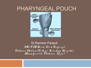
  • 7. Weakareas : LATERAL : 1. Above the superior constrictor 2. Between the superior & middle constrictors 3. Between the middle & inferior constrictors 4. Below cricopharyngeus- Killian-jamieson’s area (betn oblique & transverse fibers of cricopharyngeus muscle) POSTERIOR: 1. Laimer-Hackermann’s area (betn cricopharyngeus & superior most oesophageal circular muscle) 2. Killian’s dehiscence (betn thyropharyngeus & cricopharyngeus )
  • 8. A. Killian's triangle: Region between the thyropharyngeus & cricopharyngeus B. Laimer's triangle: Region between the cricopharyngeal and most superior esophageal circular muscle C. Killian-Jamieson's triangle: Region between the oblique and transverse fibers of the cricopharyngeal muscle
  • 9. Introduction: ī‚¨ Oesophageal diverticula – classifications ī‚¨ 1.Anatomic location : -Pharyngo-oesophageal -Middle, thoracic or mid-oesophageal -Lower or Epiphrenic ī‚¨ 2.Mechanism of origin : -Traction diverticula -Pulsion diverticula
  • 10. Traction diverticula: īą Pulling forces external to the oesophagus -Inflammatory process -Neoplastic process īą Usually anterior wall near the tracheal bifurcation īą Adhesions following surgery to fuse anterior cervical spine after trauma
  • 11. Pulsion diverticula: ī‚¨ Herniation of oesophageal mucosa & submucosa ī‚¨ Pseudodiverticulum ī‚¨ Area of weakened musculature
  • 12. Pharyngo-oesophageal diverticula: ī‚¨ Pharyngeal: -Majority arise above the cricopharyngeus muscle eg. posterior pharyngeal pulsion diverticulum (Zenker’s diverticulum) -Most frequent īą Oesophageal: -Arise below cricopharyngeus muscle -Uncommon
  • 13. Pharyngo-oesophageal diverticula: ī‚¨ Congenital or acquired: ī‚¨ Multiple or single: ī‚¨ Lateral or posterior: ī‚¨ Size of the sac may vary from 1cm -12cm or more
  • 14. ī‚¨ May present at any age ī‚¨ Most present in later lifeīƒ acquired origin ī‚¨ Normally curable unless complicated by carcinoma
  • 15. Classification of pharyngeal diverticula: ī‚¨ lateral: 1. Congenital 2. Acquired (a) Normal bulges (b) Traumatic (c) Raised intrapharyngeal pressure (pharyngocoeles) ī‚¨ Posterior: 1.Congenital 2.Acquired (a) Traumatic (b) Raised intrapharyngo-oesophageal pressure (c) Posterior pharyngeal pulsion diverticulum (Zenker’s diverticulum)
  • 16. Lateral pouches: ī‚¨ Congenital ī‚¨ Acquired 1. Normal bulge 2. Traumatic 3. Raised intrapharyngeal pressure(pharyngocoele)
  • 17. Lateral pouches: ī‚¨ Uncommon ī‚¨ Arise from the posterior faucial pillar or the pyriform fossa īą Contrast enhanced cineradiography īą Clinically –modified valsalva manoeuvre ī‚¨ Divided into Congenital and Acquired- controversial
  • 18. Congenital lateral pharyngeal diverticula: ī‚¨ Extremely rare ī‚¨ Few cases reported ī‚¨ First two decades of life ī‚¨ Recurrent infected neck swelling with previous treatment ī‚¨ Developmental defect in Branchial apparatus ī‚¨ Branchial pouch derivates ī‚¨ Diagnosis: barrium swallow ī‚¨ Treatment:excision of diverticulum
  • 19. Acquired lateral pharyngeal diverticula: ī‚¨ Aetiology-argument still continues ī‚¨ Basic defect – congenital weaknessīƒ Congenital ī‚¨ Precipitating factor -Raised intrapharyngeal pressure -Muscular laxity -Ageing īą Usually in adultīƒ Acquired
  • 20. 1.Normal bulges: ī‚¨ Frequent & incidental findings on routine barium swallow ī‚¨ Small lateral pharyngeal bulge- Asymptomatic ī‚¨ Early stage in the evolution of larger diverticula ī‚¨ Usually bilateral & asymptomaticīƒ thought as normal variants ī‚¨ Arise from-pyriform sinus or tonsillar fossa ī‚¨ Modified valsalva manoeuvre ī‚¨ More common in elderly - reduced muscular tone - loss of elasticity of tissues ī‚¨ Radiological Contrast studies: smooth,hemispherical prominences arising from the pyriform sinus or tonsillar fossaīƒ Pharyngeal ‘ears’ ī‚¨
  • 21. 2.Traumatic: ī‚¨ Self inflicted diverticula: Habitual criminals from India Repetitive introduction of piece of lead into the tonsillar fossa ī‚¨ Probably lies between the middle & superior constrictors ī‚¨ If not maintained, disappears rapidly
  • 22. 3. Pharyngoceles (Raised intra-pharyngeal pressure) ī‚¨ Large, occasionally symptomatic diverticula ī‚¨ Usually unilateral but occasionally bilateral ī‚¨ Predominantly in male (M/F=8:1) ī‚¨ First described by Wheeler (1886) ī‚¨ Arise from precursor pharyngeal ‘ears’ ī‚¨ Development 1. Frequent repetitive increase in intrapharyngeal pressure 2. Loss of muscle resilience 3. Both Lateral pharyngocele: variable location above and lateral to the cricopharyngeus
  • 23. īą Predisposing factors: Younger patients- playing wind instruments, violent sneezing, or coughing Older patients- laxity of musculature ī‚¨ Both group: intrinsic weakness in the lateral wall ī‚¨ Symptoms: Dysphagia, food regurgitation, halitosis, foul taste, nocturnal coughing, choking- food entrapment in the diverticulum Dyshphonia -Spillage into larynx or -Compression of recurrent laryngeal nerve
  • 24. ī‚¨ Signs: just anterior to SCM palpable lump, soft & compressible ī‚¨ Indirect laryngoscopy: slit like ostium in the region of the posterior faucial pillar or the pyriform sinus ī‚¨ Plain radiograph: translucency-lateral to PFS ī‚¨ Ultrasonography: ī‚¨ Cine or videofluoroscopic technique with barium: rounded, contrast lined opacity communicating with the PFS or tonsillar fossa with neck ī‚¨ Direct pharyngoscopy:search for opening in those areas ī‚¨ Treatment : Asymptomatic- no treatment, but follow up Symptomatic: excision of the diverticulum
  • 25. Posterior Pouches: ī‚¨ More common ī‚¨ Posterior pharyngeal pulsion diverticulum (zenker’s diverticulum)- most common ī‚¨ Congenital ī‚¨ Acquired 1. Traumatic pharyngeal pseudodiverticulum 2. Diverticulum resulting from raised intrapharyngo-oesophageal pressure 3. Posterior pharyngeal pulsion diverticulum(zenker’s diverticulum)
  • 26. Congenital posterior pharyngeal pouch: ī‚¨ Very rare ī‚¨ First described in infants –symtoms similar to oesophageal atresia ī‚¨ Radiological evidence of air in stomach in the absence of tracheo-oesophageal fistulaīƒ oesophageal patency ī‚¨ Whole diverticulum covered with muscle- distinguished from acquired pulsion diverticulum ī‚¨ Treatment: excision of diverticulum
  • 27. Acquired posterior pharyngeal pouch: 1.traumatic pharyngeal pseudodiverticulum: ī‚¨ Very rare condition ī‚¨ Usually presents in newborn infants but reported in adults too ī‚¨ Aetiological factor: hypopharyngeal trauma either from damage caused by the obsterician’s finger during breech delivery or blind passage of the suction tubes īą Spontaneous rupture of a retropharyngeal abscess in immunocompromised adult patient- reported case
  • 28. ī‚¨ Abdominal radiograph- air in stomachīƒ  oesophageal atresia īą Radiological appearance: irregular elongated tract originating in the pharynx & passing behind the oesophagus into the posterior mediastinum ī‚¨ Treatment : not clearly defined Conservative treatment: Deterioration of the general condition: surgical drainage of the pseudodiverticulum
  • 29. 2. Diverticulum resulting from raised intrapharyngo -oesophageal pressure: ī‚¨ Rare ī‚¨ The laimer-Hackerman area ī‚¨ Elderly people ī‚¨ Weakness of the musculature ī‚¨ Always asymptomatic ī‚¨ No treatment required ī‚¨ Vary in size depending on the peristaltic wave ī‚¨ Some discount their existence altogether
  • 30. 3. Posterior pharyngeal pulsion diverticulum: (Zenker’s diverticulum) ī‚¨ Most common ī‚¨ Many names: -Pharyngo-oesophageal pouch or diverticulum -Retropharyngeal pouch or diverticulum -Posterior pharyngeal pouch or diverticulum -Zenker’s diverticulum -Cricopharyngeal achalasia -Hypopharyngeal diverticulum Friedrich Albert von Zenker, Professor of Pathology at Erlangen University (1825– 1898), German pathologist whose name is associated with Zenker's diverticulum
  • 31. ī‚¨ Acquired, pulsion diverticulum between the thyropharyngeus and the cricopharyngeus muscle in an area of weakness called Killian’s dehiscence /triangle/hiatus ī‚¨ Described by Killian in 1907
  • 32. ī‚¨ Found almost exclusively in humans ī‚¨ Hypothesized to be secondary to the large size & relatively caudal location of the larynxīƒ oblique orientation of the pharyngeal constrictor muscles īƒ regions of weakness ī‚¨ Some animals- pig, camel, monkey & elephant ī‚¨ Theoretically- diverticulum herniates to the side of least resistance ī‚¨ ZD more prone to herniate to the left: īƒ˜ Left carotid artery located more laterallyīƒ less adherent to the adjacent prevertebral fascia īƒ˜ Cervical oesophagus slight convexity to the left
  • 33. Incidence: ī‚¨ Difficult to quantify the incidence in general population ī‚¨ Incidence of presentation to ENT specialist- 0.47 cases per 100,000 per year ī‚¨ In 1999, Incidence in Oxford region UK- 1/100,000 ī‚¨ Men affected 2-3 times more often than women ī‚¨ Usually above 50 yrs, 7th -8th decade of life ī‚¨ Affects caucasians ī‚¤ Extremely rare in Asian and African
  • 34. ī‚¨ First case described by Abraham Ludlow, surgeon from Bristol in 1764 ī‚¨ Ineffectual swallowing attempts leading to pharyngeal distension (Sir Charles Bell, 1817) ī‚¨ Early classifications of oesophageal diverticula by Zenker called this type ‘pulsion diverticula’ ī‚¨ Zenker and Von Ziemssen (1878) reviewed 22 cases between 1764-1876īƒ  symptoms & possible pathogenesis ī‚¨ Spasmodic contraction of the circular fibers at the upper end of the oesophagus (Killian, 1907)
  • 35. Aetiology: īą Unknown ī‚¨ Conflicting evidence based on anatomical, radiographic, manometric and electromyographic studies ī‚¨ Many theories: 1. Spasm of the cricopharyngeus muscle (Negus, 1950) 2. Lack of inhibitory stimuli to the cricopharyngeus (Dohlman and Mattsson, 1959) 3. The second swallow(due to pharyngeal laxity) (Wilson, 1962) 4. Neuromuscular incoordination and congenital weakness(Korkis, 1958)
  • 36. 1. Spasm of the cricopharyngeus muscle: ī‚¨ Human evolution to an erect position with larynx & cricopharyngeus moving lower down the neck, causing other constrictors to lie obliquelyīƒ Killian’s dehiscence (Negus, 1950) ī‚¨ Persistent, tonic spasm of cricopharyngeus (inflammation, stenosis, or neurological deficit)īƒ  high pressureīƒ herniation of mucosa through Killian’s dehiscence (Sutherland, 1962 and Belsey, 1996)
  • 37. 3. The second swallow (due to pharyngeal laxity): ī‚¨ Due to pharyngeal muscular laxityīƒ weak pharyngeal stripping(peristaltic) wave īƒ unable to clear the whole bolus before the cricopharyngeal sphincter contractedīƒ residue left in the pharynx ī‚¨ Second swallow needed to clear the residueīƒ against a closed sphincterīƒ high pressure īƒ  mucosal bulging, If long standingīƒ  diverticulum (Wilson, 1962)
  • 38. 2. Lack of inhibitory stimuli to the cricopharyngeus: ī‚¨ During deglutition- the larynx elevated pulling the cricopharyngeus upwards rather than stretching the muscle which normally trigger off a reflex arc resulting sphincter relaxationīƒ readiness for bolus ī‚¨ The cricopharyngeal sphincter failed to relaxīƒ increased intrapharyngeal pressure īƒ mucosal bulging posteriorly ī‚¨ Prevertebral fascia weakening with age
  • 39. 4. Neuromuscular incoordination and congenital weakness: ī‚¨ Neurological disorder in presence of congenital weaknessīƒ  diverticulum (Korkis, 1958) ī‚¨ If diverticula were acquired, they should occur more frequently in women as dysphagia is more common in women ī‚¨ But diverticula are more common in men ī‚¨ Gastro-oesophageal reflux may lead to cricopharyngeus spasm or incoordination ( Resouly, 1994)
  • 40. Risk factors: ī‚¨ Older age ī‚¨ Male gender ī‚¨ Hiatal hernia ī‚¨ Gastro-oesophageal reflux(GORD)
  • 41. īą Pulmonary complications: Aspirationīƒ  Recurrent respiratory infections, pneumonia, bronchiectasis & lung abscess īą Hoarseness: -Laryngitis(aspiration or gastric reflux) -Compression of recurrent laryngeal nerve -Carcinoma in the diverticulumīƒ vocal cord paralysis īą Belching, choking, coughing
  • 42. Symptoms: ī‚¨ Symptoms of variable severity not necessarily related to the size of the pouch ī‚¨ Longstanding and slowly progressive symptoms ī‚¨ Dysphagia- most common symptom, virtually in all pts initially for solids, then semisolid and finally liquid īą Regurgitation of undigested food- 80% patients īą Noisy deglutition (borborygmi) īą Hoarseness īą Foul taste and halitosis īą Weight loss & malnutrition īą Blood in regurgitated food contents- carcinoma ī‚¨ Pain – carcinoma īą Patients fail to respond to medication for another condition (tablets lodging in the sac)
  • 43.
  • 44. Signs: īą Usually without any specific findings, Minimal physical findings īą Emaciation or dehydration īą Soft, compressible swelling usually in the left side in anterior triangle ī‚¨ Laryingitis or Pooling of saliva in hypopharynx in I/L examination ī‚¨ Boyce’s sign (swelling in the neck that gurgles on palpation) ī‚¨ Spasm of coughing on palpation(spillage of contents into larynx) ī‚¨ Blood in regurgitated contents-carcinoma
  • 46. Investigations: ī‚¨ History and examination: virtually pathognomic ī‚¨ Confirm the diagnosis with radiological evidence 1. Barium swallow 2. Contrast videofluoroscopy 3. Plain radiography 4. Ultrasonography 5. Oesophagoscopy
  • 47. Barium swallow: ī‚¨ Internal contour examined īƒ˜ Irregular or filling defect within diverticulum: solid food remnants or carcinoma īƒ˜ Constant filling defect in lower two-third of sac-carcinoma īƒ˜ Filling defect in the neck of pouch- food and air bubbles ī‚¨ Long term radiographic follow up failed to show transient diverticulum īƒ  into full blown diverticulum
  • 48. īą Contrast video-fluoroscopy: īƒ˜ Constant monitoring of the swallowing (single shot barium swallow may miss small pouch) īƒ˜ Able to see pouch from different angles īƒ˜ Size, location, and character of the mucosal lining īƒ˜ Function of the pharyngeal muscles īƒ˜ Presence or absence of gastric reflux ī‚¨ Contrast study should include lower oesophagus & stomach – lower oesophageal carcinoma & hiatal hernia can coexist with pharyngeal pouch
  • 49. Images obtained during barium swallow videofluoroscopy demonstrating an intermediate-sized Zenker diverticulum
  • 50. ī‚¨ Plain radiograph of neck Triangular lucency in the prevertebral tissues with apex at the level of cricoid(due to air in the upper part of pouch),base has meniscus(due to fluid in the fundus) Chest x-ray of a 75-year-old patient with a 6-cm Zenker's diverticulum. A, Before barium swallow. Note the hazy soft tissue mass in the right upper lung field (arrowheads) representing the Zenker's diverticulum. B, After barium swallow in the same patient. Air-fluid level can be seen within the Zenker's diverticulum.
  • 51.
  • 52. ī‚¨ Rigid or flexible oesophagoscopy: -to assess the nature of the mucosa of the diverticulum -to exclude the presence of SCC or carcinoma in situ Care must be taken with rigid esophagoscopy to avoid perforating the Zenker diverticulum ī‚¨ Esophageal or hypopharyngeal manometry: does not add to the clinical workup ī‚¨ Ultrasonography:
  • 54.
  • 55. There are classifications based on contrast radiography, vertebral body measurements, and simple radiologic appearance, but categories are becoming increasingly complex and incorporate elements from several of the classic classification schemes. Clinical utility is not particularly high There are classifications based on contrast radiography, vertebral body measurements, and simple radiologic appearance, but categories are becoming increasingly complex and incorporate elements from several of the classic classification schemes. Clinical utility is not particularly high
  • 56. Pathology: ī‚¨ Lined with stratified squamous epithelium ī‚¨ No muscular layer exists ī‚¨ Fibrosis surrounding the diverticulum is common The fibrotic tissue limit the spread of any extravasated material from the diverticulum during endoscopic procedures īƒ  reduce likelihood of local abscess
  • 57. Complications of Zenker’s Diverticulum: 1. Oesophageal obstruction 2. Aspirarion pneumonia, bronchiectasis, lung abscess Recurrent infection 3. Compression of trachea 4. Ulceration 5. Squamous cell carcinoma: 0.4% Chronic inflammation of lining of diverticulumīƒ Ca (Sood and Newbegin, 2000) 6. Diverticulo-tracheal fistula Additional risk factor in the overall health of the elderly patient. īƒ deterioration of pulmonary function īƒ cachexia/dehydration/malnutrition secondary to “fear of eating”
  • 58. Treatment: 1.Conservative treatment: - Asymptomatic patients: No treatment but follow up -If general condition is poor and medically unfit or with minimal symptoms: No treatment 2.Pharyngeal pouch surgery: Symptomatic patients: surgery is the mainstay of treatment 1.Endoscopic surgery: 2.External approach surgery:
  • 59. Algorithm to approach a patient with pharyngeal pouch
  • 60. Surgical treatment methods: ī‚¨ External: 1. Cricopharyngeal myotomy alone 2. Diverticulectomy (Excision) 3. Diverticulopexy(Suspension) 4. Inversion īą Endoscopic: 1. Dilatation 2. Diathermy/Electrocoagulation (Dohlman’s operation) 3. Laser :Co2, KTP 4. Stapling(Endoscopic Staple Diverticulostomy)
  • 61. External approaches: 1.Diverticulectomy: ī‚¨ In 1886, Wheeler reported the first successful excision of pharyngeal pouch ī‚¨ Oesophagoscopy- openings identified ribbon gauze soaked with BIPP or proflavin packed, NG tube inserted ī‚¨ Transverse incision at the upper border of the cricoid, extending laterally to the SCM muscle(usu. left side) ī‚¨ Retract the SCM muscle and carotid sheath contents laterally, thyroid glands & cartilage retracted medially ī‚¨ Anterior belly of Omohyoid, Middle thyroid veins identified and divided
  • 62. ī‚¤ The recurrent laryngeal nerve identified, Inferior thyroid artery divided īą Diverticulum fundus grapsed with babcock forceps & the sac neck dissected free of oesophagus ī‚¤ CP sphincter & upper circular fibers of oesophagus divided posteriorly ī‚¤ Connell suturing ī‚¤ Drain ī‚¨ Especially useful: -Carcinoma in pouch: diverticulectomy + post- operative radiotherapy -Large perforation if happens during attempted endoscopic stapling
  • 63. 2. Cricopharyngeal Myotomy: ī‚¨ Richardson in 1899 perfomed first cricopharyngeal myotomy ī‚¨ Can be performed alone for small diverticulum (<2cm) or in combination to other procedure ī‚¨ Other surgical procedure combined with itīƒ decreased recurrence ī‚¨ Creating a tunnel betn circular muscle fibers & submucosa with curved artery forcepsīƒ dividing muscle betn opened forceps ī‚¨ 3-4 cm length divided, as posteriorly as possible to avoid damage to recurrent laryngeal nerve
  • 64. 3. Diveritculopexy: ī‚¨ Schmid in 1912 described the method of diverticulopexy ī‚¨ For high risk surgical candidates, CP myotomy and diverticulopexy is preferred Diverticulopexy technique: After a cricopharyngeal myotomy is performed and diverticulum freed, the sac is tacked with 2-0 silk sutures superiorly to the prevertebral fascia
  • 65. 4. Inversion: ī‚¨ First described by Girard (1896) ī‚¨ Bevan (1917) modified by placing series of purse string suture along the length of sac to obliterate it ī‚¨ Carried out in same way as for excision but After mobilisationn of pouch & CP myotomy, the pouch is invaginated into the oesophagus & its neck oversewn with interrupted catgut sutures instead of being excised
  • 66. Endoscopic treatment methods ī‚¨ In 1917 Mosher first described endoscopic approach but abandoned due to complications ī‚¨ In 1960,Dohlman and Mattsson popularised the procedure(cautery) ī‚¨ In 1984, Van Overbeek introduced use of operating microscope & CO2 LASER(15-20 W power) ī‚¨ Bent and Kuhn in 1992, used potassium titanyl phosphate laser(KTP) ī‚¨ In 1993, Collard et al introduced endoscopic stapling technique Dohlman Portrait: Gosta Dohlman, Professor of Oto-rhino-laryngology at Lund University who introduced endoscopic diathermic diverticulostomy
  • 67. Endoscopic treatment methods: 1.Dilatation: ī‚¨ Lahey in 1946 recommended cricopharyngeal dilatation ī‚¨ Early treatment method for dilating Cricopharyngeal sphincter- using bouginage or hydrostatic bag ī‚¨ Temporary relief from symptoms ī‚¨ Risk of perforation ī‚¨ Rarely used nowadays except to dilate post-operative stenosis
  • 68. 2.Endoscopic diathermy (Dohlman’s operation): ī‚¨ First described by Mosher in 1917 using scissorsīƒ abandoned due to complications ī‚¨ Dohlman and Mattsson modified and popularized in 1960 ī‚¨ Short operation lasting 30 mins ī‚¨ Can be performed under LA, if GA contraindicated (useful in elderly & GA unfit pts) ī‚¨ Doesn’t remove the pouch ī‚¨ Relieves the symptoms & restores swallowing - dividing the cricopharyngeus & widening the mouth of the diverticulum Endoscopic electrocautery technique
  • 70. 4. Endoscopic staple diverticulostomy(ESD): ī‚¨ Modified laryngoscope (Weerda bivalved laryngoscope) īƒ Visualization of the diverticulum īƒ expose common wall betn oesophageal & diverticular lumen ī‚¨ Magnified view of field-rigid 0 or 30 degree telescope with video camera ī‚¨ 2-0 silk retraction sutures through lateral edges of common wall ī‚¨ Upper blade (long beak) into oesophagus & lower blade (short beak) into neck of pouch
  • 71. ī‚¨ Suspension apparatus connected ī‚¨ Common wall(cricopharyngeal bar) divided using staplerīƒ internal cricopharyngeal myotomy ī‚¨ Stapler simultaneously cuts & staples the divided mucosal edges of common wall ī‚¨ Single lumen created without removal of pouch
  • 72.
  • 73.
  • 74. ī‚¨ Endoscopic staple diverticulostomy is superior to external as well as other, endoscopic approaches (Chang et al, 2003 ) ī‚¨ Endoscopic techniques-performed faster, short in patient stay,shorter anaesthetic time (important in elderly & medically infirm), recover more quickly ī‚¨ Simultaneously divide & “suture” with staplesīƒ reduced risk of perforation ī‚¨ No thermal damage to recurrent laryngeal nerve
  • 75. ī‚¨ Not only for ZD, but for all other hypopharyngeal and pharyngeal diverticula ī‚¨ ESD can be performed in pts with recurrence of diverticulum after external or endoscopic approaches ī‚¨ Endoscopic and external approaches are equally effective treatments (Overbeek 1994, Liang et al,1995, and Bonafede 1997) ī‚¨ The endoscopic stapling technique appears to have an improved efficacy and safety when compared with the CO2 laser technique (Miller et al,2006)
  • 76. Limitations of endoscopic techniques: ī‚¨ Exposure of the diverticulum may be difficult or impossible – kyphosis, large cervical osteophytes or small oropharyngeal opening ī‚¨ In smaller pouches(<2cm) insufficient cricopharyngeal myotomy performed ī‚¨ Pouch can be inspected throughly & biopsy taken, but complete specimen for pathological exam not obtained ī‚¨ Malignancy –endoscopic method contraindicated
  • 78. Current management in pharyngeal pouch surgery by UK Otorhinolaryngologists (Siddiq Mand Sood S, 2004) Procedures performed by consultants
  • 80. Complications of pharyngeal pouch surgery: ī‚¨ Immediate: 1. Haemorrhage: Slippage of ligature 2. Pneumothorax: Mobilisation of large pouch with adhesions 3. Surgical emphysema: Mucosal tear ī‚¨ Early: 1. Secondary haemorrhage: Usually due to infection 2. Hoarseness: Risk of damage to RLN in external approach(3-5%) 3. Wound infection or abscess: Spillage of contents during surgery or through suture line(1.5-5%) 4. Fistula: Secondary to infection(1-8%) 5. Mediastinitis: Leakage 6. Aerocele: Sup.mediastinum
  • 81. ī‚¨ Late: 1. Persistent hoarseness: Division of recurrent laryngeal nerve 2. Stricture: Excising too much mucosa 3. Recurrence: -Symptomatic relief after surgery (external or endoscopic approach)-90% in short term -All methods have recurrence -If cricopharyngeal myotomy not done- higher recurrence -Higher for endoscopic diathermy(6-7%) than diverticulectomy(0.5-4%) -Recurrences can easily be treated endoscopically than externally -If patient has recurrent symptoms after endoscopic procedure, contrast studies are rarely helpful (Jaramilo et al, 2001)
  • 82. Future and Controversies: ī‚¨ A complete understanding of the aetiology of pharyngeal pouch formation is not available ī‚¨ Further studies focused on the function of the CP muscle are likely to be fruitful ī‚¨ The final role for endoscopic procedures (with the laser or stapler) awaits further analysis and longer-term follow-up studies. ī‚¨ Flexible endoscopic cricopharyngeal myotomy (Recipi et al, 2010 ) ī‚¨ Harmonic scalpel in the treatment of Zenker's diverticulum (Fama et al, 2009)

Editor's Notes

  1. Up to 94% of patients with pharyngeal pouches are found to have GERD and/or hiatal hernias.
  2. Why treat Zenker’s? In the elderly population in particular, the presence of Zenker’s is an additional risk factor in the overall health of the elderly patient. In can cause deterioration of pulmonary function, as well as cachexia/dehydration/malnutrion secondary to “fear of eating”
  3. Thank You