2. • A 45-year-old male ….
• C/O –
Yellowish discoloration of eyes, itching & lowgrade intermittent fever since 3 months.
Swelling over the feet, abdominal distension and
abdominal pain since last month.
• Past alcoholic but had stopped since one year.
• Denied any high-risk sexual behavior.
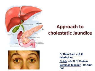
Jaundice
4. CholestasisDefinition –
Conjugated hyperbilirubinemia due to :
I. Impaired bile formation (hepatocytes)
II. Impaired bile flow (bile ducts/ductules)
Consequences• Secondary liver damage
I. Bile acid-induced hepatocyte injury
II. Secondary biliary cirrhosis
• Failure of substances secreted in bile to reach intestine
I.
II.
Bile acid deficiency in gut
Fat malabsorption/fat-soluble vitamin malabsorption
5. Towards central hepatic vein
Biliary canaliculus
Liver cell
Obstruction
C
Bilirubin,
Bile salts, &
Phospholipids
Endoplasmic reticulum
N
Sinusoidal blood
Towards Interlobular Bile duct
Screening tests that suggest cholestasis –
Color change in skin/sclera/stool/urine
Biochemical tests (Alkaline Phosphatase , Bilirubin)
6. Clinically-
Histologically-
Bile plugs (bilirubinostasis),
Feathery degeneration of
hepatocytes (cholate stasis),
Small-bile-duct destruction,
Peri cholangitis,
Portal edema,
Bile lakes and infarcts
(typically with extrahepatic
obstruction),
Finally , biliary cirrhosis.
Pruritus,
Fatigue,
Xanthomas,
Hepatic Osteodystrophy:
back pain from
osteoporosis,
Pale stools, or
steatorrhea
Evidence of fat-soluble
vitamin deficiency.
Enlarged liver with a
firm smooth non-tender
edge.
After 3–5 yrs of jaundice , liver cell failure indicated by deep jaundice, ascites, edema and
a lowered serum albumin develops. Pruritus lessens and the bleeding is not controlled by
vitamin K. Hepatic encephalopathy is terminal.
8. EVALUATION OF CHOLESTATIC JAUNDICE
• The first question -whether the cholestasis is
from intrahepatic or extrahepatic process.
CLUES TO EXTRAHEPATIC
OBSTRUCTIONS –
•Abdominal pain,
•Palpable GB or
upper abdominal mass,
•Evidence of cholangitis, and
•H/O- past biliary surgery.
CLUES TO INTRAHEPATIC
CHOLESTASIS-
Pruritus, as in primary
biliary cirrhosis (PBC)
and primary sclerosing
cholangitis (PSC) patient
9. Extrahepatic causes of cholestatic jaundice
Benign
Choledocholithiasis
Postoperative biliary
strictures
Primary sclerosing
cholangitis
Chronic Pancreatitis
AIDS cholangiopathy
Mirizzi’s Syndrome
Parasitic Disease
(Ascariasis)
Malignant
Cholangiocarcinoma
Pancreatic cancer
Gall Bladder Cancer
Ampullary Cancer
Malignant involvement
of the porta hepatis
lymph nodes
10. Intrahepatic causes of cholestatic jaundice
1) Viral Hepatitis
A. Fibrosing cholestatic hepatitis – Hep. B
&C
B. Hep.A, EBV, CMV
2) Alcoholic Hepatitis
3) Drug toxicity
A. Pure cholestasis- Anabolic &
contraceptive steroids
B. Cholestatic hepatitis- chlorpromazine,
erythromycin, Amoxiclav
C. Chronic cholestasis- chlorpromazine &
prochloperazine
4) Primary Biliary cirrhosis
5) Primary Sclerosing cholangitis
6) Vanishing Bile duct Syndrome
A. Chronic rejection of liver transplant
B. Sarcoidosis
C. Drugs
7) Non hepatobiliary Sepsis
8) Benign post-operative cholestasis
9) Para neoplastic Syndrome
10) Veno-occlusive disease
11) GVHD
12) Inherited
A. Progressive familial
intrahepatic cholestasis
B. Benign recurrent cholestasis
13) Cholestasis of pregnancy
14) Total Parenteral Nutrition
15) Infiltrative diseases
A. TB
B. Lymphoma
C. Amyloidosis
16) Infections
A. Malaria
B. Leptospirosis
11. • Risk factors1.
2.
3.
4.
5.
6.
Alcohol intake,
Medications,
Pregnancy
Sexual contact, drug abuse, needle punctures.
ICU – Sepsis, shock liver & TPN .
After BM transplantation- Veno occlusive disease or GVHD
• family history –
Benign recurrent intrahepatic cholestasis (BRIC).
• Details –
Onset, duration,
Intermittent or progressive,
Associated symptoms like dark urine, acholic stools, arthralgia,
rash, wt loss, fever, chills, and pain in RHC.
12. Clinical History & cause of cholestasis
1. Pain - duct stones, tumor or gallbladder disease.
2. Arthralgia , myalgia predating jaundice -hepatitis (viral/drug related)
3. Fever and rigors- cholangitis d/t duct stone or traumatic stricture
(Charcot’s intermittent biliary fever). Or systemic infection
4. Contaminated foods, /alcohol consumption.
5. H/O hepato-toxins - Drugs /chemicals/ occupational
6. Parenteral exposures (Bl. Transfusions, drug abuse/ tattoos, sexual
activity)
7. H/o Ulcerative colitis - ? PSC
13. Physical Examination –
1) S/o chronic liver disease, temporal & proximal muscle weakness.
2) S/o Cholesterol deposition (Xanthomas, xanthelasmas) flat or slightly raised, yellow
and soft , usually around the eyes, in the Palmar creases, below the breast and on the neck, chest or back.
3) Anemia – GI blood loss, nutritional deficiency, hypersplenism
4) Itching marks, clubbing, and lymphadenopathy.
5) Virchow’s node or sister Mary Joseph's nodule- abdominal malignancy.
6) Jugular venous distension – hepatic congestion.
7) S/o Fat soluble vitamin deficiency –
1)
2)
3)
4)
Vit.D (osteomalacia, Demineralized bone, Kyphosis, Fractures),
Vit.E (cerebellar ataxia, posterior column dysfunction, peripheral neuropathy),
Vit.K(Puncture hematoma, Spontaneous bruising )
Vit.A(night blindness, thick skin)
8) S/O Hepatic Osteoarthropathy – loss of height, back pain, collapsed
vertebrae & fractures particularly of ribs with minimal trauma.
16. Abdominal Examination
1.
Hepatomegaly
Alcoholic liver disease, primary or secondary hepatic neoplasm,
infiltrative disease, and primary biliary cirrhosis.
2.
Enlarged tender liverViral ,alcoholic hepatitis, infiltrative process, or chronic passive
congestion of liver.
3.
Murphy’s Sign –
Cholecystitis, Ascending cholangitis
4.
Enlarged gallbladder –
Non- calculous biliary obstruction
5.
Hard & nodular hepatomegaly –
? metastatic malignancy.
6.
Other abdominal masses –
Primary ca stomach or colon.
17. • Ascites + Jaundice - Cirrhosis or malignancy with peritoneal spread.
• Rectal examination and sigmoidoscopy may indicate carcinoma.
• Marked splenomegaly- Cirrhosis + portal HTN or lymphoproliferative
disease
• Stools - loose, pale, bulky and offensive , sticky to the pan & non
flushable
-Our Case –
O/E –
Pallor +, icteric , B/L pedal edema.
1 x 1 cm firm lymph node in left
axilla.
P/A –
Tender hepatomegaly - 3 cm with
a smooth surface.
-Investigations –
Total bilirubin 10 mg% (D= 4.2).
ALP (1923 IU/L) & GGT 85 IU/L- raised.
AST and ALT normal.
Albumin of 2.4 gm%.
PT INR 1.7 corrected by Vit.K suppl.
Hb 10 gm/dl .Normal PBS
Urine positive for BS & BP
18. Laboratory work up
• CBC –
Anaemia - infection, blood loss or malignant disease.
PMN leucocytosis - cholangitis or underlying neoplasm.
• LFTs1.
2.
3.
4.
Alkaline Phosphate out of proportion to ALT/AST
Albumin linked Bilirubin (δ Fraction / biliprotein)
Low albumin - chronic process (cirrhosis/ cancer)
Normal Albumin - acute process ( viral
choledocholithiasis)
5. Elevated PT – Vit K Deficiency
• RFTs- Sepsis , HRS, malignancy .
hepatitis/
19. Enzymes raised in cholestasis
1) Alkaline Phosphatase (ALP), gamma-glutamyl
transpeptidase (GGT) & 5’-nucleotidase (5’NT).
2) ALP isoenzymes are also present in bone &
placenta.
3) Increase in ALP, GGT & 5’NT hepatobiliary
origin.
4) GGT levels – Fatty liver, alcoholic liver disease.
20. Proteins:
Albumin:
Decreased – advanced cirrhosis
& signify severe hepatic dysfunction.
Usually normal - acute hepatitis
Globulins:
Non-specific elevation – Chronic liver disease.
Disproportionate elevation
1) IgG in autoimmune hepatitis,
2) IgM in PBC &
3) IgA in alcoholic liver disease.
21. Prothrombin time (PT) & (INR)
An increasing INR/PT - hepatocellular dysfunction.
May be deranged in cholestasis,
1. But due to the malabsorption of Vit. K
Rapidly corrected by Parenteral administration of Vit K.
22. Other tests:
• Serological/ replicative markers –
specific diagnosis of acute or chronic viral hepatitis.
• Anti mitochondrial antibody (AMA) for PBC (90%)
• P-ANCA in PSC (65%)
• Antinuclear factor (ANA),
• Anti-smooth muscle antibody (ASMA) &
• Anti-liver kidney microsome (LKM) antibody
• Alpha- feto protein, which is raised in HCC &
• Other Malignancies – CEA, CA19.9, PSA
• S. Ceruloplasmin for Wilson disease.
seen in
autoimmune
hepatitis;
23. • Serum drug levels
• Urine dipstic test (Ictotest) Conjugated bilirubin +ve.
• X rays- changes of
osteomalacia
• Bone mineral density
by dual energy x-ray
absorptiometry (DEXA).
24. Imaging
RUQ Ultrasound
CT scan
ERCP
PTC
Endoscopic Ultrasound
Endoscopic CT
MR cholangiography
USG abdomen –
Normal size liver & echo pattern
With intrahepatic biliary radical
dilatation (IHBRD) in left lobe,
splenomegaly (18 cm),
Normal CBD and gallbladder.
Minimal free fluid.
No focal lesions
USG
Dilated bile
ducts
Non- dilated
bile ducts
? Intra hepatic
cholestasis
CT/MRCP/
ERCP/PTC
Serologic studies
AMA
Hepatitis serologies
Hep -A, CMV, EBV
Review drugs
Negative
MRCP/ liver biopsy
AMA
Positive
Liver Biopsy
25. Imaging
USG
Limitations
First-line imaging
Inexpensive
No ionizing radiation
GB stones readily
detected.
5. Absence of biliary
dilatation suggests
intrahepatic cholestasis &
oppositely extrahepatic
cholestasis
1) Distal CBD, bowel gas,
Obesity
2) False negative – Partial
obstruction,
cirrhosis,
scarring d/t PSC
3) Except mass lesion in the
head of the pancreas, USG
usually does not identify
the type of obstruction.
1.
2.
3.
4.
27. Our Case
Other tests –
1)
2)
3)
4)
5)
Viral serologies – Ve for HIV, HBV and HCV .
Blood culture sterile.
Sputum AFB , CXR - NAD.
Weil-felix, Paul-Bunnel and Brucella serologies negative.
Ascitic fluid - Transudative no cells & negative ADA.
Provisional diagnosisAlcoholic liver disease with ? Biliary malignancy
A contrast CT abdomen –
Multiple ill-defined nonenhancing lesions in the
liver, largest 1.4 cm x 1.0 x 1.0 cm rounded lesion (? necrotic lymph node)
at the porta hepatis with IHBRD seen above this level in left lobe. Multiple
small para-aortic, periportal and mesenteric lymph nodes present.
28. CT Scan
1) Localizes level of the
obstruction, in about
90% cases.
2) First choice in
lymphoma,
for retroperitoneal
lymph node
involvement
Gallbladder
Dilated bile
ducts
Mass in
head of
the
pancreas
Dilated bile ducts and gallbladder
29. When clinical suspicion is supported by CT or USG,
1) MRCP
Noninvasive screening ,rapid and comfortable.
Failed or incomplete conventional ERCP.
Variant biliary duct anatomy/ congenital duct abnormalities.
Post operative anatomy where ERCP would be difficult.
Evaluating changes of chronic pancreatitis or sclerosing cholangitis.
Distal CBD Stone
PSC
30. Direct cholangiography (PTC and ERCP)
Direct visualization
99% sensitivity & specificity.
Therapeutic interventions .
ERCP is the procedure of choice in suspected
ampullary or duodenal lesions in ca pancreas & when
gallstone
obstruction
is
suspected,
where
sphincterotomy & stone extraction can be
implemented.
34. Biliary stricture due to
cholangiocarcinoma
Alk phos = 669 IU Bili = 17.5 mg/dl
AST =
68 IU
ALT = 38 IU
Bile duct obstruction
from chronic pancreatitis
35. • PTC is preferred when obstructing lesion is high
• C/I- Marked ascites and coagulopathy.
• PTC and ERCP may be used together across a difficult
obstruction.
• Sometimes hepatobiliary scintigraphy, may help
evaluating biliary leaks & congenital malformations.
• Endoscopic CT & MRCP –
Biliary obstruction, specially
transplantation.
in
setting
of
in
liver
37. Liver Biopsy
Major indications –
Contraindications –
1)
2)
3)
4)
1) Bleeding tendencies,
chronic hepatitis,
cirrhosis,
Unexplained abnormal LFT,
hepatosplenomegaly of
unknown etiology,
5) suspected infiltrative /
Granulomatous disease
Complications
1) Minor – Prolonged RHC pain,
(6%).
2) Major - intra-abdominal
bleeding, mortality ( 0.01%).
2) INR>1.5 or PT >3 sec
above the control,
3) Severe thrombocytopenia
4) Marked ascites .
Transjugular liver biopsy
or no biopsy at all are
alternatives.
38. Cholestatic liver disease is broadly categorized as
extra-hepatic or intrahepatic.
Common
hepatic duct
Liver
Gallbladder
Intrahepatic
Perihilar
Distal
extrahepatic
Common
bile duct
Ampulla
Of Vater
Duodenum
39. CHOLEDOCHOLITHIASIS
• Mechanism of jaundice –
i. Impaction & edema of the common duct (Mirizzi syndrome)
ii. Direct inflammation of porta hepatis.
• CBD stones retained after cholecystectomy may produce jaundice
in the immediate postoperative period or even several years later .
• Pain (biliary colic or from acute pancreatitis).
• Rapid rise & rapid decline within 72 hours in aminotransferases .
• If Cholangitis in choledocholithiasis –
Fever with chills, abdominal pain, & jaundice. ( Charcot’s triad )
40. BENIGN STRICTURES OF THE BILE DUCTS
• In adults most common after surgery.
• PSC -multiple or diffuse strictures.
• Chronic alcoholic pancreatitis- a long stricture in the
intrapancreatic portion of the common duct.
• Ampullary stenosis - trauma during passage of a stone
& AIDS.
• Cholangitis - frequent in benign than in malignant one.
41. NEOPLASTIC OBSTRUCTION
• Pancreatic carcinoma commonest
• Other tumors Cholangiocarcinoma, ampullary
tumors, and carcinoma of GB
• Abdominal pain
• Loss of appetite and weight ,
• Progressive deep & painless
jaundice.
• Klatskin’s tumor
Macro cystic adenocarcinoma of
the pancreatic head.
42. • Tumors producing complete obstruction of CBD may be
accompanied by marked, palpable dilatation of the gallbladder
(Courvoisier’s law).
• Ampullary tumors produce intermittent jaundice because of
sloughing and partial relief of the block.
• Highest surgical cure of all tumors presenting as painless jaundice.
• Metastatic cancer may obstruct the bile duct, as may lymphoma.
• Hepatocellular carcinoma rupture into the biliary system throwing
tumor emboli obstructing common duct.
• Compression by adjacent tumor/ peribiliary lymph node
infiltrated by lymphoma, or metastatic ca breast.
• Direct infiltration by lymphoma.
43.
44. UNCOMMON CAUSES OF OBSTRUCTIVE JAUNDICE
• Choledochal cyst .
• Duodenal diverticulum
• Hemobilia, (biliary colic, jaundice & GI bleeding).
• Ascaris ,liver flukes (Fasciola, Clonorchis or Opisthorchis).
• Secondary sclerosing cholangitis
infections in immunodeficiency.
d/t
opportunistic
• Cryptosporidium parvum, cytomegalovirus (CMV), and
Microsporidia most frequently found.
46. Drug-induced cholestasis.
Direct hepato-toxic
Idiosyncratic
• Clinically mimic viral hepatitis or biliary tract disease.
• Serum-sickness-like features (rash , arthralgia,& eosinophilia)
• Only practical approach is to eliminate the drug and monitor.
Antimicrobial agents
Augmentin , cloxacillin,
erythromycin, ethambutol,
dapsone, fluconazole,
griseofulvin, ketoconazole
Cardiovascular agents
Disopyramide β-blockers,
ACE inhibitors, ticlopidine,
warfarin, methyldopa
Endocrine agents
Sulfonylureas, estrogens,
tamoxifen, androgens,
niacin, OCPs, anabolic
steroids
HAART-
Immunosuppressive agents –
Azathioprine, cyclosporine,
gold salts, NSAIDs
Psychopharmacologic
agents
Tricyclic
antidepressants, BZDs,
Phenothiazines,
Phenytoin, halothane
Zidovudine,
Protease Inhibitors
( Indinavir, Ritonavir)
47. Alcoholic hepatitis.
Marked tender hepatomegaly & e/o liver cell
failure .
Viral hepatitis.
Acute phase of viral hepatitis;
Most commonly hepatitis A, Hepatitis C, and
hepatitis E.
Though jaundice may be profound up to 6
months, complete recovery is the rule.
48. AIDS related cholangiopathy
• Cryptosporidium most frequent .
• Rarely
Microsporidia,
CMV,
Mycobacterium avium complex, and
Cyclospora
• Papillary stenosis if CD4 <100
• Elevated ALP (mean 800 IU/L).
• Jaundice unusual, If present, suggests
other disorders, like drug, alcohol
abuse or neoplasm
49. Primary biliary cirrhosis.
Autoimmune chronic non- suppurative cholangitis
AMA positivity in 95% & ANA 30% cases.
Progressive destruction of interlobular bile ducts(medium & small).
Elevated ALP, IgM, and cholesterol & later on bilirubin.
Predominantly middle aged female.
Fatigue, pruritus, hyper pigmentation, Xanthomas.
Majority have associated autoimmune disorders
(Sjögren’s syndrome, scleroderma, and arthritis).
Biopsy is diagnostic .
UDCA- only drug, prolongs survival & improves biochemical
abnormalities.
53. Idiopathic adulthood ductopenia.
Rare and defined by the presence of ductopenia (decrease of bile
ducts in >50% of the portal triads) and cholestasis in the absence of
known cholestatic liver disease. A diagnosis of exclusion.
Histology similar to primary biliary cirrhosis.
UDCA may result in biochemical improvement.
Autoimmune hepatitis.
Females (70%)
Anti LKM 1 , ANA and ASMA & hyper gammaglobulinemia in 80%.
Associated autoimmune disorders include arthritis, rash, thyroiditis,
Sjögren’s syndrome, and ulcerative colitis.
55. Decompensated chronic liver disease.
Jaundice may occur in chronic hepatitis or cirrhosis.
Other evidence of severe liver cell dysfunction is
present & jaundice is prognostically a grave sign.
Lymphoma.
3% and 10% cases of lymphoma develop jaundice .
Fatty liver.
Middle aged women with obesity, diabetes, and Hyper
lipidemia and a variety of other medical problems.
Cholestasis seen in about 5% cases.
56. Granulomatous hepatitis.
• Common cause of cholestatic liver disease.
• Sarcoidosis, infection (TB and fungal, esp. histoplasmosis),
hypersensitivity reaction, malignancies, IBDs, and as a feature of
other chronic liver disease.
• Pathologically,
Granulomas are nodular infiltrates consisting of aggregates of
epithelioid cells or macrophages with a rim of mononuclear cells/
Giant cells .
• Clinically, often asymptomatic, or Nonspecific symptoms .
• Routine bacterial & fungal blood culture, may be required.
• Benign course, with spontaneous recovery in most .
58. Sarcoidosis.
Systemic disease characterized by non- caseating
granuloma of multiple organs.
70% have hepatic granulomas. Portal granuloma result in
cholestasis & destruction of interlobular bile ducts.
Elevated alkaline phosphatase most characteristic
abnormality & reduced with corticosteroids.
Concomitant intrathoracic disease, pulmonary symptoms,
and significant anemia/ leucopenia.
59. Bacterial infection (sepsis)
• Most commonly gram-negative bacteria,
• also Staphylococcus aureus
streptococcal pneumonia.
in
TSS
&
• Rarely leptospira, clostridium & borrelia.
• Massive ductular dilatation & retained bile at
the interface of hepatic parenchyma & portal
tracts, (cholangitis lenta).
60. Total parental nutrition.
• When >60% calories as carbohydrates given > 3-4 weeks.
• Gallbladder stasis is almost universal & thereby gall stones.
• No oral feeding
that stimulate bile flow
Diminished release of hormones
Diminished bile flow
• Direct oxidant stress to the liver.
• If TPN cannot be discontinued, it should be cycled around
10 hrs/day. Keeping glucose <6 g/kg/day and lipid <2
g/kg/day.
• Recently UDCA is found helpful.
61. Benign recurrent intrahepatic cholestasis (BRIC).
• Characterized by...
Recurrent epi. of jaundice & pruritus,+ Symptom-free intervals.
Biochemical signs of cholestasis.
Histologically- canalicular stasis, normal bile ducts and absence of
inflammation and fibrosis.
• Sporadic or familial forms (chromosome 18)- Progressive familial
intrahepatic cholestasis (PFIC types 1-3) .
• GGT is normal with high alkaline phosphatase.
• Cholestatic episodes may last for many months.
• The episodes in BRIC eventually resolve without morphological
sequelae
62. Cholestasis of pregnancy.
Recurrently in 2nd & 3rd trimester of pregnancy &
resolves after delivery.
? inherited & Contraceptive drugs are a risk factor.
Biochemical cholestasis with pruritus, & jaundice.
Histologically similar to BRIC.
Increased risk of premature delivery or stillbirths.
UDCA has been used with success.
63. Sickle cell anemia.
Causes of jaundice –
Viral hepatitis,
Choledocholithiasis,
Hepatic sickle cell crisis
Hepatic sickle cell crisisSevere RUQ pain, fever,
leukocytosis, jaundice, tender
hepatomegaly, and moderate
elevation of alkaline
phosphatase.
Resolution followed by persistent
cholestatic jaundice for several
weeks
Postoperative jaundice.
• Prevalence - 17%
• Causes –
1.
2.
3.
4.
Sepsis,
Drugs or anesthetic-induced
hepatitis,
Obstruction from
pancreatitis,
choledocholithiasis,
Or direct injury to the biliary
tree.
• “Benign postoperative
cholestatic jaundice”
occurs between post op
day 1 to 10.
64. OTHER INTRAHEPATIC CAUSES OF
CHRONIC CHOLESTASIS
•
•
•
•
•
•
•
•
•
Nodular regenerative hyperplasia (NRH),
Bone marrow transplant (BMT),
Connective tissue diseases (CTD),
Felty’s syndrome,
Mastocytosis,
Hypereosinophilic syndrome,
Hyperthyroidism,
Space occupying lesions.
Para neoplastic syndromes of Hodgkin’s, Medullary thyroid
Ca, RCC, Renal sarcoma, T cell lymphoma, Prostate Ca, Many
GI malignancies.
• Stauffer’s Syndrome – Intrahepatic cholestasis in RCC
65. SUMMARY
EVALUATION OF CHOLESTASIS AND/OR JAUNDICE
1) Suspect cholestasis on history, physical exam, lab.
1) Look for clues to mechanical obstruction of ducts
and/or mass lesions (radiologic studies).
1) Visualize, diagnose and treat mechanical obstruction.
Consider intrahepatic cholestasis, obtain liver biopsy.
66. What happened to our patient…
Histopathology of lymph node - caseating granulomatous lymphadenitis.
Liver biopsy - consistent with tuberculosis, with periportal epitheloid
granulomas.
The patient was started on AKT.
He became afebrile on ATT with regression of jaundice and
constitutional symptoms.
After completing six months of AKT, complete resolution of jaundice.
Repeat ultrasound showed normal liver with no IHBRD or focal lesions.
67. Treatment of cholestatic jaundice …
Medical management:
Obstructive Jaundice :
• UDCA
Key Principle is Decompression
• DietMCT, Fat soluble vitamin
supplementations & calcium
• Pruritus –
Cholestyramine, antihistaminic, phenobarbitone
• Bone disease –
Vit.D, Bisphopshonates
1)
When cholangitis IVF, Antibiotics,
Decompression
2)
Stones Remove stones vs. stent vs
Drainage (ERCP /PTC/ surgery)
3)
Benign stricture Endoscopic dilatation/ stent vs
drainage catheter
4)
Cancer Stent vs drainage +/- resection