
Recommended
More Related Content
What's hot
What's hot (20)
Viewers also liked
Viewers also liked (10)
Similar to 4 Levels of Protein Structure Organization
Similar to 4 Levels of Protein Structure Organization (20)
More from ranjani n
More from ranjani n (17)
Recently uploaded
Recently uploaded (20)
4 Levels of Protein Structure Organization
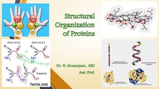
- 1. Dr. N. Sivaranjani , MD Asst. Prof.
- 2. Proteins are the polymers of L- α amino acids. Native protein structure is essential for the biological function of a protein Levels of structural organization of proteins : Primary structure Secondary structure Tertiary structure Quaternary structure Loss of structure results in loss of biological function
- 3. 4 Levels of Protein Organization 1. Primary structure: The linear sequence of amino acids forming the backbone of proteins. 2. Secondary structure: The spatial arrangement of protein by twisting of the polypeptide chain. 3. Tertiary structure: The three dimensional structure of a functional protein. 4.Quaternary structure: proteins are composed of two or more polypeptide chains referred to as subunits. The spatial arrangement of these subunits.
- 4. Proteins are formed of A.A. linked together by the following types of bonds Covalent bonds Non Covalent bonds Peptide bond Disulfide bond Hydrogen bond Electrostatic bond / ionic interactions Van der Waals interactions Hydrophobic interactions Strong bonds in protein structure
- 5. Covalent bond Disulfide bond Peptide bond
- 6. Non-covalent Bonds in Proteins
- 7. Primary structure The Number and Linear sequence of amino acids in a protein is called the primary structure. Presence of specific amino acids at a specific number is very significant for a particular function of a protein. Every polypeptide chain has unique a.a sequence, dictated by genes. Genetic diseases – proteins with abnormal amino acid sequences Bonds -- only covalent bonds – Peptide bond , disulfide bonds
- 8. Peptide bond formation • α-COOH group of one a.a (with side chain R1) forms a covalent peptide bond with α-NH2 group of another a.a ( with the side chain R2) by removal of a molecule of water. • It result in Dipeptide ( i.e. Two amino acids linked by one peptide bond). • Tripeptide (3 a.a linked by 2 peptide bond) • < 50 a.a - “Peptides”, • > 50 a.a - “Proteins”
- 9. Formation of Peptide Bond Amino acid 1 Amino acid 2 H2O H H H N H C R O C O H H H N H C R O C H C R C O H H N H O + Amino gr Carboxyl gr H C R C O N H O N terminal C terminal Peptide bond
- 10. Naming of Peptide bond H3N-Glutamate – cysteine - methionine – glycine-COO N- terminal C- terminal Glu – cys – met- Gly glutamyl – cystenyl – methionyl-glycine Amino acid sequence is read from N-terminal to C-terminal. Protein synthesis occurs in the same way. N-terminal a.a – “ yl” ; C- terminal a.a – “ine” .
- 11. Characteristics of peptide bond • Partial double bond character (distance is 1.32Å which is midway b/w single bond 1.49 Å and double bond 1.27Å) • Rigid and planar • Always in Trans configuration • Side chains are free to rotate on either side of the peptide bond • The angles of rotation known as Ramachandran angle – determine the spatial orientation of the peptide chain Dr. GN Ramachandran
- 12. Importance of Primary structure • Higher levels of organization of Proteins are dependent on the primary structure • Even a single amino acid change (mutation) in the linear sequence – cause disease • Eg:- Sickle Cell Anemia HbA (normal Hemoglobin) the 6th amino acid in the beta chain is glutamic acid - it is changed to valine in HbS (sickle cell anemia)
- 13. 2 Interchain disulphide bonds – A chain 7th cysteine and B chain 7th cysteine A chain 20th cysteine and B chain 19th cysteine 1 Intrachain disulphide bond - between 6th and 11th cysteine residues of A chain 86 amino acids C-peptide 33 21 30 51 amino acids
- 14. Determination of Primary structure of Protein Identification of amino acids with regard to their quality, quantity and sequence in a protein structure
- 15. • Steps for Determining the Primary Structure Steps Technique I. Determination of amino acid composition in a protein Complete hydrolysis of protein Pronase enzyme Separation and estimation of amino acids Chromatographic technique II. Degradation of protein into smaller fragments Liberation of polypeptides Urea or guanidine hydrochloride Determination of the number of polypeptide Dansyl chloride chains in a protein Breakdown of polypeptides into fragments Enzymatic cleavage Chemical cleavage III. Determination of amino acid sequence Sanger's reagent Edman's reagent Nowadays, DNA sequencing is used to determine the amino acid sequence.
- 16. Determination of amino acid composition 1. Hydrolysis of protein • Pronase- mixture of non-specific proteolytic enzymes that causes complete hydrolysis of proteins. 2. Separation and estimation of amino acids: The mixture of a.a`s liberated by protein hydrolysis can be determined by chromatographic techniques , by using Ninhydrin.
- 17. Degradation of protein or polypeptide into smaller fragments • Liberation of polypeptides • Urea or Guanidine hydrochloride - disrupts the non-covalent bonds and dissociates the protein into polypeptide units. • Disulfide linkages b/w the polypeptide broken- Performic acid • Number of polypeptides • Dansyl chloride (1-dimethyl amino naphthalene 5-sulfonyl chloride) – Specifically binds with N-terminal amino acids to form dansyl polypeptides which on hydrolysis yield N-terminal dansyl amino acid. • Number of Dansyl AA = Number of Polypeptide chains in a Protein
- 18. Breakdown of polypeptides into fragments • Polypeptides are degraded into smaller peptides - enzymatic or chemical methods • Enzymatic cleavage: exhibit specificity in cleaving the peptide bonds • Chemical cleavage: Cyanogen bromide (CNBr ) commonly used • Attacks Carbonyl side contributed by Methionine AA. Enzymes Specific action N terminal AA is identified by Trypsin -commonly used Lysine and Arginine Chymotrypsin Phe,Tyr, Trp, or Leu C-terminal AA is identified by Carboxypeptidase A Acidic / neutral AA Carboxypeptidase B Proline
- 19. Determination of the amino acid sequence • Sangers reagent: 1-fluoro-2,4-dinitrobenzene (FDNB) • FDNB specifically binds with N-terminal amino acids to form a Dinitrophenyl (DNP) derivative of peptide • This is on hydrolysis yields DNP – amino acids ( N-terminal) and free amino acids from the rest of the peptide chain. • Used for identification of N terminal AA • DNP-AA – identified by Chromatography
- 20. Edmans reagent: Phenyl isothiocyanate Reacts with N-terminal amino acid of peptide to form a phenyl thiocarbamyl derivative Phenyl thiohydantoin (PTH)-amino acid is liberated - Identified by chromatography Edman's reagent would then react with the second amino acid which now has the alpha amino group Useful in sequencing first 10-30 amino acids Label first AA Release of 1st AA Label 2nd AA Polypeptide chain Release of 2nd AA
- 21. For identifying the amino-terminal residue The Edman’s degradation Reveals the entire sequence of a peptide Sanger’s degradation
- 22. Sequenator • This is an automatic machine to determine the amino acid sequence in a polypeptide • It is based on the principle of Edmans degradation . • Amino acids are determined sequentially from N-terminal end • The PTH-amino acid liberated is identified by HPLC. • Sequenator takes about 2 hours to determine each amino acid.
- 23. Reverse sequencing technique • It is the genetic material (DNA) which ultimately determines the sequence of amino acids in a polypeptide chain • By analyzing the nucleotide sequence of DNA that codes for protein, it is possible to translate the nucleotide sequence into amino acid sequence. • Demerits - fails to identify the disulfide bonds and changes that occur in the amino acids after the protein is synthesized.
- 24. Secondary structure • The spatial arrangement of protein by twisting and folding of the polypeptide chain is called Secondary structure. • The amino acids are located close to each other in their sequence. • Two types of secondary structures :- α-Helix and β-sheet
- 25. Alpha helix • Most common and Stable conformation • Spiral structure - Polypeptide bonds form the back-bone and Side chain extend outwards • Stabilized by H bonding b/w carbonyl oxygen and amide hydrogen. • Right handed helix • Amino acids per turn – 3.6 • Hemoglobin, Myoglobin -Abundant , Chymotrypsin – absent • Proline – disrupts the α-helix H bonding The hydrogen bonds are individually weak but collectively, strong enough to stabilize the helix
- 26. β-pleated sheet (β-sheets) • The surfaces of β – sheets appear “pleated”, and these structures are, therefore, often called “ β – pleated sheets”. • Polypeptide chains – fully extended • distance between adjacent a.a -3.5Å • Stabilized by hydrogen bonds 2 types Parallel Anti -Parallel strands in a sheet run in an opp. direction - Silk fibroin C C N C C N run in the same direction – Flavodoxin Carbonic anhydrase – contains both N N
- 28. • -helix – protein turns like a spiral • Keratin, fibrous proteins structures are almost entirely alpha helical (hair, nails, horns) -sheet – protein extended –Globular protein
- 29. Super secondary structures • Some of the common structural motifs combining α – helices and β – sheets • found in globular proteins These are :- • β- α- β unit • Greek key • β-meander Beta barrel beta-alpha-beta motif Greek key motif β-meander motif
- 32. Tertiary structure • Peptide chain with its secondary structure ,may be further folded & twisted about itself forming 3D arrangement of a functional protein. • It is a compact structure - hydrophobic side chains held interior ,hydrophilic groups - surface of the protein molecule. • Vander Waal's forces , disulfide bonds(-S-S ), electrostatic bonds and hydrophobic interactions contribute to the stability of tertiary structure of proteins. The function of a protein depends on its tertiary structure. If this is disrupted, it loses its activity Eg:- active / catalytic site of an enzyme
- 33. Domains • A domain represents compact functional unit of protein . • A polypeptide with 200 amino acids normally consists of two or more domains • Part of protein that can fold into a stable structure independently • Domains are usually connected with relatively flexible areas of protein • Different domains can impart different functions to proteins
- 34. Chaperones / Hsp Chaperone proteins participate in the folding of proteins. The hsp 70 ( 70 – kDa heat shock protein ) family of chaperones binds short segments of hydrophobic AAs in newly synthesized polypeptides. Chaperones prevent aggregation of polypeptide chain thus giving a chance for formation of secondary and tertiary structures. Neurological diseases result from altered protein folding - Alzheimer's disease (β – Amyloid protein, a misfolded protein is formed in brain) Prion diseases (altered secondary –tertiary structure)
- 35. Quaternary structure • Many proteins have single polypeptide chains – Monomeric proteins • Proteins having more than one polypeptide chains have this structure – Oligomeric proteins • Each polypeptide chain is called subunits or monomers. • Subunits either function independently or may work co-operateively (Hb) • Dimer – 2 polypeptide chains, Trimer , Tetramer – 3,4 polypeptide chains. • The protein function is lost if subunits dissociate • Bonds- non covalent interactions
- 36. • Hemoglobin • 2 globin subunits • 2 globin subunits Other Examples:– Immunoglobulin – 2 heavy chains and 2 light chains Lactate Dehydrogenase (LDH) - Tetramer Creatine Kinase (CK) - Dimer
- 37. • Study of Higher Levels of Protein Structure • The higher levels of protein structure may be studied by techniques using :- • X-ray diffraction, • Ultraviolet light spectroscopy, • Optical rotatory dispersion, • Circular dichroism, • Nuclear Magnetic resonance (NMR).
- 38. Structure-Function Relationship • The functions of proteins are maintained because of their ability to recognize and interact with a variety of molecules. Three dimensional structural conformation provides and maintains the functional characteristics Three dimensional structure is dependent on the 1⁰ structure Difference in the 1⁰ structure causes loss of protein function
- 39. Heme Transport proteins Hemoglobin Hb has a quaternary structure with 2 α and 2β Tetrameric protein – each Monomer has a Heme unit which binds the oxygen Binding of oxygen to one Heme facilitates oxygen binding by other subunits In Primary structure any one amino acid replacement by another can cause abnormalities in its function. Eg:- Sickle cell Anemia :- In 6th position of β-chain of Hemoglobin GLUTAMIC ACID is replaced by VALINE.
- 40. Structural proteins Collagen Main fibrous component of skin, bone, tendon, cartilage and teeth. Triple helix – every 3rd a.a is Glycine. Proline, Lysine, Hydroxyl Proline, hydroxyl Lysine Strength of collagen depends upon its structure (triple helix) In vitamin C deficiency - Collagen becomes weak as hydroxylation of proline and lysine does not occur. This causes reduced H-bonding and consequently weakness of collagen.
- 41. Enzymes Enzyme catalysis needs precise binding of the substrate to the active site of the enzymes. This depends on structural conformation of the active sites - precisely oriented for substrate binding.
- 42. Biologically important peptides Peptide name and the No of amino acids Origin Functions Glucagon - 29 amino acids α cells of Pancreas Increases Blood glucose Adrenocorticotropic hormone – 39 a.a Hypothalamus Stimulate and secrete the steroid hormone corticotropin by anterior pituitary gland Oxytocin - 9 a.a Posterior pituitary Stimulates uterine contractions Vasopressin – 9 a.a Posterior pituitary Increases blood pressure and has an antidiuretic action Angiotensin II – 8 a.a Liver Stimulates the release of aldosterone from adrenal gland ACE inhibitors – used in Rx of HTN Glutathione (GSH) -3 a.a Antioxidant
- 43. Biologically important peptides When 10 or less number of amino acids are joined together, it is called an Oligopeptide -Some of them are biologically active Glutathione, TRH, Encephalins, Oxytocin , Vasopressin, Angiotensin I & II, Gramicidin S. Polypeptide hormones (more than 10 amino acids) – Insulin , Glucagon , Growth hormone, Gastrin, ACTH etc.
- 44. Glutathione (GSH) GSH is a Tripeptide ( gama glutamyl – cysteinyl – glycine) Gluamate – Cysteine - Glycine SH Reduced glutathione (GSH) – active form Oxidized glutathione (G-S-S-G) – inactive form Functions of Glutathione • Membrane stabilization - Reduced glutathione is essential for maintaining the normal structure of RBCs.
- 45. Biological antioxidants – it suppresses H2O2 and organic peroxides, (lipid peroxides) to H2O by Glutathione Peroxidase. Maintenance of biological activity of protein – helps to keep E in active site Prevention of Met Hb – keeps Fe in Ferrous state in Hb Role in detoxification - Glutathione plays a key role in detoxification of compounds by conjugation Absorption of amino acids – small intestinal mucosal cell and proximal renal tubules - ɣ glutamyl cycle Disorder – Hemolytic Anemia
- 46. Thyrotropin releasing hormone (TRH) – 3 amino acids Hypothalamus PGA-Glu-His-Pro-NH2 Glu is modified to form pyroglutamic acid (cyclization of N terminal Glutamate). Proline – carboxyl gr of proline is amidated It stimulates the release of TSH from antr. pituitary
- 47. Nanopeptide Oxytocin – 9 amino acids Posterior pituitary It stimulates uterine contractions. Vasopressin / Anti diuretic hormone – 9 amino acids Posterior pituitary It facilitate water reabsorption in DCT and Collecting ducts of kidney – decreasing the Urine output. Diabetes insipidus – decreased ADH synthesis
- 48. Pentapeptide Enkephalins – 5 amino acids Met-enkephalin - Tyr-Gly-Gly-Phe-Met. Leu-enkephalin - Tyr-Gly-Gly-Phe-Leu. CNS Analgesic action – dec. pain
- 49. Angiotensinogen – 400 a.a Synthesized in Liver Renin (Kidney) Angiotensin I (10 amino acids) Angiotensin Converting Enzyme (ACE) Angiotensin II (8 amino acids) It has hypertensive effect – inc. BP Vasoconstriction Stimulates the release of aldosterone from adrenal medulla Na retention Octapeptide Lungs ACE inhibitors – Captopril, Enalapril are used in the Rx of Hypertension
- 50. Carnosine Dipeptide – βAlanine – Histidine Activates myosine ATPase activity – muscle contraction Chelates Cu2+ and facilitates absorption of Cu Aspartame Dipeptide – Aspartate and Phenylalanine Artificial sweetner
- 51. Primary Secondary Tertiary Quaternary S T R U C T U R E P R O C E S S Linear 1D sequence of AAs Folding 2D Packing 3D Interaction of 2 or more subunits