Trujillo 2016, El estetoscopio tiene pantalla de laennec a rus 2016
•
3 likes•1,371 views

Trujillo 2016, El estetoscopio tiene pantalla de laennec a rus 2016

Recommended
More Related Content
What's hot
What's hot (20)
Viewers also liked
Viewers also liked (20)
Similar to Trujillo 2016, El estetoscopio tiene pantalla de laennec a rus 2016
Similar to Trujillo 2016, El estetoscopio tiene pantalla de laennec a rus 2016 (20)
More from Luis Vargas
More from Luis Vargas (17)
Recently uploaded
Recently uploaded (20)
Trujillo 2016, El estetoscopio tiene pantalla de laennec a rus 2016
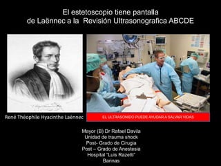
- 1. El estetoscopio tiene pantalla de Laënnec a la Revisión Ultrasonografica ABCDE Mayor (B) Dr Rafael Davila Unidad de trauma shock Post- Grado de Cirugia Post – Grado de Anestesia Hospital “Luis Razetti” Barinas René Théophile Hyacinthe Laënnec EL ULTRASONIDO PUEDE AYUDAR A SALVAR VIDAS
- 2. En los ultimos doscientos años la bata blanca y el estetoscopio son los símbolos de referencia de la profesión y arte de la medicina el estetoscopio ha sido una maravillosa herramienta para adquirir datos objetivos de los sujetos sanos y enfermos por 198 años
- 3. Por centurias los medicos han escuchado y tratado de entender los sonidos producidos por el cuerpo humano inicialmente lo hacían pegando el oído al cuerpo y se denomino AUSCULTACION Referencias a la auscultación de los sonidos respiratorios se encuentran: Papiros Egipcios (1500 AC) Vedas Hindúes ( 1400-1200AC) Escritos Hipocráticos ( 500 AC) Caelius Aureliaus (50AC) Da Vinci, Ambrose Pare, Harvey, Morggni mencionan la Auscultation.
- 4. Laennec desarrolla el estetoscopio Hasta 1815 se practicaba la auscultación apoyando el oído directamente al pecho. Esto tenía varios inconvenientes entre ellos la dificultad de percibir ruidos en pacientes obesos y el atropello al recato de mujeres. 1816 pectoriloquia, egofonía, crepitación, estertor “I rolled a quire of paper into a kind of cylinder and applied one end of it to the region of the heart and the other to my ear, and was not a little surprised and pleased to find that I could thereby perceive the action of the heart in a manner much more clear and distinct than I had ever of been able to do by the immediate application of my ear.”
- 5. MANUAL FOR THE USE OF THE STETHOSCOPE. JUST published by Benjamin Perkins, k Co. MANUAL FOR THE USE OF THE STETHOSCOPE being a short Treatise on investigating Diseases of Chest. From the French of M. Collin, •with an Introduction and Plates. By a Fellow of the Mass. Med. Soc. The Stethoscope may also be obtained as above in the most approved form. Jan. 20. 1829
- 6. EXCERPTS FROM "NEW STETHOSCOPE" BY NICHOLAS COMINS To the Editor of The London Medical Gazette, August 12, 1829 ------------------------------------------------------------------------------- Description of Comins' stethoscope: "It consists of two tubes, (A,B), each 7 inches in length, and 5/8ths of an inch in diameter, except at the part to be applied to the thorax, where the diameter of the aperture is and inch and a half. These pieces are united by a perforated joint, (C,D), three inches in length, at right angles to their extremities : and the two pieces of which the joint consists, being united like the joint of a flute, permit the limbs of the cylinder to form any angle. The upper end of the instrument is provided with an ear-piece, (E), sufficiently large and concave to envelop the ear. Its central portion, (F), being angular and moveable, admits the extremity of the cylinder as nearly as convenient to the meatus auditorius externus. The ear-piece can, by means of a moveable joint, (G,H), be placed laterally with respect to the extremity of the tube. The moveable joints, (C,D--G,H), were they formed of brass, could, by a simple contrivance, be rendered in any position air-tight. But as this, and perhaps every substance that would deviate from the homogeneousness of the cylinder, would injure the sound, external securities arenecessary. The following has been devised : each joint is covered by a metallic ferrule, (iii) 5/8ths of an inch in length, and 6/8ths of an inch in diameter, except at one extremity, (J), where it is reflected inwards, at right angles, and where the diameter reaches about 3/16ths of an inch. Within the ferrule, and in contact with the reflected extremity, is placed a small flat ring, (K), which is secured through an aperture in the ferrule, (L), at three points, to one side of the joint, (M.) The other end of the ferrule is screwed also at three points to the other side of the joint, (N). The ferrule, and the inclosed ring, are by this contrivance permitted each to move freely with respect to each other ; while with respect to the joint, they preclude the possibility of its opening, or of its not being air-tight. Should friction, however, eventually cause it to become too free, the screws can be withdrawn from the pieces, (D,G), and by means of silk coiled in the depressions, (O,O), the joint can again be rendered air-tight. The piece of ivory that is screwed to the lower extremity of the cylinder is externally somewhat concave, in order that it may be more advantageously applied to the thorax. The ivory ferrules, and the pieces to which they are screwed, and each marked with corresponding circles..... " Earliest known description of a binaural stethoscope: ".....It is surprising that the discoverer of mediate auscultation had not suggested a flexible instrument. But Laennec, like the immortal Archimides, grappled with great ideas in unexplored regions of thought. Contented with the acquisition of all the knowledge attainable by the stethoscope, he despised the drudgery connected with the minutiae of mechanical invention ; directed his thoughts to great pursuits ; and permitted the instrument to be modified by the humblest of labou Caelius aureliaus (c.500BC rers in the field of science. It had occurred to the writer that both ears might be simultaneously and advantageously employed in stethoscopic examinations. The instrument (Comins') adapted to this purpose consists of a tube, connected at its middle at right angles to the cylinder, to be apllied to the patient, and connected at its movable extremities to two tubes, moveable also on teh principle that has been described. It admits of easy adaption at once of the patient, and to both ears."
- 7. El estetoscopio evoluciona y llega hasta el estetoscopio electronico RAPPAPORT LITTMANN 19601892 BINOMIAL RESONANTE
- 9. Estetoscopio con transferenciaEstetoscopio con oximetría y ECG
- 10. El estetoscopio, símbolo de la medicina a punto de desaparecer ¿Está obsoleto el estetoscopio, un símbolo de la medicina?
- 11. INICIO DEL USO DEL ULTRASONIDO
- 12. 1794: Lazzaro Spallanzani fisiólogo hace el primer estudio de la física del ultrasonido para deducir como los murciélagos usan el efecto Doopler para navegar por eco colocación
- 13. 1826: Jean Daniel Colladon – Físico Usa una campana de iglesia bajo el agua para probar como el sonido viaja mas rápido a través del agua que el aire (primer traductor).
- 14. 1880: Pierre & Jacques Curie Descubren el efecto Piezo - eléctrico Pierre Curie Jacques Curie
- 15. 1915: Paul Langevin –FISICO en 1912 posterior al hundimiento del Titanic inventa el Hidrófono que es el primer traductor para detectar Icebergs y luego submarinos en la primera guerra mundial
- 16. 1942: Karl Dussik, Neurólogo y Psiquiatra de la Universidad de Viena usa el ultrasonido para el diagnostico de tumores cerebrales
- 17. 1948: George Ludwig M.D. Internista Es el primero en describir el uso del ultrasonido para el diagnostico de cálculos biliares
- 18. 1950: Douglas Howry & Joseph Holmes Pioneros en el uso del ultrasonido en 2D y modo B en la Univ. de Colorado
- 19. 1958: Ian Donald Pionero del uso del ultrasonido en OBSTETRICIA - GINECOLOGIA
- 20. Difusión del uso del Ultrasonido en las especialidades medicas Sports Medicine Internal Medicine Anesthesiology Critical Care Emergency Medicine Physiatry /PMR Surgery Cardiology OB-GYN Peds Emerg Med General Pediatrics • No produce eventos adversos. • Es operador dependiente. • Requiere entrenamiento. • Hace diagnostico. • Diagnostico en nivel básico de atencion. • Permite monitorización del paciente. • Metodo accesible. • Barato.
- 22. En 1990 aparece el eco compacto y se desarrolla su uso en áreas de combate en Irak para decidir en el frente de batalla en forma precoz la necesidad de tratamiento Quirurgico En el año 2000 el eco se convierte en una laptop
- 23. NEJM Feb 2006; 354(6); 548-551 ¿RIP? Physical Exam Vs. CXR as Gold Standard: N=52; Sensitivity 47-69% /Specificity 58- 75% Wipf et al. Diagnosing Pneumonia by Physical Exam: Relevant or Relic? Arch Intern Med 1999 Point-of-Care Ultrasound as a stethoscope replacement?
- 28. 2004: Tom: Cruise “Yo compre una maquina de ultrasonido para Katie”
- 29. William Richard David Zar Universidad de Washington-Louis han transformado un teléfono con Windows Mobile en ecógrafo portátil 2009 Carolina del Sur
- 32. 2010 - 2013
- 33. La organización Mundial de la Salud (WHO) estima que mas del 75% de la población mundial no tiene acceso a ningún método de diagnostico por imágenes
- 34. The Lung not suitable for Ultrasound Los pulmones son el principal obstáculo para el uso del ultrasonido a nivel toraxico Harrisons principles of Internal Medicine 1992 Simply wrong
- 35. 1989: Daniel Lichtenstein pionero ultrasonido de atencion puntual pulmonar en UCI «Ultrasonido es el estetoscopio real»
- 37. Ecografía en pacientes criticos o Ecografía focalizada • No es un estudio de ecografía convencional • No es un estudio ecográfico por órganos como los estudios de ecocardiografía o ecografía hepato bilio pancreatica que realizan los radiólogos eco grafistas Es el uso de la ecografía como un estetoscopio visual para contestar preguntas clínicas concretas
- 38. La respuesta rapida a estas preguntas permite modificar de manera inmediata una conducta terapéutica y brindar un beneficio inmediato al enfermo ¿este paciente hipotenso hipovolemia? ¿tiene un aneurisma abdominal roto? ¿hay taponamiento cardiaco? ¿tiene el paciente traumatizado liquido libre en cavidad? ¿hay derrame pericárdico o derrame pleural? Tiene trombosis venosa profunda? ¿tiene un TEP? ¿esta bien colocado el tubo orotraqueal? ¿necesita guiar la toma de una vía central o una vía periférica? ¿Tiene hipertensión endocraneana? ¿Estoy sobre hidratando el paciente? ¿le falta volumen al paciente?
- 39. Estudio limitado y focalizado para responder preguntas clínicas concretas • No invasivo • Sin radiación • Rápido • Barato • accesible • Permite monitorizar el enfermo • Operador dependiente • Difícil en obesos • Enfisema subcutáneo Sirve como método diagnostico y de monitoreo
- 40. 2002 2007 2010 Uso de la ecografia en escenarios criticos
- 41. 2010 2010 2012 Uso de la ecografia en escenarios criticos con protocolos integrados del ABCDE del ACLS y TRAUMA con un examen de cabeza a pie
- 42. 2008 Dr. Luca en Milán contacta por teléfono y mail a todos los que han trabajado en ecografia critica • FAST • EFAST • FATE • FEEL • BLUE • APH “Improving Primary, Emergency, and Critical Care Medicine, by incorporating 'point-of-care' Ultrasound into Clinical Practice”. Promote and spread “point- of-care” ultrasound into clinical practice, in order to improve primary, emergency and critical care provided in all extra-hospital and in- hospital scenarios.
- 44. 2012 2013 2013
- 45. 2014 Entrenamiento basado en la resolución de problemas clinicos ayudados por la ecografia
- 46. FAST ABCDE método asociado al examen físico como una variante sofisticada del estetoscopio integrado a los protocolos del ACLS; PHTLS ATLS; AMLS, FEEL, BLUE, RUSH El estetoscopio tiene pantalla
- 47. Permeabilidad y causas obstructivas A performance ventilatorio y causas de disnea o hipoxemia B hemodinamia y causas de shock C Status neurológico y causas coma o signos focales D Excluye causas ocultas E POLMONAR US TEJIDOS BLANDOS US USMC: ultrasonido mejora manejo del paciente critico 1. “ABCDE” evaluación primaria. 2. “ABCD” resucitacion. 3. “Cabeza a pie” evaluación secundaria. 4. Transporte UCI / cuidados definitivos. 5. Evaluación continua. Neri L Storti E, Lichtenstein D. Toward an Ultrasound Curriculum in Critical Care Med 2007 FAST ABCDE evaluación primaria
- 50. 1. Aprender a realizar el examen 2. Reconocer y diferenciar la anatomía normal en US. 3. Diagnosticar anormalidades comunes. 4. Integrar la información sonografica en la toma de decisiones clínicas. 5. Incorporar la informacion sonografica en los procedimientos de emergencia.
- 51. Soluciones: Recurso de aprendizaje disponible en el internet www.SinaiEM.US llevando la tecnología junto a la cama para mejorar la atencion del paciente www.hqmeded.com
- 53. El ultrasonido llego para quedarse ¿y usted donde va a estar?
- 55. Soluciones: Aprendizaje por simulación de esta Tecnología : permite entrenamiento por reconocimiento de modelos en forma intensiva a través de videos y simulación de casos
- 56. • Tiene una presunción , duda o no sabe que esta pasando. • Hace una observación. • Ve el problema. • Resuelve el problema. • Reevalúa la situación.
- 57. Si puedes verlo para que oírlo , además puedes reevaluar cada vez que quieras y ver el resultado de tus medidas terapéuticas
- 58. El futuro es ahora
- 60. La invasión de un ejercito puede resistirse pero cuando una idea llega no hay como resistirla Víctor Hugo