SlideShare a Scribd company logo
1 of 219
RED AND WHITE LESIONS
OF ORAL CAVITY
BY
Dr. REVATH VYAS
PG III YEAR
Color of oral mucosa depends on…
Concentration and dilatation of blood vessels in the
underlying connective tissue
Degree of keratinisation
Amount of melanin pigment in epithelium
Thickness of epithelium
Orban’s Oral Histology & Embryology 11th edition.
1) Hyper keratosis
2) Acanthosis
3) Intra and extracellular accumulation of fluid in the epithelium (i.e.
Leukoedema)
4) Necrosis of the oral epithelium
5) Microbes, particularly fungi, produce whitish pseudomembranes
6) Reduced vascularity in the underlying lamina propria.
Why abnormally white?
Why abnormally red ?
Atrophic epithelium
Reduction in the number of epithelial cells
Increased vascularisation
Blood vessel enlargement
Presence of blood in the tissue
Increased hemoconcentration
Bengel, Veltman, Loevy, Taschini - Differential diagnosis of diseases of the oral mucosa
Burket’s oral medicine – 10th edn.
Causes of red lesions
Crispian Scully - Oral & Maxillofacial Medicine; 2004.
CAUSES
Inflammatory
Reactive
Atrophic/
Erosive
Neoplasms
Purpura
Vascular
CLASSIFICATIONS
Acute
Chronic
Localised
Generalised
Developmental
Pathologic
CLASSFICATION ACCORDING TO Wood & Goaz, V edition
CLASSIFICATIONACORDINGTORAVIKIRANONGOLE2ND EDITION
CLASSFICATION ACCORDING TO BURKET’S 11th edition
INFECTIOUS DISEASES
ORAL CANDIDIASIS
HAIRY LEUKOPLAKIA
A) ORAL CANDIDIASIS
(Candidosis, Moniliasis, Thrush)
1) Opportunistic infection affecting the oral mucosa.
2) Caused by the yeast candida albicans.
3) Predisposing factors convert candida from the normal
commensal flora (saprophytic stage) to a pathogenic organism
(Parasitic stage).
Etiology:
Candida species responsible for candidal infections are
c. albicans
c. tropicalis and 80% of the species
c. glabrata
Yeasts penetration of epithelial cells is facilitated by their production of lipases
CLINICAL FINDINGS
PSEUDOMEMBRANOUS CANDIDIASIS
1) Acute form is recognized as classic candida infection
2) Affects patients medicated with antibiotics, immunosuppressant drugs or a disease
3) Oral lesions are soft, white, slightly elevated plaques occurring on the buccal mucosa,
tongue, palate, gingiva and floor of the mouth.
4) “Curdly white” Plaques can be wiped away with guaze, leaves an inflamed, sometimes
bleeding area.
5) Clinical presentations of acute and chronic are indistinguishable.
6) Chronic form associated with HIV infections and with steroid inhalers.
ERYTHEMATOUS CANDIDIASIS
1) Previously referred as atrophic candidiasis (Antibiotic sore mouth or
Antibiotic stomatitis)
2) Occurs as a sequelae to a course of broad spectrum antibiotics,
inhalation steroids smoking and diseases.
3) Lesions appear red or erythematous
4) May occur at any site
5) Only variety of oral candidiasis, which is consistently painful.
CHRONIC PLAQUE-TYPE AND NODULAR CANDIDIASIS
(Chronic Hyperplastic candidiasis)
1) Chronic-plaque type replaces candidal leukoplakia
2) Characterized by a white plaque.
3) Firm, white persistent plaques, usually on the lips, tongue and cheeks
4) Chronic plaque-type and nodular candidiasis have been associated with
malignant transformation.
CANDIDA – ASSOCIATED LESIONS
I. DENTURE STOMATITIS
1) Most prevalent site is palatal mucosa
2) Classified into three different types
Type I – Localized to minor erythematous sites
Type II – affects a major part of the denture covered
mucosa
Type III – has a granular mucosa
II. ANGULAR CHEILITIS
Characterized by erythema, fissuring, scaling
Saliva tends to pool in, keeping the area moist & favoring
yeast infection
Candidal infection involving perioral skin - creates a
clinical pattern
CHEILOCANDIDIASIS
CHEILOCANDIDIASIS
A diffuse form of chronic candidiasis characterized by pain, swelling,
erythema with focal ulcerations, and crusting is called
cheilocandidiasis.
It represents a secondary candidal infection superimposed on areas
of trauma from mechanical or solar factors.
Cheilocandidiasis and juvenile juxtavermilion candidiasis refer to
more extensive and often desquamative lesions affecting the full
width of the lip, even extending into adjacent lesions.
They are associated with habitual lip sucking, prolonged dental
appointments, sunlight, and chronic candida infection. Differential
diagnoses of these lesion include perioral erythema and Id reaction
(localized sterile vesiculopapular rash).
Etiology
Infection
(bacterial, fungal)
Nutritional
deficiency
Reduced vertical
dimension
Long-term
denture wearers
Shafer, Hine, Levy. Textbook of Oral Pathology, 6th edn;2009.
III. MEDIAN RHOMBOID GLOSSITIS
1) Characterized by a erythematous lesion in the centre of the posterior
part of the dorsum of the tongue.
2) Lesion has a oval configuration
3) Resulting from atrophy of the filiform papillae and lobulated.
4) Smokers, denture-wearers and patients using inhalation steroids have
an increased risk
5) Asymptomatic
6) Management is restricted to a reduction in predisposing factors.
III. MEDIAN RHOMBOID GLOSSITIS
Brocq : 1914
Prevalence: < 1%
Males > Females
Synonyms
Median candidiasis of the tongue
Atypical/ paramedial rhomboid glossitis
Central papillary atrophy
Posterior lingual papillary atrophy
Posterior midline atrophic candidiasis
Etiology
Embryological
Inflammatory
Immunological
Infectious
Associated with: Tobacco smoking
Denture wearing
Trauma
Int J Oral Surg 1984;13(5):411-415.
Oral Surg Oral Med Oral Pathol 1996;81(4):379-80.
Med Oral Patol Oral Cir Bucal 2005;10:123-7.
Kissing lesion
Investigations
Cytosmear
Fungal culture
Biopsy:
Candidal hyphae > 85% of lesions
Management of median rhomboid
glossitis
Topical antifungal: clotrimazole troches
fluconazole
Topical corticosteroid: fluocinonide 0.05%
clobetasol 0.05%
Bruch JM, Treister NS. Clinical Oral Medicine and Pathology.
SECONDARY ORAL CANDIDIASIS
1) Accompanied by systemic mucocutaneous candidiasis and other immune deficiencies
2) Chronic mucocutaneous candidiasis
a) Also affect the skin, the nail bed, genital mucosa
b) Face and scalp may be involved
c) Occur as part of
1) Endocrine disorders – Hyperparathyroidism and Addisons disease
2) Myeloperoxidase deficiency
3) Chediak – Higashi syndrome,
4) Severe combined immunodeficiency syndrome characterized by a defect in the
function of the cell-mediated immunity
5) Thymoma is a neoplasm of thymic epithelial cells
6) Chronic familial mucocutaneous candidiasis an inherited disorder, occurs early in
life.
ORAL CANDIDIASIS ASSOCIATED WITH HIV
1) 90% of AIDS patients have oral candidiasis
2) Most common types
a) Pseudomembranous candidiasis
b) Erythematous Candidiasis
c) Angular cheilitis and
d) Chronic hyperplastic candidiasis
DIAGNOSIS AND LABORATORY FINDINGS
Smear:- useful in Pseudomembranous oral candidiasis and angular cheilitis
Swab:- Taken by rubbing cotton-tipped swabs over the lesional tissue.
Imprint Culture: Sterile (plastic) foam pads submerged in sabouraud broth placed on
the infected surface for 60 seconds. Cultivated at 37oC.
Result is expressed as colony forming units per
cubicmillimeter. (CFU/mm3)
Valuable in erythematous candidiasis and denture stomatitis.
Salivary culture technique:- Used to get an adequate quantification of candida.
Histopathological Examination:- Chronic plaque-type and nodular candidiasis,
identify the presence of epithelial dysplasia.
Treatment of Denture stomatitis
Eliminate predisposing factors
Improve denture hygiene
Denture should be stored in antimicrobial solutions
Type III denture stomatitis treated with surgical
excision
Treatment of Angular cheilitis
 Topical treatment with miconazole
 A mild steroid ointment suppress the inflammation
 A moisturizing cream prevent new fissure formation
Treatment of Median rhomboid glossitis
Topical antifungal: clotrimazole troches
fluconazole
Topical corticosteroid: fluocinonide 0.05%
clobetasol 0.05%
Bruch JM, Treister NS. Clinical Oral Medicine and Pathology.
ANTIFUNGALS
Topical antifungals are usually the drug of choice for
uncomplicated, localized candidiasis in patients with
normal immune function.
Systemic antifungals are usually indicated in cases of
disseminated disease and/or in immunocompromised
patients.
Dosage form/ strength Indication
Miconazole cream 2% Angular cheilitis
Clotrimazole cream 1% Angular cheilitis
Ketoconazole cream 2% (Prescription) Angular cheilitis
Nystatin ointment 100,000 units/gram
(Prescription)
Angular cheilitis
Nystatin topical powder 100,000 units/gram
(Prescription)
Denture stomatitis
Nystatin oral suspension 100,000 units/gram
(Prescription)
Intraoral candidiasis
Betamethasone dipropionate clotrimazole
cream (Prescription)
Chronic angular cheilitis
Clotrimazole troches 10 mg (Prescription) Intraoral candidiasis
Amphotericin B 100 mg/ml (Prescription) Intraoral candidiasis
Generic name Formulation
Amphotericin B 100 mg/ml oral suspension
Clotrimazole 10 mg troche
Fluconazole 100 mg tablet
10 mg/ml oral suspension
40 mg/ml oral suspension
Itraconazole 100 mg capsule
10 mg/ml oral suspension
Ketoconazole 200 mg tablet
Nystatin 100,000 units/ml oral suspension
200,000 units/ml pastille
500,000 units/ml tablet
100,000 units/ml vaginal tablet
ANTIFUNGALS
Medication should be continued for at least 48 hours after the
disappearance of clinical signs of candidiasis along with complete
healing and the absence of mucosal erythema.
Some sources recommend drug therapy should be continued for 10–
14 days regardless of the disappearance of clinical signs of
candidiasis.
SURGERY
May be advised for hyperplastic and nodular types of candidiasis.
(Cryotherapy, CO2 laser therapy, and cold-knife surgery).
Small lesions may be allowed for primary healing
Large lesions are closed by skin grafts after surgery.
But there are chances of reccurrence.
Long term and intermittent anti fungal therapy may lead to
complete or partial resolution.
Prognosis
1) Prognosis is good if predisposing factors are reduced or
eliminated.
2) Persistant chronic plaque-type and nodular candidiasis
increased risk for malignant transformation.
3) Patients with severe immunosuppression may encounter
disseminating candidiasis with a fatal course.
HAIRY LEUKOPLAKIA
1) Oral hairy Leukoplakia is a corrugated white lesion that usually occurs on the
lateral or ventral surfaces of the tongue in patients with severe
immunodeficiency
2) Second most common HIV associated lesion
3) Marker of disease activity associated with low CD4+ T-lymphocyte counts.
4) Not pathognomonic for HIV since other immunodeficiencies, such as
immunosuppressive drugs and cancer chemotherapy are also associated with
HL. Also occur in patients undergoing prolonged steroid theropy.
5) It was first reported by Greenspan and coworkers in 1984 in young
homosexual males.
Epidemiology:
1) Prevalence may be as high as 80%
2) The prevalence in children is lower 2%
3) More frequently encountered in men
4) Correlation between smoking and HL has also been
observed.
Etiology and Pathogenesis:
1) Strongly associated with Epstein-Barrvirus
with low levels of CD4+ T-lymphocytes
2) Antiviral medication, which prevents EBV
replication, support the fact that EBV is an
etiological factor.
Clinical Findings:
1) Most commonly involves the lateral border of the
tongue but may extend to the ventral or dorsal
surfaces.
2) Occasionally located on the buccal mucosa.
3) Typical clinical appearance is vertical white folds
oriented as a palisade along the borders of the tongue
4) Lesions on the tongue are corrugated and have a
shaggy or frayed appearance
5) May also be displayed as white and somewhat
elevated plaque, which cannot be scraped off
6) It is often bilateral.
7) Asymptomatic
Diagnosis:
Diagnosis of HL based on clinical characteristics, a
histopathologic examination and detection of EBV
Pathology:
1) Histopathology of HL is characterized by
a) Severe hyperkeratosis with an irregular surface
b) Acanthosis with superficial edema
c) corrugations and often thin tufts (“hairs”) may be seen
projecting from the surface
d) numerous koilocytic cells (virally affected “balloon” cells)
in the spinous layer.
e) Pyknotic nuclei
2) Characteristic microscopic feature is homogenous viral
nuclear inclusions with a residual rim of normal chromatin
3) Candida hyphae surrounded by polymorphonuclear
granulocytes - common feature.
4) EBV can be detected by in-situ hybridization or by
immunohistochemistry.
Differential Diagnosis
It is important to differentiate HL from other clinically similar entities such as
1) Hyperplastic candidiasis
2) Idiopathic leukoplakia
3) Leukoplakia induced by tongue chewing
4) Tobacco associated leukoplakia
5) Lichen planus
6) Lupus Erythematosus
7) White sponge nevus
8) Verrucous Leukoplakia
Treatment and Prognosis
1) No treatment is indicated
2) Condition disappears when antiviral medications such as
zidovudine, acyclovir, or ganacyclovir are used in the
treatment of the HIV infection
3) Topical application of podophyllin resin 25% solution (or)
tretinoin short-term resolution of the lesions.
4) HL is not related to increased risk of malignant
transformation.
PREMALIGNANT
LESIONS
LEUKOPLAKIA
ERYTHROPLAKIA
OSMF
ORAL LEUKOPLAKIA
- A Keratotic plaque occurring on mucous membranes
- considered a premalignant lesion.
Definition:
WHO defined leukoplakia as “a white patch or plaque that
cannot be characterised clinically or pathologically as any other
disease”
Oral leukoplakia is defined as a predominantly white
lesion of the oral mucosa that cannot be characterized as any
other definable lesion.
Etiology and Pathogenesis:
Etiologic factors of Leukoplakia
1. Tobacco products
2. Ethanol
3. Hot, Cold, Spicy and acidic foods and beverages
4. Alcoholic mouth rinse
5. Occlusal trauma
6. Sharp edges of prostheses or teeth
7. Actinic radiation
8. Syphilis
9. Presence of candida albicans
10. Presence of viruses
Leukoplakia commences as a protective reaction against a chronic irritant.
Epidemiology:
1. Prevalence is 2.6%
2. Over 50yrs of age
3. In men
Clinical Findings:
1. Asymptomatic
2. Lip vermilion, buccal mucosa, mandibular gingiva, tongue, floor of the mouth,
hard palate, maxillary gingiva, Lip mucosa and soft palate.
3. Borders may be distinct or indistinct, smoothly contoured or ragged.
4. Solitary or multiple plaques scattered through the mouth
5. Floor of the mouth and the lateral borders of the tongue are high-risk sites for
malignant transformation
6. It is divided into
a. Homogenous type and
b. Non-Homogenous type
7. Classification:
1. Homogenous Leukoplakia
a. White, well-demarcated plaque with an identical reaction
pattern throughout the entire lesion.
b. No red component
c. A smooth thin surface to a leathery appearance with surface
fissures as cracked mud.
d. Asymptomatic
2. Speckled Leukoplakia:
Composed of white and red flecks of fine or coarse
variety.
3. Erythroleukoplakia:
a) Combination of red and white patches, segregation of
the red and white components.
4. Verrucous leukoplakia:-
a) Large white verrucous areas to small nodular
structures.
b) Verrucous, papillary (nodular) or exophytic components
c) More aggressive proliferation pattern, recurrence rate
are designated proliferative verrucous leukoplakia
(PVL)
d) PVL common in women
e) lower gingiva is a predilection site.
f) Malignant potential is high in PVL.
Diagnosis:
1. The cause, such as a sharp tooth cusp or restoration, should be
eliminated.
2. If healing does not occur in 2 weeks, biopsy is essential.
Pathology:
1. Hyperkeratosis without any other features is compatible with
homogenous oral leukoplakia.
2. Histological features include hyperkeratosis, atrophy, hyperplasia with
or without dysplasia.
3. Carcinoma-in-situ or intraepithelial carcinoma
4. Invasive squamous cell carcinoma
5. Leukoplakia may also be divided
Reversible leukoplakia
Irreversible leukoplakia
6. Malignant potential:
Ranges between 3% and 7%.
Lesions in the floor of the mouth, ventral surface of the
tongue, margins of the tongue and retromolar region have a
higher risk.
High-Risk Leukoplakias
1. Red component
2. Raised component
3. Presence in high-risk oval
4. Tobacco and alcohol use
5. Non smoker and un known etiology
6. Non reversible type
7. Microscopic atypia
Differential Diagnosis:
The keratotic varieties considered in the differential diagnosis are
1) Lichen planus
2) Leukoedema
3) Cheek-biting lesion
4) Smokeless tobacco lesion
5) Lupus erythematosus
6) Hyperplastic or hypertrophic candidiasis
7) Hairy leukoplakia
8) Electrogalvanic or mercury contact allergy
9) Verrucous or squamous cell carcinoma
10) Verruca vulgaris
11) White sponge nevus.
Management of Leukoplakia:
1. Discontinue the habits
2. Cold-knife surgical excision, laser surgery, widely used to eradicate leukoplakias
3. Low-risk lesions – identify and eliminate the local and chronic causative irritants
4. Chemoprevention in the form of topical bleomycin
5. Systemically administered antioxidants such as retinoic acid, β carotene, and
vitamins C & E have been administered
6. Lesions are large and widespread – stripping procedures with free grafts.
7. Reexamine every 3 months for the first year.
8. Following 5 years of no relapse, self examination.
Recurrence after surgical treatment 10%–35% of cases.
Nonsurgical advantages.
Minimal adverse effects to patients
Large area of the oral mucosa
Patients with medical problems due to surgical risks.
Easy application
Relative low cost.
91
Carotenoids
1.Beta-Carotene
Group of extremely hydrophobic molecules with little or no
solubility in water.
Betacarotene is a vitamin A precursor.
Found in Dark green, orange or yellowish vegetables, such as
spinach, carrots, sweet potato, mango, papaya, and oranges.
92
A Review of the Nonsurgical Treatment of Oral
Leukoplakia International Journal of Dentistry 2010
Lycopene
This is a fat-soluble red pigment
Fruit vegetables( tomatoes).
Most efficient biological antioxidizing agent.
Antioxidant
It binds to chemical species that react with oxygen.
Modify intercellular exchange junctions.
This is considered to be an anticancer mechanism
Lycopene inhibits human neoplastic cellular growth by interfering
with growth factor receptor signaling.
93
A Review of the Nonsurgical Treatment of Oral
Leukoplakia International Journal of Dentistry 2010
Vitamins
L-Ascorbic Acid (Vitamin C).
Citrous fruits as kiwi, strawberries, papaya, and mango.
The current US recommanded dose is 100–120 mg/per day for adults.
140 mg/day smokers because they have low concentration of L-AA in
serum leukocytes.
94
A Review of the Nonsurgical Treatment of Oral
Leukoplakia International Journal of Dentistry 2010
α-Tocoferol (Vitamin E)
α-Tocoferol (AT) is most active form of vitamin E.
Plant oil, margarine, and green leaves.
The recommended dose is 10 mg/day men
8 mg/day women.
Absorption rate is reduced consumption exceeds 30mg/day.
α-Tocoferol is an effective antioxidant at high levels of oxygen.
Protects cell membrane from lipidic peroxidation.
95
A Review of the Nonsurgical Treatment of Oral
Leukoplakia International Journal of Dentistry 2010
Retinoic Acid (Vitamin A)
Retinoic acid is obtained from carotene and animal products such as
meat, milk, and eggs.
In the intestine retinoic acid is converted into retinal and retinol.
Retinoids are transported by plasma proteins.
Hepatic metabolization cytochrome P- 450.
Hypervitaminosis  consumption exceeds storage capacity of liver
13-cRA was used for the first time against acne in 1969.
96
A Review of the Nonsurgical Treatment of Oral
Leukoplakia International Journal of Dentistry 2010
Retinoids affect
Keratin production
Expression of growth factors and kinases
Oncogenesis
Apoptosis
Production of the collagen matrix
Immunologic and inflammatory response
Cellular differentiation
Embrionary morphogenesis and
Carcinogenesis
97
A Review of the Nonsurgical Treatment of Oral
Leukoplakia International Journal of Dentistry 2010
Fenretinide
Fenretinide (4-HPR) or N-(4-hydroxyphenyl retinamide) is a vitamin A analogue.
Synthesized in United States 1960.
Less toxic than many other vitamin A analogues.
98
A Review of the Nonsurgical Treatment of Oral
Leukoplakia International Journal of Dentistry 2010
Photodynamic Therapy
(PDT) is a noninvasive method for the treatment of premalignant
lesions and head and neck Cancers
photosensitising drug (photosensitiser), oxygen, and visible light
99
A Review of the Nonsurgical Treatment of Oral
Leukoplakia International Journal of Dentistry 2010
ERYTHROPLAKIA
Pre-malignant lesion
Prevalence: 0.02 – 0.83%
India: 0.2%
Female: male = 1:1.04
Well-demarcated, fiery red patch
The word is an adaptation of the French term “erythroplasie de Queyrat”,
which was originally used by Queyrat to describe a precancerous red
lesion that develops on the glans penis.
Oral erythroplakia is clinically and histopathologically similar to the genital
process with a comparable premalignant tendency..
Oral Oncol 2005;41:551-61.
Definitions
WHO 1978 Bright red velvety plaques which cannot be characterized
clinically or pathologically as being due to any other condition.
Axell et al
1984
Lesions of the oral mucosa that present as bright red patches or
plaques that cannot be characterized clinically or pathologically
as any other condition.
Axell et al
1986
Lesions of the oral mucosa that present as red areas and cannot
be diagnosed as any other definable lesion.
Pindborg et al
1997 (WHO)
A fiery red patch that cannot be characterized clinically or
pathologically as any other definable lesion.
Crit Rev Oral Biol Med 2003;14(1):47-62.
Provisional diagnosis
A provisional diagnosis of oral erythroplakia is made when a lesion at
clinical examination cannot be clearly diagnosed as any other disease
of the oral mucosa with red appearance.
Definitive diagnosis
A definitive diagnosis of oral erythroplakia is made as a result of
identification, and if possible elimination, of suspected etiological
factors and, in the case of persistent lesions, histopathological
examination.
J Oral Pathol Med 1996;25:49-54.
Etiologyand Pathogenesis:
1) Etiology is uncertain, most cases are associated with heavy smoking.
2) Majority of erythroplakias exhibit premalignant and malignant changes.
3) EP is often regraded as the earliest sign of asymptomatic oral cancer.
4) EP’s reddish color results from the absence of a surface keratin layer, connective
tissue papillae project close to the surface, general failure of the epithelial cells
to achieve significant maturation (keratinization)
1) Appears as a velvety red or granular red macule (patch) that may be
slightly raised.
2) Occurs predominantly in older men, in the 6th and 7th decades of life.
3) More commonly seen on the floor of the mouth, the ventral tongue,
the soft palate, and the tonsillar fauces, all prime areas for the
development of carcinoma
4) Multiple lesions may be present.
Clinical features
5) Commonly described as erythematous plaques with a soft velvety texture.
6) Asymptomatic
7) This painless lesion varies greatly in size, and borders may be well
circumscribed or may blend imperceptibly with the surrounding normal
mucosa.
8) Three different clinical appearances described by shear.
a) the homogenous form, red in appearance
b) patches of Erythroplakia and leukoplakia
c) Speckled erythroplakia or granular erythroplakia
9) Homogenous EP is generally much more aggressive than
leukoplakia or speckled EP
10) The floor of the mouth is the most common site in men,
mandibular gingival-alveolar mucosa – mandibular
sulcus is most common site in women.
11) Retromolar region is the second most common site in
both genders.
Most asymptomatic malignant erythroplakic
lesions are small; 84% are ≤ 2 cm in diameter,
and 42% are ≤ 1 cm.
Burket’s Oral Medicine – 11th edn.
Diagnosis:
The provisional diagnosis is based on the clinical observation of a white or red patch. If
trauma is suspected, sharp tooth cusp or restoration, should be eliminated. If healing
doesn’t occur in 2 weeks, biopsy is essential.
Histopathology:
1) 80 to 90% of cases of erythroplakia are histopathologically show severe epithelial
dysplasia, carcinoma-in-situ or invasive carcinoma
2) Epithelial dysplasia is defined as a precancerous lesion of stratified squamous
epithelium charaterized by cellular atypia and loss of normal maturation.
3) Carcinoma in situ is defined as a lesion in which the full thickness of squamous
epithelium shows the cellular features of carcinoma without stromal invasion
Criteria used for DiagnosingEpithelialDysplasia
Differential Diagnosis:
The lesions to be considered in the differential diagnosis of
Erythroplakia are:
1) Traumatic erythema
2) Atrophic candidiasis
3) Purpuric macule (early stage)
4) Macular hemangioma
5) Contact allergy
6) Other infection
7) Localized gingivitis
8) Kaposis sarcoma (early stage)
Treatment and Prognosis:
1) If a red lesion persists for more than 14 days biopsy is mandatory
2) Epithelial dysplasia or carcinoma in situ warrants complete removal of
the lesion
3) Alcohol and smoking are well established risk factors
- advice the patients to discontinue the habits.
4) Cold knife surgical excision, as well as laser surgery widely used to
eradicate erythroplakias
5) Follow-up of Erythroplakia:- Reexamine the premalignant site irrespective
of surgical excision every 3 months for the first year. Follow-up intervals
may be extended to once every 6 months.
Management of oral Leukoplakia / Erythroplakia
Adapted from vander waal et al
ORAL SUBMUCOUS FIBROSIS
Chronic insidious disease caused by arecanut use, and is
associated with both significant morbidity and an increased
risk for malignancy.
History:
1. First described by schwartz in 1952 coined the term
“atrophia idiopathica (trophica) mucosae oris”
2. In 1953, Joshi from Mumbai redesignated the condition as
oral submucous fibrosis.
Etiology and Pathogenesis:
1. Arecoline a primary etiologic factor.
2. Arecoline affecting the metabolism of collagen leads to an
increased fibrosis.
3. Evidence of a genetic predisposition.
Clinical Features:
EARLY OSF (Prodromal Symptoms)
1. Burning sensation in the mouth
2. Ulcerations or generalized inflammation
3. Excessive salivation
4. Defective gustatory sensation
5. Dryness of the mouth
6. Appearance of small vesicles in the cheek and palate.
7. Petechiae on the tongue, labial and buccal mucosa
8. Pain where submucous fibrotic bands are developing
ADVANCED OSF:
1. White marbling
2. Fibrous bands appear beneath an atrophic epithelium
3. In the buccal mucosa run in a vertical direction
4. Circular band around the entire rima oris (mouth orifice)
5. Impairment of tongue movement
6. Difficulty in opening the mouth
7. Inability to whistle or blow out a candle
8. Difficulty in swallowing
9. Increased fibrosis interferes with speech
10. 25% patients exhibit oral leukoplakias.
Diagnosis:
For the diagnosis of OSF
a. Palpable fibrous bands
b. Mucosal texture feels tough and leathery
c. Blanching of mucosa
d. Histo pathological features
atrophic epithelium
loss of rete ridges and
juxta epithelial hyalinization of laminapropria
Pathology:
Histo pathologic features of OSF are
1. Fine fibrils of collagen
2. Edema
3. Hypertrophic fibroblasts
4. Dilated and congested blood vessels
5. Infiltration of neutrophilic and eosinophilic granulocytes
6. Down-regulation of fibroblasts
7. Epithelial atrophy
8. Loss of rete pegs
9. Early signs of hyalinization
10. Epithelial dysplasia appears in 7-26% of OSF tissues.
Management:
1. Elimination of the habit
2. Management strategies are
A. Nutritional support:
high protein and calories, vitamin B complex and
minerals
B. Immunomodulatory drugs:
1. Local and systemic application of glucocorticoids
and placental extract
2. Glucocorticoids act as immunosuppressive agents.
3. Also prevent or suppress inflammatory reactions.
C. Physiotherapy:
1. Heat has been commonly used. In the form of hot rinses,
lukewarm water, or short wave and microwave
diathermy.
2. Microwave diathermy seems superior to short wave.
D. Local Drug Delivery
1. Local injections of dexamethasone, hyaluronidase and
placental extract
2. Hyaluronidase
Breaks down hyaluronic acid
Lowers the viscosity of the intercellular cement substances
Decreases collagen formation
3. Chymotrypsin, acts as a proteolytic and anti inflammatory agent.
3. Placental extracts:
a. Local injections as well as parental form
b. Placenta acts as a biogenous stimulator
c. Other actions of placental extract are
1. Anti-inflammatory
2. Increase in blood circulation
3. Arrest of tissue growth stagnation, metabolic
degenerative conditions and lowered immunity
response
d. Used as a local nutrient.
E. Combined Therapy:
1. Local injections of chymotrypsin, hyaluronidase and
dexamethasone
2. Combined therapy with nylidrin hydrochloride,
vitamins D, E and B complex, iodine, placental extract,
local and systemic corticosteroids and physiotherapy
- success rate of 62% in OSMF
F. Surgical Management:
1. Submucosal resection of fibrotic bands and replacement
with a partial-thickness skin or mucosal graft
2. Bilateral temporalis myotomy
3. New treatment regimen composed of surgical excision of
the fibrotic bands with submucosal placement of fresh
human placental grafts, followed by local injections of
dexamethasone for advanced cases.
IMMUNOPATHOLOGIC
DISEASES
ORAL LICHENPLANUS
DRUG INDUCED LICHENOID REACTION
LICHENOID REACTION OF GVHD
LUPUS ERYTHEMATOSIS
ORAL LICHEN PLANUS
INTRODUCTION:
Chronic inflammatory disease that affects the skin and the mucous
membrane.
First described by Erasmus wilson in 1869.
One of the most common oral diseases.
The exact cause is unknown, but the immunologic system plays a
leading role in the pathogenesis.
Oral Lichenoid reactions include the following
disorders:
1) Lichen planus
2) Lichenoid contact reactions
3) Lichenoid drug eruptions
4) Lichenoid reactions of graft-versus-host disease (GVHD)
EtiologyandPathogenesis
1. Is not known
2. Immune system has a primary role supported by the subepithelial band formed
infiltrate dominated by T-lymphocytes and macrophages and the degeneration
of basal cells known as liquefaction degeneration.
3. Autoreactive T-lymphocytes may be of primary importance for the development
of oral lichen planus.
Epidemiology:
1. Prevalence is 0.5 – 2.2%
2. Commonly seen in women than men.
3. Mean age at the time of diagnosis is approximately 55 years.
Lavanya N, Jayanthi P, Rao UK, Ranganathan K.
Oral lichen planus: An update on pathogenesis and treatment.
J Oral Maxillofac Pathol 2011;15:127-32.
ClinicalFindings:
1. Cutaneous lesions encountered in 15% of patients with OLP.
2. Classic appearance of skin lesions consists of pruritic erythematous to
violaceous papules which are flat topped, small, angular only a few millimeters
in diameter.
3. Larger plaques, covered by a fine, glistening scale.
4. Surface is covered by characteristic, very fine grayish-white lines, called
wickham’s striae
5. Occur in a bilaterally symmetrical pattern, on the flexor surfaces of the wrist and
forearms, the inner aspect of the knees and thighs, and the trunk.
Primary symptom is a severe pruritis
Trauma may aggravate the disease, referred to as a Koebner
phenomenon.
Most frequent extra oral mucosal site is the genital mucosa.
Classic skin lesions described as
Purplish
Polygonal
Planar
Pruritic
Papules
Plaques
OralManifestations:
• Classically characterized by lesion consisting of radiating
white or gray, velvety, thread – like papules in a linear,
annular, or retiform arrangement
• A tiny white elevated dot present at the intersection of the
white lines, known as the striae of wickham.
• OLP may contain both red and white components with
different textures.
a. Reticulum
b. Papules
c. Plaque-like
d. Bullous
e. Erythematous
f. Ulcerative
A. Reticular form:
1. Characterized by fine, white lines or striae
2. Striae form a network also show annular
(circular) patterns.
3. A peripheral erythematous zone
4. Most frequently observed bilaterally in the
buccal mucosa.
B. Papular form:
1. Present in the initial phase of the disease.
2. Characterized by small white dots
C. Plaque form:
1. Homogeneous well-demarcated white plaque, surrounded by
striae.
2. Similar to homogeneous oral leukoplakias
3. Most often encountered in smokers
D) Bullous form:
1. Unusual
2. Clinically resemble erosive lichen planus
E) Erythematous (Atrophic) OLP:
1. Characterized by homogeneous red area
2. Present in the buccal mucosa or palate, striae are
frequently seen in the periphery
3. Exclusively affecting attached gingiva presents as
desquamative gingivitis.
F) Ulcerative form:
1. Disabling form of OLP
2. Fibrin-coated ulcers are surrounded by an erythematous zone
frequently displaying radiating white striae
3. Complains of a smarting sensation
Diagnosis
1. Lichenoid contact reactions most often detected on the buccal mucosa
and lateral border of the tongue.
2. Oral Lichenoid drug eruptions
- Patient’s drug history
- Withdrawal of the drug causes resolution of the lesions
3. Oral Graft Versus Host Disease
- Lesion is usually generalized in GVHD
- Frequently seen simultaneously with xerostomia, presence of localized
skin involvement and liver dysfunction
4. Discoid Lupus Erythematosus
- Striae in DLE are typically more prominent with a more
marked hyperkeratinization may terminate against a sharp
demarcation.
5. Leukoplakia
- Leukoplakia is not featured with papular or reticular
elements.
6. Mucous membrane pemphigoid
- In pemphigoid lesions, the epithelium is easily detached
from the connective tissue by a probe (Nikolsky’s
phenomenon)
7. Erythema Multiforme
- In EM, the lesions do not typically appear with reticular
or papular elements.
Pathology
Histopathologic features
1. Areas of hyper para keratosis or hyper ortho keratosis
2. Thickening of the granular cell layer
3. Saw-toothed appearance to retepegs
4. “Liquefaction degeneration” or necrosis of the basal cell
layer.
5. An eosinophilic band may be seen just beneath the
basement membrane and represent fibrin covering the
lamina propria.
6. A dense subepithelial band-shaped infiltrate of
lymphocytes and macrophages is also characteristic of the
disease.
MANAGEMENT OF ORAL LICHEN PLANUS
I PHARMACOLOGICAL MODALITIES
A) CORTICOSTEROIDS
1. Ability to modulate inflammation and immune response
2. Topical midpotency corticosteroids triamcinolone acetonide,
high-potency fluorinated corticosteroids fluocinonide
acetonide, betamethasone and superpotent corticosteroids
clobetasol are used
3. Wide spread forms of OLP - potent and superpotent
corticosteroids mouth washes and intralesional injections.
4. Antifungal therapy
5. Systemic corticosteroids for recalcitrant erosive or
erythematous LP
6. Systemic prednisolone (40-80mg for 5-7 days)
B. CALCINEURIN INHIBITORS
Calcineurin is inhibited by Cyclosporine, tacrolimus and
pimecrolimus.
1) Cyclosporine
1. An immunosuppressant used widely in post-allogenic
organ transplant.
2. Used as a mouthrinse or topically with adhesive bases
in OLP
3. Expensive
4. Reserved for highly recalcitrant cases of OLP
5. Causes gingival hyperplasia
2. TACROLIMUS:
1. Immunosuppressive agent for the treatment of atopic dermatitis
2. Successfully used in recalcitrant OLP cases
3. Has a potential cancer risk from the prolonged use
4. Recommended for short periods.
3. PIMECROLIMUS:
1. 1% topical cream used for OLP
2. Has significant anti-inflammatory and immunomodulatory
capabilities.
C. RETINOIDS
1. Topical retionoids - tretinoin, isotretinoin and fenretinide
2. Has immunomodulatory properties and effective in OLP
3. Reversal of white striae
4. Systemic retinoids used in severe lichen planus.
D. DAPSONE
1. Antibacterial agent
2. Used in the treatment of leprosy
3. Used for skin diseases (acts as an anti inflammatory agent)
4. Common untoward effect is hemolysis.
E) MYCOPHENOLATES
1. Used to treat psoriasis
2. Mycophenolate mofetil is a immunosuppressive drug used in organ
transplant
3. To treat severe cases of OLP
4. Expensive
F) LOW-DOSE, LOW MOLECULAR WEIGHT HEPARIN (ENOXAPARIN)
1. Devoid of anticoagulant properties inhibits T-lymphocyte heparanase
activity
2. Simple, effective and safe treatment for OLP
3. Injected subcutaneously has no side effects.
G. EFALIZUMAB
1. Is a recombinant humanized monoclonal antibody
2. An immunosuppressant in the treatment of psoriasis
3. Causes improvement in OLP
4. Once a week as a subcutaneous injection.
IINON-PHARMACOLOGICALMODALITIES
A. PUVA THERAPY:
1. Uses photochemotherapy with 8-methoxypsoralen and long wave
ultraviolet light (PUVA)
2. Methoxypsoralen is given orally, followed by administration of 2
hours of UV radiation intraorally in the affected sites.
3. Successfully used in the treatment of severe cases of OLP.
B. PHOTODYNAMIC THERAPY
1. Uses a photosensitizing compound like methylene blue.
2. It may reverse the hyper proliferation and inflammation of lichen
planus.
C. LASER THERAPY:
1. Surgical management using cryosurgery and different
types of lasers have been tried.
2. All types of laser destroy the superficial epithelium
containing the target keratinocytes by protein
denaturation.
3. Effectiveness is yet to be proven.
Lavanya N, Jayanthi P, Rao UK, Ranganathan K.
Oral lichen planus: An update on pathogenesis and treatment.
J Oral Maxillofac Pathol 2011;15:127-32.
DRUG-INDUCED LICHENOID REACTIONS
Lichenoid reactions and Lichen planus exhibit similar histopathologic features.
Lichenoid reactions were differentiated from Lichen planus on the basis of
1) The administration of a drug, contact with a metal, use of a food flavoring or
systemic disease
2) Resolution when the drug or other factor was eliminated
Clinically, lichenoid reactions may exhibit the classic appearance of lichen planus, but
atypical presentations are seen.
LICHENOID REACTIONS
DRUG-INDUCED LICHENOID REACTIONS
Clinically, there is often little to distinguish drug-induced lichenoid reactions from
lichen planus.
Lichenoid lesions that include the lip, symmetric in distribution and also involve skin
are more likely to be drug-related.
Histopathologically, Lichenoid drug eruptions may show a deep as well as superficial
perivascular lymphocytic infiltrate rather than the classic band like infiltrate of
lichenplanus.
Drug-induced Lichenoid reactions may resolve promptly when the offending drug is
eliminated.
Important causes of lichenoid reactions
Gold therapy
Nonsteroidal anti-inflammatory drugs (NSAIDS), Diuretics
Other antihypertensives
Oral hypoglycemic agents of the sulfonyl urea
DrugsandMaterialsimplicatedinOralLichenoidreactions
Category Drugs or Materials
Antimicrobials Dapsone
Ketoconazole
Para-aminosalicylic acid
Sodium amino salicylate
Streptomycin
Sulfamethaxazole
Tetracycline
Antiparasitics Antimony compounds (stibophen,
stibocaptate)
Organic arsenicals
Chloroquine
Pyrimethamine
Quinacrine
Anti hypertensives ACE inhibitors
Chlorothiazide
Hydrochlorothiazide
Labetalol
Mecurial Diuretics
Methyldopa
Practolol
Antiarthritics Aurothioglucose
Colloidal gold (Europe only)
Gold sodium thiomalate, thiosulfate
Anxiolytics Lorazepam
NSAIDS Fenclofenac
Ibuprofen
Naproxen
Phenylbutazene
Oral hypoglycemic agents Chlorpropamide
Tolazamide
Tolbutamide
Uricosuric agents Allopurinol
Miscellaneous drugs Iodides
Pencillamine
Quinidine Sulfate
Chemicals, dental restorative
materials
Substituted Paraphenylene-
diamines used in color film
developers, dental casting alloys
and amalgam restorations
EtiologyandPathogenesis:
1) Drugs and their metabolites act as haptens trigger a lichenoid reaction
2) Pencillin and gold may bind directly to self-protiens, presented by
antigen-presenting cells and perceived as foreign by specific T-
lymphocytes, similar to a delayed hypersensitivity reaction.
3) DILRs may result from poor drug metabolism
Clinical Findings:-
1) Unilateral
2) Ulcerative reaction pattern
3) Oral lichen planus and lichenoid reactions should be considered ,
clinically indistinguishable.
4) Lichenoid drug eruptions appear similar to Lichen ruber planus and
may be severely pruritic.
Diagnosis:
1) Oral lesions comprise a white reticulum (or) papules
concurrently with erythematous and ulcerative lesions
2) A correct diagnosis is easier when a patient develops DILR
after starting a new drug.
Management:
1) Discontinuance of the drug and symptomatic treatment
with topical steroids.
LICHENOIDREACTIONSOFGRAFTVERSUS HOSTDISEASE
Complex multisystem immunologic phenomenon characterized by the
interaction of immunocompetent cells from one individual (the donor) to a
host (the recipient
Etiology and Pathogenesis:
1) Major cause is allogenic hematopoietic cell transplantation
Clinical Findings:
1) Occur in both acute and chronic GVHD
2) Lesions of GVHD are indistinguishable from the lesions of OLP.
3) The lesions of GVHD associated with a more widespread involvement
of the oral mucosa
4) Involves the cheeks, tongue, lips and gingivae
5) A fine reticular network of white striae that resembles OLP is seen
6) Complain of a burning sensation of the oral mucosa.
7) Xerostemia
Clinical Manifestations
1) Skin lesions present with pruritic maculopapular and mobiliform
rash, affecting the palms and soles
2) Violaceous scaly papules and plaques, generalized erythroderma,
bulla formation.
3) A toxic epidermal necrolysis like epidermal desquamation may
occur.
Diagnosis:
Presence of systemic GVHD facilitates the diagnosis of oral mucosal changes of
chronic oral GVHD
Differential Diagnosis:
The differential diagnosis of GVHD includes
1) Candidiasis
2) Toxic reactions to chemotherapeutic drugs
3) Unusual viral infections (herpes virus, cytomegalovirus)
4) Lichenoid drug eruptions
Management:
1) Same as that of OLP, topical steroid preparations,
fluocinonide and clobetasol gel.
2) Patients with a history of oral GHVD should be examined
for oral malignancies as part of the medical follow-up
procedure.
ALLERGIC REACTIONS
Lichenoid contact reactions
Reactions to dentifrice and chlorhexidine
LICHENOID CONTACT REACTIONS
Delayed hyper sensitivity reaction to constituents derived
from dental materials likes mercury (Hg)
Clinical Findings:
1. Delayed hypersensitivity reaction to amalgam fillings.
2. Same reaction patterns as seen in OLP
3. Major clinical difference between OLP and LCR is the
extensions of lesions
4. Non symptomatic
Diagnosis:
Replacement of the dental material may assist to discriminate
between LCR and OLP.
Management:
1. Replacement of dental materials result in cure in 90% of
the cases
2. Lesions expected to heal within 1 to 2 months.
B. REACTIONS TO DENTIFRICE AND
CHLORHEXIDINE
1. Rare
2. Allergic reactions include flavor additives such as
carvone and cinnamon or preservatives
3. Clinical manifestations – fiery red edematous
gingiva, include both ulcerations and white
lesions
4. Involve other sites labial, buccal and tongue
mucosa.
5. Healing seen after withdrawal of the allergen –
containing agent.
TOXIC REACTIONS
Reactions to smokeless tobacco
Smoker’s palate
A. Reactions to Smokeless Tobacco
1. Smokeless tobacco divided into three different groups;
chewing tobacco, moist snuff and dry snuff.
2. Mildest form, the lesion noted as wrinkles at the site of
application, high consumers may display a white and
leathery lesion.
3. Gingival retractions most common adverse reaction seen in
smokeless tobacco habit.
4. Smokeless tobacco products contain nitrosamines polycyclic
hydrocarbons, aldehydes, heavy metals, and polonium 210
B. SMOKER’S PALATE
1. Dark brown pigmentations of the oral mucosa and as
white leathered lesions of the palate.
2. Red dots can be observed representing orifices of
accessory salivary glands.
3. More prevalent in men
4. Common in high consumers of pipe tobacco,
cigarettes and Reverse smoking
5. Etiology related to the high temperature.
REACTIONS TO
MECHANICAL TRAUMA
MORSICATIO
FRICTIONAL KERATOSIS
A. MORSICATIO
1. Habitual chewing
2. Seen in the buccal and lip mucosa
3. Three times more common in women
4. Management is by assurance
B. FRICTIONAL KERATOSIS
1. Characterized by a white lesion without any red
elements
2. Obesrved in areas subjected to increased friction
Etiology and Pathogenesis:
1. Increased abrasion stimulates the epithelium to respond with an
increased production of keratin.
2. Regarded as a physiologic response to trauma
3. Predisposing factors are smoking and alcohol
Clinical Findings:
1. Seen in edentulous areas of the alveolar ridge
2. Asymptomatic
Management:
1. No surgical intervention
2. Reduce predisposing factors.
OTHER RED AND WHITE
LESIONS
Benign Migratory Glossitis (Geographic Tongue)
Leukoedema
White sponge Nevus
Hairy tongue
A. GEOGRAPHIC STOMATITIS
1831 – Rayer
1955 – Cooke : “erythema migrans”
Prevalence : 1 – 2.5%
Age : 20-29 years
Females > Males
Hacettepe Diş Hekimliği Fakültesi Dergisi 2008;32(2):73-8.
Pediatr Dentistry 1992;14(6):392-6.
Benign inflammatory condition of the mucosa with map-like areas of
erythema which are not constant in size, shape or location.
Oral Surg Oral Med Oral Pathol 1993;76(4):476-9.
Synonyms
Erythema migrans
Erythema circinata migrans
Benign migratory glossitis
Stomatitis areata migrans
Annulus migrans
Cooke’s disease
Continental tongue
Marginal exfoliative glossitis
Transitory benign plaques of the tongue
Wandering rash of the tongue
Exfoliatio areata linguae
Lingua geographica
Am J Med 2002;113:751-5.
Etiopathogenesis
Genetic (B15, DR7, DRW6 and CW6)
Nutritional deficiency
Psychological disturbances
Hormonal disturbances
Allergies
Associated with - Fissured tongue (50%)
- Psoriasis (5%)
- Reiter’s syndrome
- Juvenile diabetes
- Atopic dermatitis
- Pityriasis rubra pilaris
Pediatr Dentistry 1992;14(6):392-6.
Syndromes associated
Reiter’s syndrome
Robinow’s syndrome
Aarskog syndrome
Down’s syndrome
Foetal hydantoin syndrome
The Internet Journal of Family Practice 2011;9(2).
Geographic tongue
Geographic stomatitis
Ectopic geographic tongue
Geographic lip
Annulus migrans
Weathers et al (1974):
All geographic lesions do not migrate
erythema circinata persistans
erythema circinata migrans
Hacettepe Diş Hekimliği Fakültesi Dergisi 2008;32(2):73-8.
Classification (Hume)
Type 1: Lesions on the dorsum, lateral borders and tip of tongue
with possible extension to undersurface.
Type 2: Geographic tongue, accompanied by geographic lesions
elsewhere in the mouth.
Type 3: Atypical tongue lesions whether or not accompanied by
lesions elsewhere in the mouth.
Type 4: Geographic lesions elsewhere in the mouth, without the
presence of a geographic tongue.
Hacettepe Diş Hekimliği Fakültesi Dergisi 2008;32(2):73-8.
Management
No causal treatment
May regress with time
Benzydamine hydrochloride 0.15% spray or mouthwash (Tantum)
Diphenhydramine HCl suspension (Benadryl®)
Topical steroids
Zinc
Vitamin B
Pediatr Dentistry 1992;14(6):392-6.
B. LEUKOEDEMA
A white and veil-like alteration of the oral mucosa considered a
normal variant.
Etiology and Pathogenesis:
Not clear
Clinical Findings:
1. Bilaterally in the buccal mucosa
2. Less clinically evident after stretching the mucosa
3. Asymptomatic
Diagnosis:
Gentle stretching results in a temporary disappearance
Management:
1. No treatment
C. WHITE SPONGE NEVUS
1. First described by cannon in 1935
2. An autosomal dominant disorder
Etiology and Pathogenesis:
Mutations in genes coding for epithelial keratin
Epidemiology:
1. Rare disorder one in 200,000.
2. May be present at birth or appear in child hood or young adult life.
Clinical Findings:
1. A white lesion with an elevated and irregular surface comprising fissures
or plaque formations
2. Affected sites are the buccal mucosa
3. Mucosa appears to be swollen much like a sponge or chamois leather.
4. Asymptomatic
Management:
1. Asymptomatic, therefore no treatment
2. Totally benign condition
D. HAIRY TONGUE
Etiology and Pathogenesis:
1. Unknown
2. Predisposing factors neglected oral hygiene, shift in the
microflora, antibiotics, immunosuppressive drugs, oral
candidiasis, excessive alcohol consumption, oral
inactivity and therapeutic radiation.
Clinical Findings:
1. Impaired desquamation of the filiform papillae leads to the hairy-like
clinical appearance
2. Elongated papillae
3. Lengths of more than 15mm have been reported in hairy tongue.
4. Found in the posterior one third of the tongue.
5. Colors from white to black
Management:
Reduction or elimination of predisposing factors
removal of the elongated filiform papillae.
 THANK YOU 

More Related Content

What's hot

PERIAPICAL DISEASES
PERIAPICAL DISEASESPERIAPICAL DISEASES
PERIAPICAL DISEASESAshok Kumar
 
Odontogenic Keratocyst (OKC)
Odontogenic Keratocyst (OKC)Odontogenic Keratocyst (OKC)
Odontogenic Keratocyst (OKC)Avinandan Jana
 
Radicular cyst or Periapical cyst
Radicular cyst or Periapical cystRadicular cyst or Periapical cyst
Radicular cyst or Periapical cystdrabbasnaseem
 
Early childhood caries
Early childhood cariesEarly childhood caries
Early childhood cariesshayonisen2012
 
4.furcation involvement and its treatment
4.furcation involvement and its treatment4.furcation involvement and its treatment
4.furcation involvement and its treatmentpunitnaidu07
 
DEAN’S FLUOROSIS INDEX 1943 (PUBLIC HEALTH DENTISTRY)
DEAN’S FLUOROSIS INDEX 1943 (PUBLIC HEALTH DENTISTRY)DEAN’S FLUOROSIS INDEX 1943 (PUBLIC HEALTH DENTISTRY)
DEAN’S FLUOROSIS INDEX 1943 (PUBLIC HEALTH DENTISTRY)Jeban Sahu
 
Amelogenesis Imperfecta
Amelogenesis ImperfectaAmelogenesis Imperfecta
Amelogenesis Imperfectashabeel pn
 
Isolation in dentistry
Isolation in dentistryIsolation in dentistry
Isolation in dentistryPiyush Verma
 
Gngival enlargement
Gngival enlargement Gngival enlargement
Gngival enlargement Parth Thakkar
 
Obturating materials for primary tooth
Obturating materials for primary toothObturating materials for primary tooth
Obturating materials for primary toothjhansi mutyala
 
Acute Necrotising Ulcerative Gingivitis
Acute Necrotising Ulcerative GingivitisAcute Necrotising Ulcerative Gingivitis
Acute Necrotising Ulcerative Gingivitisshabeel pn
 

What's hot (20)

PERIAPICAL DISEASES
PERIAPICAL DISEASESPERIAPICAL DISEASES
PERIAPICAL DISEASES
 
Dental splinting
Dental splintingDental splinting
Dental splinting
 
Odontogenic Keratocyst (OKC)
Odontogenic Keratocyst (OKC)Odontogenic Keratocyst (OKC)
Odontogenic Keratocyst (OKC)
 
Radicular cyst or Periapical cyst
Radicular cyst or Periapical cystRadicular cyst or Periapical cyst
Radicular cyst or Periapical cyst
 
Pulpitis
PulpitisPulpitis
Pulpitis
 
Early childhood caries
Early childhood cariesEarly childhood caries
Early childhood caries
 
Dentinogenesis Imperfecta
Dentinogenesis ImperfectaDentinogenesis Imperfecta
Dentinogenesis Imperfecta
 
4.furcation involvement and its treatment
4.furcation involvement and its treatment4.furcation involvement and its treatment
4.furcation involvement and its treatment
 
Transalveolar Extraction
Transalveolar ExtractionTransalveolar Extraction
Transalveolar Extraction
 
Enamel hypoplasia ppt
Enamel hypoplasia pptEnamel hypoplasia ppt
Enamel hypoplasia ppt
 
Impaction
Impaction Impaction
Impaction
 
Osteomyelitis of jaw
Osteomyelitis of jawOsteomyelitis of jaw
Osteomyelitis of jaw
 
DEAN’S FLUOROSIS INDEX 1943 (PUBLIC HEALTH DENTISTRY)
DEAN’S FLUOROSIS INDEX 1943 (PUBLIC HEALTH DENTISTRY)DEAN’S FLUOROSIS INDEX 1943 (PUBLIC HEALTH DENTISTRY)
DEAN’S FLUOROSIS INDEX 1943 (PUBLIC HEALTH DENTISTRY)
 
Amelogenesis Imperfecta
Amelogenesis ImperfectaAmelogenesis Imperfecta
Amelogenesis Imperfecta
 
Isolation in dentistry
Isolation in dentistryIsolation in dentistry
Isolation in dentistry
 
Gngival enlargement
Gngival enlargement Gngival enlargement
Gngival enlargement
 
Obturating materials for primary tooth
Obturating materials for primary toothObturating materials for primary tooth
Obturating materials for primary tooth
 
Pulpotomy
Pulpotomy Pulpotomy
Pulpotomy
 
Space maintainer
Space maintainerSpace maintainer
Space maintainer
 
Acute Necrotising Ulcerative Gingivitis
Acute Necrotising Ulcerative GingivitisAcute Necrotising Ulcerative Gingivitis
Acute Necrotising Ulcerative Gingivitis
 

Viewers also liked (7)

Pulp diseases
Pulp diseasesPulp diseases
Pulp diseases
 
Red and White Lesions
Red and White LesionsRed and White Lesions
Red and White Lesions
 
Red lesion of oral mucosa
Red lesion of oral mucosa Red lesion of oral mucosa
Red lesion of oral mucosa
 
Leukoplakia final 1
Leukoplakia final 1Leukoplakia final 1
Leukoplakia final 1
 
White lesions (2)
White lesions (2)White lesions (2)
White lesions (2)
 
Oral manifestations of systemic diseases
Oral manifestations of systemic diseasesOral manifestations of systemic diseases
Oral manifestations of systemic diseases
 
Candidiasis
CandidiasisCandidiasis
Candidiasis
 

Similar to red and white lesions of oral cavity

Oral candidiosis: A Review
Oral candidiosis: A ReviewOral candidiosis: A Review
Oral candidiosis: A ReviewZiad Abdul Majid
 
Oral mycotic infections
Oral mycotic infectionsOral mycotic infections
Oral mycotic infectionsPrashant Munde
 
11- gingival and periodontal diseses.pdf
11- gingival and periodontal diseses.pdf11- gingival and periodontal diseses.pdf
11- gingival and periodontal diseses.pdfEslam Elghazouly
 
Fungal Infections of Oral Mucosa
Fungal Infections of Oral MucosaFungal Infections of Oral Mucosa
Fungal Infections of Oral MucosaHadi Munib
 
Candidal infections of the oral cavity
Candidal infections of the oral cavityCandidal infections of the oral cavity
Candidal infections of the oral cavityArsalan Wahid Malik
 
3rd lecture fungal infection
3rd lecture fungal infection3rd lecture fungal infection
3rd lecture fungal infectionLama K Banna
 
2. Infectiodddddddddddddddddddddddn.pptx
2. Infectiodddddddddddddddddddddddn.pptx2. Infectiodddddddddddddddddddddddn.pptx
2. Infectiodddddddddddddddddddddddn.pptxMuhammadSdiq2
 
1 FUNGAL DISEASES OF ORAL CAVITY.pptx
1 FUNGAL DISEASES OF ORAL CAVITY.pptx1 FUNGAL DISEASES OF ORAL CAVITY.pptx
1 FUNGAL DISEASES OF ORAL CAVITY.pptxJishnu Kinkor Goswami
 
Oral manf of systemic disease
Oral manf of systemic diseaseOral manf of systemic disease
Oral manf of systemic diseaseMusa Abusabha
 
navya Desquamative Gingivitis.pptx
navya Desquamative Gingivitis.pptxnavya Desquamative Gingivitis.pptx
navya Desquamative Gingivitis.pptxAshokKp4
 
Diseases of oral cavity and ludwig’s angina ug,18.07.16 dr.davis thomas
Diseases of oral cavity and ludwig’s angina ug,18.07.16  dr.davis thomasDiseases of oral cavity and ludwig’s angina ug,18.07.16  dr.davis thomas
Diseases of oral cavity and ludwig’s angina ug,18.07.16 dr.davis thomasophthalmgmcri
 

Similar to red and white lesions of oral cavity (20)

Oral candidiosis: A Review
Oral candidiosis: A ReviewOral candidiosis: A Review
Oral candidiosis: A Review
 
Oral mycotic infections
Oral mycotic infectionsOral mycotic infections
Oral mycotic infections
 
Fungal infections
Fungal infectionsFungal infections
Fungal infections
 
Oral candidiasis
Oral candidiasis Oral candidiasis
Oral candidiasis
 
Oral candidiasis
Oral candidiasis Oral candidiasis
Oral candidiasis
 
11- gingival and periodontal diseses.pdf
11- gingival and periodontal diseses.pdf11- gingival and periodontal diseses.pdf
11- gingival and periodontal diseses.pdf
 
Fungal Infections of Oral Mucosa
Fungal Infections of Oral MucosaFungal Infections of Oral Mucosa
Fungal Infections of Oral Mucosa
 
Fungal infections lec
Fungal infections lecFungal infections lec
Fungal infections lec
 
Candidal infections of the oral cavity
Candidal infections of the oral cavityCandidal infections of the oral cavity
Candidal infections of the oral cavity
 
3rd lecture fungal infection
3rd lecture fungal infection3rd lecture fungal infection
3rd lecture fungal infection
 
Candidiasis.pptx
Candidiasis.pptxCandidiasis.pptx
Candidiasis.pptx
 
Vesiculo bullous diseases
Vesiculo bullous diseasesVesiculo bullous diseases
Vesiculo bullous diseases
 
2. Infectiodddddddddddddddddddddddn.pptx
2. Infectiodddddddddddddddddddddddn.pptx2. Infectiodddddddddddddddddddddddn.pptx
2. Infectiodddddddddddddddddddddddn.pptx
 
1 FUNGAL DISEASES OF ORAL CAVITY.pptx
1 FUNGAL DISEASES OF ORAL CAVITY.pptx1 FUNGAL DISEASES OF ORAL CAVITY.pptx
1 FUNGAL DISEASES OF ORAL CAVITY.pptx
 
Part i head and neck pathology
Part i  head and neck pathologyPart i  head and neck pathology
Part i head and neck pathology
 
Oral manf of systemic disease
Oral manf of systemic diseaseOral manf of systemic disease
Oral manf of systemic disease
 
navya Desquamative Gingivitis.pptx
navya Desquamative Gingivitis.pptxnavya Desquamative Gingivitis.pptx
navya Desquamative Gingivitis.pptx
 
Red Lesions #1.pptx
Red Lesions #1.pptxRed Lesions #1.pptx
Red Lesions #1.pptx
 
Diseases of oral cavity and ludwig’s angina ug,18.07.16 dr.davis thomas
Diseases of oral cavity and ludwig’s angina ug,18.07.16  dr.davis thomasDiseases of oral cavity and ludwig’s angina ug,18.07.16  dr.davis thomas
Diseases of oral cavity and ludwig’s angina ug,18.07.16 dr.davis thomas
 
Diseases of oral cavity and ludwig’s angina ug,18.07.16 dr.davis thomas
Diseases of oral cavity and ludwig’s angina ug,18.07.16  dr.davis thomasDiseases of oral cavity and ludwig’s angina ug,18.07.16  dr.davis thomas
Diseases of oral cavity and ludwig’s angina ug,18.07.16 dr.davis thomas
 

More from Revath Vyas Devulapalli (14)

periapical radiopacities
periapical radiopacitiesperiapical radiopacities
periapical radiopacities
 
Cysts of jaws pathogenesis
Cysts of jaws pathogenesisCysts of jaws pathogenesis
Cysts of jaws pathogenesis
 
Development of face by revath
Development of face by revathDevelopment of face by revath
Development of face by revath
 
Vascular supply of head and neck
Vascular supply of head and neckVascular supply of head and neck
Vascular supply of head and neck
 
Examination of the oral cavity
Examination of the oral cavityExamination of the oral cavity
Examination of the oral cavity
 
Nuclear imaging
Nuclear imagingNuclear imaging
Nuclear imaging
 
Calcium and phosphate METABOLISM
Calcium and phosphate METABOLISMCalcium and phosphate METABOLISM
Calcium and phosphate METABOLISM
 
Digital imaging IN DENTISTRY
Digital imaging IN DENTISTRYDigital imaging IN DENTISTRY
Digital imaging IN DENTISTRY
 
Panaromic radiography
Panaromic radiographyPanaromic radiography
Panaromic radiography
 
Ideal radiography
Ideal radiographyIdeal radiography
Ideal radiography
 
NTRUHS MDS question papers from 2002-2014 Oral medicine
NTRUHS MDS question papers from 2002-2014 Oral medicineNTRUHS MDS question papers from 2002-2014 Oral medicine
NTRUHS MDS question papers from 2002-2014 Oral medicine
 
forensic odontology by Dr. Revath Vyas Devulapalli
forensic odontology by Dr. Revath Vyas Devulapalliforensic odontology by Dr. Revath Vyas Devulapalli
forensic odontology by Dr. Revath Vyas Devulapalli
 
Revath nanilu
Revath naniluRevath nanilu
Revath nanilu
 
Ntr uhs mds question papers.....
Ntr uhs mds question papers.....Ntr uhs mds question papers.....
Ntr uhs mds question papers.....
 

Recently uploaded

Top Quality Call Girl Service Kalyanpur 6378878445 Available Call Girls Any Time
Top Quality Call Girl Service Kalyanpur 6378878445 Available Call Girls Any TimeTop Quality Call Girl Service Kalyanpur 6378878445 Available Call Girls Any Time
Top Quality Call Girl Service Kalyanpur 6378878445 Available Call Girls Any TimeCall Girls Delhi
 
Coimbatore Call Girls in Thudiyalur : 7427069034 High Profile Model Escorts |...
Coimbatore Call Girls in Thudiyalur : 7427069034 High Profile Model Escorts |...Coimbatore Call Girls in Thudiyalur : 7427069034 High Profile Model Escorts |...
Coimbatore Call Girls in Thudiyalur : 7427069034 High Profile Model Escorts |...chennailover
 
Call Girls Rishikesh Just Call 9667172968 Top Class Call Girl Service Available
Call Girls Rishikesh Just Call 9667172968 Top Class Call Girl Service AvailableCall Girls Rishikesh Just Call 9667172968 Top Class Call Girl Service Available
Call Girls Rishikesh Just Call 9667172968 Top Class Call Girl Service Availableperfect solution
 
(Low Rate RASHMI ) Rate Of Call Girls Jaipur ❣ 8445551418 ❣ Elite Models & Ce...
(Low Rate RASHMI ) Rate Of Call Girls Jaipur ❣ 8445551418 ❣ Elite Models & Ce...(Low Rate RASHMI ) Rate Of Call Girls Jaipur ❣ 8445551418 ❣ Elite Models & Ce...
(Low Rate RASHMI ) Rate Of Call Girls Jaipur ❣ 8445551418 ❣ Elite Models & Ce...parulsinha
 
Call Girls Jaipur Just Call 9521753030 Top Class Call Girl Service Available
Call Girls Jaipur Just Call 9521753030 Top Class Call Girl Service AvailableCall Girls Jaipur Just Call 9521753030 Top Class Call Girl Service Available
Call Girls Jaipur Just Call 9521753030 Top Class Call Girl Service AvailableJanvi Singh
 
Call Girls Varanasi Just Call 8250077686 Top Class Call Girl Service Available
Call Girls Varanasi Just Call 8250077686 Top Class Call Girl Service AvailableCall Girls Varanasi Just Call 8250077686 Top Class Call Girl Service Available
Call Girls Varanasi Just Call 8250077686 Top Class Call Girl Service AvailableDipal Arora
 
Top Rated Pune Call Girls (DIPAL) ⟟ 8250077686 ⟟ Call Me For Genuine Sex Serv...
Top Rated Pune Call Girls (DIPAL) ⟟ 8250077686 ⟟ Call Me For Genuine Sex Serv...Top Rated Pune Call Girls (DIPAL) ⟟ 8250077686 ⟟ Call Me For Genuine Sex Serv...
Top Rated Pune Call Girls (DIPAL) ⟟ 8250077686 ⟟ Call Me For Genuine Sex Serv...Dipal Arora
 
Call Girls in Delhi Triveni Complex Escort Service(🔝))/WhatsApp 97111⇛47426
Call Girls in Delhi Triveni Complex Escort Service(🔝))/WhatsApp 97111⇛47426Call Girls in Delhi Triveni Complex Escort Service(🔝))/WhatsApp 97111⇛47426
Call Girls in Delhi Triveni Complex Escort Service(🔝))/WhatsApp 97111⇛47426jennyeacort
 
Premium Call Girls In Jaipur {8445551418} ❤️VVIP SEEMA Call Girl in Jaipur Ra...
Premium Call Girls In Jaipur {8445551418} ❤️VVIP SEEMA Call Girl in Jaipur Ra...Premium Call Girls In Jaipur {8445551418} ❤️VVIP SEEMA Call Girl in Jaipur Ra...
Premium Call Girls In Jaipur {8445551418} ❤️VVIP SEEMA Call Girl in Jaipur Ra...parulsinha
 
Mumbai ] (Call Girls) in Mumbai 10k @ I'm VIP Independent Escorts Girls 98333...
Mumbai ] (Call Girls) in Mumbai 10k @ I'm VIP Independent Escorts Girls 98333...Mumbai ] (Call Girls) in Mumbai 10k @ I'm VIP Independent Escorts Girls 98333...
Mumbai ] (Call Girls) in Mumbai 10k @ I'm VIP Independent Escorts Girls 98333...Ishani Gupta
 
Call Girls Madurai Just Call 9630942363 Top Class Call Girl Service Available
Call Girls Madurai Just Call 9630942363 Top Class Call Girl Service AvailableCall Girls Madurai Just Call 9630942363 Top Class Call Girl Service Available
Call Girls Madurai Just Call 9630942363 Top Class Call Girl Service AvailableGENUINE ESCORT AGENCY
 
💚Call Girls In Amritsar 💯Anvi 📲🔝8725944379🔝Amritsar Call Girl No💰Advance Cash...
💚Call Girls In Amritsar 💯Anvi 📲🔝8725944379🔝Amritsar Call Girl No💰Advance Cash...💚Call Girls In Amritsar 💯Anvi 📲🔝8725944379🔝Amritsar Call Girl No💰Advance Cash...
💚Call Girls In Amritsar 💯Anvi 📲🔝8725944379🔝Amritsar Call Girl No💰Advance Cash...Sheetaleventcompany
 
Top Rated Hyderabad Call Girls Chintal ⟟ 9332606886 ⟟ Call Me For Genuine Se...
Top Rated  Hyderabad Call Girls Chintal ⟟ 9332606886 ⟟ Call Me For Genuine Se...Top Rated  Hyderabad Call Girls Chintal ⟟ 9332606886 ⟟ Call Me For Genuine Se...
Top Rated Hyderabad Call Girls Chintal ⟟ 9332606886 ⟟ Call Me For Genuine Se...chandars293
 
9630942363 Genuine Call Girls In Ahmedabad Gujarat Call Girls Service
9630942363 Genuine Call Girls In Ahmedabad Gujarat Call Girls Service9630942363 Genuine Call Girls In Ahmedabad Gujarat Call Girls Service
9630942363 Genuine Call Girls In Ahmedabad Gujarat Call Girls ServiceGENUINE ESCORT AGENCY
 
💕SONAM KUMAR💕Premium Call Girls Jaipur ↘️9257276172 ↙️One Night Stand With Lo...
💕SONAM KUMAR💕Premium Call Girls Jaipur ↘️9257276172 ↙️One Night Stand With Lo...💕SONAM KUMAR💕Premium Call Girls Jaipur ↘️9257276172 ↙️One Night Stand With Lo...
💕SONAM KUMAR💕Premium Call Girls Jaipur ↘️9257276172 ↙️One Night Stand With Lo...khalifaescort01
 
Top Rated Call Girls Kerala ☎ 8250092165👄 Delivery in 20 Mins Near Me
Top Rated Call Girls Kerala ☎ 8250092165👄 Delivery in 20 Mins Near MeTop Rated Call Girls Kerala ☎ 8250092165👄 Delivery in 20 Mins Near Me
Top Rated Call Girls Kerala ☎ 8250092165👄 Delivery in 20 Mins Near Mechennailover
 
Call Girl in Indore 8827247818 {LowPrice} ❤️ (ahana) Indore Call Girls * UPA...
Call Girl in Indore 8827247818 {LowPrice} ❤️ (ahana) Indore Call Girls  * UPA...Call Girl in Indore 8827247818 {LowPrice} ❤️ (ahana) Indore Call Girls  * UPA...
Call Girl in Indore 8827247818 {LowPrice} ❤️ (ahana) Indore Call Girls * UPA...mahaiklolahd
 
Call Girls Mumbai Just Call 8250077686 Top Class Call Girl Service Available
Call Girls Mumbai Just Call 8250077686 Top Class Call Girl Service AvailableCall Girls Mumbai Just Call 8250077686 Top Class Call Girl Service Available
Call Girls Mumbai Just Call 8250077686 Top Class Call Girl Service AvailableDipal Arora
 

Recently uploaded (20)

Top Quality Call Girl Service Kalyanpur 6378878445 Available Call Girls Any Time
Top Quality Call Girl Service Kalyanpur 6378878445 Available Call Girls Any TimeTop Quality Call Girl Service Kalyanpur 6378878445 Available Call Girls Any Time
Top Quality Call Girl Service Kalyanpur 6378878445 Available Call Girls Any Time
 
Coimbatore Call Girls in Thudiyalur : 7427069034 High Profile Model Escorts |...
Coimbatore Call Girls in Thudiyalur : 7427069034 High Profile Model Escorts |...Coimbatore Call Girls in Thudiyalur : 7427069034 High Profile Model Escorts |...
Coimbatore Call Girls in Thudiyalur : 7427069034 High Profile Model Escorts |...
 
Call Girls Rishikesh Just Call 9667172968 Top Class Call Girl Service Available
Call Girls Rishikesh Just Call 9667172968 Top Class Call Girl Service AvailableCall Girls Rishikesh Just Call 9667172968 Top Class Call Girl Service Available
Call Girls Rishikesh Just Call 9667172968 Top Class Call Girl Service Available
 
(Low Rate RASHMI ) Rate Of Call Girls Jaipur ❣ 8445551418 ❣ Elite Models & Ce...
(Low Rate RASHMI ) Rate Of Call Girls Jaipur ❣ 8445551418 ❣ Elite Models & Ce...(Low Rate RASHMI ) Rate Of Call Girls Jaipur ❣ 8445551418 ❣ Elite Models & Ce...
(Low Rate RASHMI ) Rate Of Call Girls Jaipur ❣ 8445551418 ❣ Elite Models & Ce...
 
Call Girls Jaipur Just Call 9521753030 Top Class Call Girl Service Available
Call Girls Jaipur Just Call 9521753030 Top Class Call Girl Service AvailableCall Girls Jaipur Just Call 9521753030 Top Class Call Girl Service Available
Call Girls Jaipur Just Call 9521753030 Top Class Call Girl Service Available
 
Call Girls Varanasi Just Call 8250077686 Top Class Call Girl Service Available
Call Girls Varanasi Just Call 8250077686 Top Class Call Girl Service AvailableCall Girls Varanasi Just Call 8250077686 Top Class Call Girl Service Available
Call Girls Varanasi Just Call 8250077686 Top Class Call Girl Service Available
 
Top Rated Pune Call Girls (DIPAL) ⟟ 8250077686 ⟟ Call Me For Genuine Sex Serv...
Top Rated Pune Call Girls (DIPAL) ⟟ 8250077686 ⟟ Call Me For Genuine Sex Serv...Top Rated Pune Call Girls (DIPAL) ⟟ 8250077686 ⟟ Call Me For Genuine Sex Serv...
Top Rated Pune Call Girls (DIPAL) ⟟ 8250077686 ⟟ Call Me For Genuine Sex Serv...
 
Call Girls in Delhi Triveni Complex Escort Service(🔝))/WhatsApp 97111⇛47426
Call Girls in Delhi Triveni Complex Escort Service(🔝))/WhatsApp 97111⇛47426Call Girls in Delhi Triveni Complex Escort Service(🔝))/WhatsApp 97111⇛47426
Call Girls in Delhi Triveni Complex Escort Service(🔝))/WhatsApp 97111⇛47426
 
Premium Call Girls In Jaipur {8445551418} ❤️VVIP SEEMA Call Girl in Jaipur Ra...
Premium Call Girls In Jaipur {8445551418} ❤️VVIP SEEMA Call Girl in Jaipur Ra...Premium Call Girls In Jaipur {8445551418} ❤️VVIP SEEMA Call Girl in Jaipur Ra...
Premium Call Girls In Jaipur {8445551418} ❤️VVIP SEEMA Call Girl in Jaipur Ra...
 
Mumbai ] (Call Girls) in Mumbai 10k @ I'm VIP Independent Escorts Girls 98333...
Mumbai ] (Call Girls) in Mumbai 10k @ I'm VIP Independent Escorts Girls 98333...Mumbai ] (Call Girls) in Mumbai 10k @ I'm VIP Independent Escorts Girls 98333...
Mumbai ] (Call Girls) in Mumbai 10k @ I'm VIP Independent Escorts Girls 98333...
 
Call Girls Madurai Just Call 9630942363 Top Class Call Girl Service Available
Call Girls Madurai Just Call 9630942363 Top Class Call Girl Service AvailableCall Girls Madurai Just Call 9630942363 Top Class Call Girl Service Available
Call Girls Madurai Just Call 9630942363 Top Class Call Girl Service Available
 
💚Call Girls In Amritsar 💯Anvi 📲🔝8725944379🔝Amritsar Call Girl No💰Advance Cash...
💚Call Girls In Amritsar 💯Anvi 📲🔝8725944379🔝Amritsar Call Girl No💰Advance Cash...💚Call Girls In Amritsar 💯Anvi 📲🔝8725944379🔝Amritsar Call Girl No💰Advance Cash...
💚Call Girls In Amritsar 💯Anvi 📲🔝8725944379🔝Amritsar Call Girl No💰Advance Cash...
 
Top Rated Hyderabad Call Girls Chintal ⟟ 9332606886 ⟟ Call Me For Genuine Se...
Top Rated  Hyderabad Call Girls Chintal ⟟ 9332606886 ⟟ Call Me For Genuine Se...Top Rated  Hyderabad Call Girls Chintal ⟟ 9332606886 ⟟ Call Me For Genuine Se...
Top Rated Hyderabad Call Girls Chintal ⟟ 9332606886 ⟟ Call Me For Genuine Se...
 
Call Girls in Gagan Vihar (delhi) call me [🔝 9953056974 🔝] escort service 24X7
Call Girls in Gagan Vihar (delhi) call me [🔝  9953056974 🔝] escort service 24X7Call Girls in Gagan Vihar (delhi) call me [🔝  9953056974 🔝] escort service 24X7
Call Girls in Gagan Vihar (delhi) call me [🔝 9953056974 🔝] escort service 24X7
 
9630942363 Genuine Call Girls In Ahmedabad Gujarat Call Girls Service
9630942363 Genuine Call Girls In Ahmedabad Gujarat Call Girls Service9630942363 Genuine Call Girls In Ahmedabad Gujarat Call Girls Service
9630942363 Genuine Call Girls In Ahmedabad Gujarat Call Girls Service
 
💕SONAM KUMAR💕Premium Call Girls Jaipur ↘️9257276172 ↙️One Night Stand With Lo...
💕SONAM KUMAR💕Premium Call Girls Jaipur ↘️9257276172 ↙️One Night Stand With Lo...💕SONAM KUMAR💕Premium Call Girls Jaipur ↘️9257276172 ↙️One Night Stand With Lo...
💕SONAM KUMAR💕Premium Call Girls Jaipur ↘️9257276172 ↙️One Night Stand With Lo...
 
🌹Attapur⬅️ Vip Call Girls Hyderabad 📱9352852248 Book Well Trand Call Girls In...
🌹Attapur⬅️ Vip Call Girls Hyderabad 📱9352852248 Book Well Trand Call Girls In...🌹Attapur⬅️ Vip Call Girls Hyderabad 📱9352852248 Book Well Trand Call Girls In...
🌹Attapur⬅️ Vip Call Girls Hyderabad 📱9352852248 Book Well Trand Call Girls In...
 
Top Rated Call Girls Kerala ☎ 8250092165👄 Delivery in 20 Mins Near Me
Top Rated Call Girls Kerala ☎ 8250092165👄 Delivery in 20 Mins Near MeTop Rated Call Girls Kerala ☎ 8250092165👄 Delivery in 20 Mins Near Me
Top Rated Call Girls Kerala ☎ 8250092165👄 Delivery in 20 Mins Near Me
 
Call Girl in Indore 8827247818 {LowPrice} ❤️ (ahana) Indore Call Girls * UPA...
Call Girl in Indore 8827247818 {LowPrice} ❤️ (ahana) Indore Call Girls  * UPA...Call Girl in Indore 8827247818 {LowPrice} ❤️ (ahana) Indore Call Girls  * UPA...
Call Girl in Indore 8827247818 {LowPrice} ❤️ (ahana) Indore Call Girls * UPA...
 
Call Girls Mumbai Just Call 8250077686 Top Class Call Girl Service Available
Call Girls Mumbai Just Call 8250077686 Top Class Call Girl Service AvailableCall Girls Mumbai Just Call 8250077686 Top Class Call Girl Service Available
Call Girls Mumbai Just Call 8250077686 Top Class Call Girl Service Available
 

red and white lesions of oral cavity

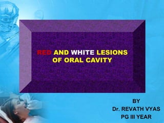
  • 1. RED AND WHITE LESIONS OF ORAL CAVITY BY Dr. REVATH VYAS PG III YEAR
  • 2. Color of oral mucosa depends on… Concentration and dilatation of blood vessels in the underlying connective tissue Degree of keratinisation Amount of melanin pigment in epithelium Thickness of epithelium Orban’s Oral Histology & Embryology 11th edition.
  • 3. 1) Hyper keratosis 2) Acanthosis 3) Intra and extracellular accumulation of fluid in the epithelium (i.e. Leukoedema) 4) Necrosis of the oral epithelium 5) Microbes, particularly fungi, produce whitish pseudomembranes 6) Reduced vascularity in the underlying lamina propria. Why abnormally white?
  • 4.
  • 5. Why abnormally red ? Atrophic epithelium Reduction in the number of epithelial cells Increased vascularisation Blood vessel enlargement Presence of blood in the tissue Increased hemoconcentration Bengel, Veltman, Loevy, Taschini - Differential diagnosis of diseases of the oral mucosa Burket’s oral medicine – 10th edn.
  • 6.
  • 7. Causes of red lesions Crispian Scully - Oral & Maxillofacial Medicine; 2004. CAUSES Inflammatory Reactive Atrophic/ Erosive Neoplasms Purpura Vascular
  • 10. CLASSFICATION ACCORDING TO Wood & Goaz, V edition
  • 12. CLASSFICATION ACCORDING TO BURKET’S 11th edition
  • 14. A) ORAL CANDIDIASIS (Candidosis, Moniliasis, Thrush) 1) Opportunistic infection affecting the oral mucosa. 2) Caused by the yeast candida albicans. 3) Predisposing factors convert candida from the normal commensal flora (saprophytic stage) to a pathogenic organism (Parasitic stage).
  • 15.
  • 16. Etiology: Candida species responsible for candidal infections are c. albicans c. tropicalis and 80% of the species c. glabrata Yeasts penetration of epithelial cells is facilitated by their production of lipases
  • 17.
  • 18. CLINICAL FINDINGS PSEUDOMEMBRANOUS CANDIDIASIS 1) Acute form is recognized as classic candida infection 2) Affects patients medicated with antibiotics, immunosuppressant drugs or a disease 3) Oral lesions are soft, white, slightly elevated plaques occurring on the buccal mucosa, tongue, palate, gingiva and floor of the mouth. 4) “Curdly white” Plaques can be wiped away with guaze, leaves an inflamed, sometimes bleeding area. 5) Clinical presentations of acute and chronic are indistinguishable. 6) Chronic form associated with HIV infections and with steroid inhalers.
  • 19.
  • 20. ERYTHEMATOUS CANDIDIASIS 1) Previously referred as atrophic candidiasis (Antibiotic sore mouth or Antibiotic stomatitis) 2) Occurs as a sequelae to a course of broad spectrum antibiotics, inhalation steroids smoking and diseases. 3) Lesions appear red or erythematous 4) May occur at any site 5) Only variety of oral candidiasis, which is consistently painful.
  • 21.
  • 22. CHRONIC PLAQUE-TYPE AND NODULAR CANDIDIASIS (Chronic Hyperplastic candidiasis) 1) Chronic-plaque type replaces candidal leukoplakia 2) Characterized by a white plaque. 3) Firm, white persistent plaques, usually on the lips, tongue and cheeks 4) Chronic plaque-type and nodular candidiasis have been associated with malignant transformation.
  • 23.
  • 24. CANDIDA – ASSOCIATED LESIONS I. DENTURE STOMATITIS 1) Most prevalent site is palatal mucosa 2) Classified into three different types Type I – Localized to minor erythematous sites Type II – affects a major part of the denture covered mucosa Type III – has a granular mucosa
  • 25.
  • 26. II. ANGULAR CHEILITIS Characterized by erythema, fissuring, scaling Saliva tends to pool in, keeping the area moist & favoring yeast infection Candidal infection involving perioral skin - creates a clinical pattern CHEILOCANDIDIASIS
  • 27. CHEILOCANDIDIASIS A diffuse form of chronic candidiasis characterized by pain, swelling, erythema with focal ulcerations, and crusting is called cheilocandidiasis. It represents a secondary candidal infection superimposed on areas of trauma from mechanical or solar factors.
  • 28. Cheilocandidiasis and juvenile juxtavermilion candidiasis refer to more extensive and often desquamative lesions affecting the full width of the lip, even extending into adjacent lesions. They are associated with habitual lip sucking, prolonged dental appointments, sunlight, and chronic candida infection. Differential diagnoses of these lesion include perioral erythema and Id reaction (localized sterile vesiculopapular rash).
  • 29. Etiology Infection (bacterial, fungal) Nutritional deficiency Reduced vertical dimension Long-term denture wearers Shafer, Hine, Levy. Textbook of Oral Pathology, 6th edn;2009.
  • 30.
  • 31. III. MEDIAN RHOMBOID GLOSSITIS 1) Characterized by a erythematous lesion in the centre of the posterior part of the dorsum of the tongue. 2) Lesion has a oval configuration 3) Resulting from atrophy of the filiform papillae and lobulated. 4) Smokers, denture-wearers and patients using inhalation steroids have an increased risk 5) Asymptomatic 6) Management is restricted to a reduction in predisposing factors.
  • 32. III. MEDIAN RHOMBOID GLOSSITIS Brocq : 1914 Prevalence: < 1% Males > Females
  • 33. Synonyms Median candidiasis of the tongue Atypical/ paramedial rhomboid glossitis Central papillary atrophy Posterior lingual papillary atrophy Posterior midline atrophic candidiasis
  • 34. Etiology Embryological Inflammatory Immunological Infectious Associated with: Tobacco smoking Denture wearing Trauma Int J Oral Surg 1984;13(5):411-415. Oral Surg Oral Med Oral Pathol 1996;81(4):379-80.
  • 35. Med Oral Patol Oral Cir Bucal 2005;10:123-7.
  • 36.
  • 39. Management of median rhomboid glossitis Topical antifungal: clotrimazole troches fluconazole Topical corticosteroid: fluocinonide 0.05% clobetasol 0.05% Bruch JM, Treister NS. Clinical Oral Medicine and Pathology.
  • 40. SECONDARY ORAL CANDIDIASIS 1) Accompanied by systemic mucocutaneous candidiasis and other immune deficiencies 2) Chronic mucocutaneous candidiasis a) Also affect the skin, the nail bed, genital mucosa b) Face and scalp may be involved c) Occur as part of 1) Endocrine disorders – Hyperparathyroidism and Addisons disease 2) Myeloperoxidase deficiency 3) Chediak – Higashi syndrome, 4) Severe combined immunodeficiency syndrome characterized by a defect in the function of the cell-mediated immunity 5) Thymoma is a neoplasm of thymic epithelial cells 6) Chronic familial mucocutaneous candidiasis an inherited disorder, occurs early in life.
  • 41.
  • 42.
  • 43. ORAL CANDIDIASIS ASSOCIATED WITH HIV 1) 90% of AIDS patients have oral candidiasis 2) Most common types a) Pseudomembranous candidiasis b) Erythematous Candidiasis c) Angular cheilitis and d) Chronic hyperplastic candidiasis
  • 44.
  • 45.
  • 46. DIAGNOSIS AND LABORATORY FINDINGS Smear:- useful in Pseudomembranous oral candidiasis and angular cheilitis Swab:- Taken by rubbing cotton-tipped swabs over the lesional tissue. Imprint Culture: Sterile (plastic) foam pads submerged in sabouraud broth placed on the infected surface for 60 seconds. Cultivated at 37oC. Result is expressed as colony forming units per cubicmillimeter. (CFU/mm3) Valuable in erythematous candidiasis and denture stomatitis. Salivary culture technique:- Used to get an adequate quantification of candida. Histopathological Examination:- Chronic plaque-type and nodular candidiasis, identify the presence of epithelial dysplasia.
  • 47.
  • 48. Treatment of Denture stomatitis Eliminate predisposing factors Improve denture hygiene Denture should be stored in antimicrobial solutions Type III denture stomatitis treated with surgical excision
  • 49. Treatment of Angular cheilitis  Topical treatment with miconazole  A mild steroid ointment suppress the inflammation  A moisturizing cream prevent new fissure formation
  • 50. Treatment of Median rhomboid glossitis Topical antifungal: clotrimazole troches fluconazole Topical corticosteroid: fluocinonide 0.05% clobetasol 0.05% Bruch JM, Treister NS. Clinical Oral Medicine and Pathology.
  • 51. ANTIFUNGALS Topical antifungals are usually the drug of choice for uncomplicated, localized candidiasis in patients with normal immune function. Systemic antifungals are usually indicated in cases of disseminated disease and/or in immunocompromised patients.
  • 52.
  • 53. Dosage form/ strength Indication Miconazole cream 2% Angular cheilitis Clotrimazole cream 1% Angular cheilitis Ketoconazole cream 2% (Prescription) Angular cheilitis Nystatin ointment 100,000 units/gram (Prescription) Angular cheilitis Nystatin topical powder 100,000 units/gram (Prescription) Denture stomatitis Nystatin oral suspension 100,000 units/gram (Prescription) Intraoral candidiasis Betamethasone dipropionate clotrimazole cream (Prescription) Chronic angular cheilitis Clotrimazole troches 10 mg (Prescription) Intraoral candidiasis Amphotericin B 100 mg/ml (Prescription) Intraoral candidiasis
  • 54. Generic name Formulation Amphotericin B 100 mg/ml oral suspension Clotrimazole 10 mg troche Fluconazole 100 mg tablet 10 mg/ml oral suspension 40 mg/ml oral suspension Itraconazole 100 mg capsule 10 mg/ml oral suspension Ketoconazole 200 mg tablet Nystatin 100,000 units/ml oral suspension 200,000 units/ml pastille 500,000 units/ml tablet 100,000 units/ml vaginal tablet
  • 55. ANTIFUNGALS Medication should be continued for at least 48 hours after the disappearance of clinical signs of candidiasis along with complete healing and the absence of mucosal erythema. Some sources recommend drug therapy should be continued for 10– 14 days regardless of the disappearance of clinical signs of candidiasis.
  • 56. SURGERY May be advised for hyperplastic and nodular types of candidiasis. (Cryotherapy, CO2 laser therapy, and cold-knife surgery). Small lesions may be allowed for primary healing Large lesions are closed by skin grafts after surgery. But there are chances of reccurrence. Long term and intermittent anti fungal therapy may lead to complete or partial resolution.
  • 57. Prognosis 1) Prognosis is good if predisposing factors are reduced or eliminated. 2) Persistant chronic plaque-type and nodular candidiasis increased risk for malignant transformation. 3) Patients with severe immunosuppression may encounter disseminating candidiasis with a fatal course.
  • 58.
  • 59.
  • 60. HAIRY LEUKOPLAKIA 1) Oral hairy Leukoplakia is a corrugated white lesion that usually occurs on the lateral or ventral surfaces of the tongue in patients with severe immunodeficiency 2) Second most common HIV associated lesion 3) Marker of disease activity associated with low CD4+ T-lymphocyte counts. 4) Not pathognomonic for HIV since other immunodeficiencies, such as immunosuppressive drugs and cancer chemotherapy are also associated with HL. Also occur in patients undergoing prolonged steroid theropy. 5) It was first reported by Greenspan and coworkers in 1984 in young homosexual males.
  • 61. Epidemiology: 1) Prevalence may be as high as 80% 2) The prevalence in children is lower 2% 3) More frequently encountered in men 4) Correlation between smoking and HL has also been observed. Etiology and Pathogenesis: 1) Strongly associated with Epstein-Barrvirus with low levels of CD4+ T-lymphocytes 2) Antiviral medication, which prevents EBV replication, support the fact that EBV is an etiological factor.
  • 62. Clinical Findings: 1) Most commonly involves the lateral border of the tongue but may extend to the ventral or dorsal surfaces. 2) Occasionally located on the buccal mucosa. 3) Typical clinical appearance is vertical white folds oriented as a palisade along the borders of the tongue 4) Lesions on the tongue are corrugated and have a shaggy or frayed appearance 5) May also be displayed as white and somewhat elevated plaque, which cannot be scraped off 6) It is often bilateral. 7) Asymptomatic
  • 63.
  • 64.
  • 65. Diagnosis: Diagnosis of HL based on clinical characteristics, a histopathologic examination and detection of EBV
  • 66. Pathology: 1) Histopathology of HL is characterized by a) Severe hyperkeratosis with an irregular surface b) Acanthosis with superficial edema c) corrugations and often thin tufts (“hairs”) may be seen projecting from the surface d) numerous koilocytic cells (virally affected “balloon” cells) in the spinous layer. e) Pyknotic nuclei
  • 67. 2) Characteristic microscopic feature is homogenous viral nuclear inclusions with a residual rim of normal chromatin 3) Candida hyphae surrounded by polymorphonuclear granulocytes - common feature. 4) EBV can be detected by in-situ hybridization or by immunohistochemistry.
  • 68.
  • 69. Differential Diagnosis It is important to differentiate HL from other clinically similar entities such as 1) Hyperplastic candidiasis 2) Idiopathic leukoplakia 3) Leukoplakia induced by tongue chewing 4) Tobacco associated leukoplakia 5) Lichen planus 6) Lupus Erythematosus 7) White sponge nevus 8) Verrucous Leukoplakia
  • 70. Treatment and Prognosis 1) No treatment is indicated 2) Condition disappears when antiviral medications such as zidovudine, acyclovir, or ganacyclovir are used in the treatment of the HIV infection 3) Topical application of podophyllin resin 25% solution (or) tretinoin short-term resolution of the lesions. 4) HL is not related to increased risk of malignant transformation.
  • 72. ORAL LEUKOPLAKIA - A Keratotic plaque occurring on mucous membranes - considered a premalignant lesion. Definition: WHO defined leukoplakia as “a white patch or plaque that cannot be characterised clinically or pathologically as any other disease” Oral leukoplakia is defined as a predominantly white lesion of the oral mucosa that cannot be characterized as any other definable lesion.
  • 73. Etiology and Pathogenesis: Etiologic factors of Leukoplakia 1. Tobacco products 2. Ethanol 3. Hot, Cold, Spicy and acidic foods and beverages 4. Alcoholic mouth rinse 5. Occlusal trauma 6. Sharp edges of prostheses or teeth 7. Actinic radiation 8. Syphilis 9. Presence of candida albicans 10. Presence of viruses Leukoplakia commences as a protective reaction against a chronic irritant.
  • 74. Epidemiology: 1. Prevalence is 2.6% 2. Over 50yrs of age 3. In men Clinical Findings: 1. Asymptomatic 2. Lip vermilion, buccal mucosa, mandibular gingiva, tongue, floor of the mouth, hard palate, maxillary gingiva, Lip mucosa and soft palate. 3. Borders may be distinct or indistinct, smoothly contoured or ragged. 4. Solitary or multiple plaques scattered through the mouth 5. Floor of the mouth and the lateral borders of the tongue are high-risk sites for malignant transformation
  • 75. 6. It is divided into a. Homogenous type and b. Non-Homogenous type 7. Classification: 1. Homogenous Leukoplakia a. White, well-demarcated plaque with an identical reaction pattern throughout the entire lesion. b. No red component c. A smooth thin surface to a leathery appearance with surface fissures as cracked mud. d. Asymptomatic
  • 76.
  • 77.
  • 78.
  • 79. 2. Speckled Leukoplakia: Composed of white and red flecks of fine or coarse variety.
  • 80. 3. Erythroleukoplakia: a) Combination of red and white patches, segregation of the red and white components.
  • 81. 4. Verrucous leukoplakia:- a) Large white verrucous areas to small nodular structures. b) Verrucous, papillary (nodular) or exophytic components c) More aggressive proliferation pattern, recurrence rate are designated proliferative verrucous leukoplakia (PVL) d) PVL common in women e) lower gingiva is a predilection site. f) Malignant potential is high in PVL.
  • 82.
  • 83.
  • 84. Diagnosis: 1. The cause, such as a sharp tooth cusp or restoration, should be eliminated. 2. If healing does not occur in 2 weeks, biopsy is essential. Pathology: 1. Hyperkeratosis without any other features is compatible with homogenous oral leukoplakia. 2. Histological features include hyperkeratosis, atrophy, hyperplasia with or without dysplasia. 3. Carcinoma-in-situ or intraepithelial carcinoma 4. Invasive squamous cell carcinoma
  • 85. 5. Leukoplakia may also be divided Reversible leukoplakia Irreversible leukoplakia 6. Malignant potential: Ranges between 3% and 7%. Lesions in the floor of the mouth, ventral surface of the tongue, margins of the tongue and retromolar region have a higher risk.
  • 86.
  • 87. High-Risk Leukoplakias 1. Red component 2. Raised component 3. Presence in high-risk oval 4. Tobacco and alcohol use 5. Non smoker and un known etiology 6. Non reversible type 7. Microscopic atypia
  • 88. Differential Diagnosis: The keratotic varieties considered in the differential diagnosis are 1) Lichen planus 2) Leukoedema 3) Cheek-biting lesion 4) Smokeless tobacco lesion 5) Lupus erythematosus 6) Hyperplastic or hypertrophic candidiasis 7) Hairy leukoplakia 8) Electrogalvanic or mercury contact allergy 9) Verrucous or squamous cell carcinoma 10) Verruca vulgaris 11) White sponge nevus.
  • 89. Management of Leukoplakia: 1. Discontinue the habits 2. Cold-knife surgical excision, laser surgery, widely used to eradicate leukoplakias 3. Low-risk lesions – identify and eliminate the local and chronic causative irritants 4. Chemoprevention in the form of topical bleomycin 5. Systemically administered antioxidants such as retinoic acid, β carotene, and vitamins C & E have been administered 6. Lesions are large and widespread – stripping procedures with free grafts. 7. Reexamine every 3 months for the first year. 8. Following 5 years of no relapse, self examination.
  • 90.
  • 91. Recurrence after surgical treatment 10%–35% of cases. Nonsurgical advantages. Minimal adverse effects to patients Large area of the oral mucosa Patients with medical problems due to surgical risks. Easy application Relative low cost. 91
  • 92. Carotenoids 1.Beta-Carotene Group of extremely hydrophobic molecules with little or no solubility in water. Betacarotene is a vitamin A precursor. Found in Dark green, orange or yellowish vegetables, such as spinach, carrots, sweet potato, mango, papaya, and oranges. 92 A Review of the Nonsurgical Treatment of Oral Leukoplakia International Journal of Dentistry 2010
  • 93. Lycopene This is a fat-soluble red pigment Fruit vegetables( tomatoes). Most efficient biological antioxidizing agent. Antioxidant It binds to chemical species that react with oxygen. Modify intercellular exchange junctions. This is considered to be an anticancer mechanism Lycopene inhibits human neoplastic cellular growth by interfering with growth factor receptor signaling. 93 A Review of the Nonsurgical Treatment of Oral Leukoplakia International Journal of Dentistry 2010
  • 94. Vitamins L-Ascorbic Acid (Vitamin C). Citrous fruits as kiwi, strawberries, papaya, and mango. The current US recommanded dose is 100–120 mg/per day for adults. 140 mg/day smokers because they have low concentration of L-AA in serum leukocytes. 94 A Review of the Nonsurgical Treatment of Oral Leukoplakia International Journal of Dentistry 2010
  • 95. α-Tocoferol (Vitamin E) α-Tocoferol (AT) is most active form of vitamin E. Plant oil, margarine, and green leaves. The recommended dose is 10 mg/day men 8 mg/day women. Absorption rate is reduced consumption exceeds 30mg/day. α-Tocoferol is an effective antioxidant at high levels of oxygen. Protects cell membrane from lipidic peroxidation. 95 A Review of the Nonsurgical Treatment of Oral Leukoplakia International Journal of Dentistry 2010
  • 96. Retinoic Acid (Vitamin A) Retinoic acid is obtained from carotene and animal products such as meat, milk, and eggs. In the intestine retinoic acid is converted into retinal and retinol. Retinoids are transported by plasma proteins. Hepatic metabolization cytochrome P- 450. Hypervitaminosis  consumption exceeds storage capacity of liver 13-cRA was used for the first time against acne in 1969. 96 A Review of the Nonsurgical Treatment of Oral Leukoplakia International Journal of Dentistry 2010
  • 97. Retinoids affect Keratin production Expression of growth factors and kinases Oncogenesis Apoptosis Production of the collagen matrix Immunologic and inflammatory response Cellular differentiation Embrionary morphogenesis and Carcinogenesis 97 A Review of the Nonsurgical Treatment of Oral Leukoplakia International Journal of Dentistry 2010
  • 98. Fenretinide Fenretinide (4-HPR) or N-(4-hydroxyphenyl retinamide) is a vitamin A analogue. Synthesized in United States 1960. Less toxic than many other vitamin A analogues. 98 A Review of the Nonsurgical Treatment of Oral Leukoplakia International Journal of Dentistry 2010
  • 99. Photodynamic Therapy (PDT) is a noninvasive method for the treatment of premalignant lesions and head and neck Cancers photosensitising drug (photosensitiser), oxygen, and visible light 99 A Review of the Nonsurgical Treatment of Oral Leukoplakia International Journal of Dentistry 2010
  • 100. ERYTHROPLAKIA Pre-malignant lesion Prevalence: 0.02 – 0.83% India: 0.2% Female: male = 1:1.04 Well-demarcated, fiery red patch
  • 101. The word is an adaptation of the French term “erythroplasie de Queyrat”, which was originally used by Queyrat to describe a precancerous red lesion that develops on the glans penis. Oral erythroplakia is clinically and histopathologically similar to the genital process with a comparable premalignant tendency.. Oral Oncol 2005;41:551-61.
  • 102. Definitions WHO 1978 Bright red velvety plaques which cannot be characterized clinically or pathologically as being due to any other condition. Axell et al 1984 Lesions of the oral mucosa that present as bright red patches or plaques that cannot be characterized clinically or pathologically as any other condition. Axell et al 1986 Lesions of the oral mucosa that present as red areas and cannot be diagnosed as any other definable lesion. Pindborg et al 1997 (WHO) A fiery red patch that cannot be characterized clinically or pathologically as any other definable lesion. Crit Rev Oral Biol Med 2003;14(1):47-62.
  • 103. Provisional diagnosis A provisional diagnosis of oral erythroplakia is made when a lesion at clinical examination cannot be clearly diagnosed as any other disease of the oral mucosa with red appearance. Definitive diagnosis A definitive diagnosis of oral erythroplakia is made as a result of identification, and if possible elimination, of suspected etiological factors and, in the case of persistent lesions, histopathological examination. J Oral Pathol Med 1996;25:49-54.
  • 104. Etiologyand Pathogenesis: 1) Etiology is uncertain, most cases are associated with heavy smoking. 2) Majority of erythroplakias exhibit premalignant and malignant changes. 3) EP is often regraded as the earliest sign of asymptomatic oral cancer. 4) EP’s reddish color results from the absence of a surface keratin layer, connective tissue papillae project close to the surface, general failure of the epithelial cells to achieve significant maturation (keratinization)
  • 105. 1) Appears as a velvety red or granular red macule (patch) that may be slightly raised. 2) Occurs predominantly in older men, in the 6th and 7th decades of life. 3) More commonly seen on the floor of the mouth, the ventral tongue, the soft palate, and the tonsillar fauces, all prime areas for the development of carcinoma 4) Multiple lesions may be present. Clinical features
  • 106. 5) Commonly described as erythematous plaques with a soft velvety texture. 6) Asymptomatic 7) This painless lesion varies greatly in size, and borders may be well circumscribed or may blend imperceptibly with the surrounding normal mucosa. 8) Three different clinical appearances described by shear. a) the homogenous form, red in appearance b) patches of Erythroplakia and leukoplakia c) Speckled erythroplakia or granular erythroplakia
  • 107. 9) Homogenous EP is generally much more aggressive than leukoplakia or speckled EP 10) The floor of the mouth is the most common site in men, mandibular gingival-alveolar mucosa – mandibular sulcus is most common site in women. 11) Retromolar region is the second most common site in both genders.
  • 108.
  • 109. Most asymptomatic malignant erythroplakic lesions are small; 84% are ≤ 2 cm in diameter, and 42% are ≤ 1 cm. Burket’s Oral Medicine – 11th edn.
  • 110.
  • 111. Diagnosis: The provisional diagnosis is based on the clinical observation of a white or red patch. If trauma is suspected, sharp tooth cusp or restoration, should be eliminated. If healing doesn’t occur in 2 weeks, biopsy is essential. Histopathology: 1) 80 to 90% of cases of erythroplakia are histopathologically show severe epithelial dysplasia, carcinoma-in-situ or invasive carcinoma 2) Epithelial dysplasia is defined as a precancerous lesion of stratified squamous epithelium charaterized by cellular atypia and loss of normal maturation. 3) Carcinoma in situ is defined as a lesion in which the full thickness of squamous epithelium shows the cellular features of carcinoma without stromal invasion
  • 112. Criteria used for DiagnosingEpithelialDysplasia
  • 113. Differential Diagnosis: The lesions to be considered in the differential diagnosis of Erythroplakia are: 1) Traumatic erythema 2) Atrophic candidiasis 3) Purpuric macule (early stage) 4) Macular hemangioma 5) Contact allergy 6) Other infection 7) Localized gingivitis 8) Kaposis sarcoma (early stage)
  • 114. Treatment and Prognosis: 1) If a red lesion persists for more than 14 days biopsy is mandatory 2) Epithelial dysplasia or carcinoma in situ warrants complete removal of the lesion 3) Alcohol and smoking are well established risk factors - advice the patients to discontinue the habits. 4) Cold knife surgical excision, as well as laser surgery widely used to eradicate erythroplakias 5) Follow-up of Erythroplakia:- Reexamine the premalignant site irrespective of surgical excision every 3 months for the first year. Follow-up intervals may be extended to once every 6 months.
  • 115. Management of oral Leukoplakia / Erythroplakia Adapted from vander waal et al
  • 116. ORAL SUBMUCOUS FIBROSIS Chronic insidious disease caused by arecanut use, and is associated with both significant morbidity and an increased risk for malignancy. History: 1. First described by schwartz in 1952 coined the term “atrophia idiopathica (trophica) mucosae oris” 2. In 1953, Joshi from Mumbai redesignated the condition as oral submucous fibrosis.
  • 117. Etiology and Pathogenesis: 1. Arecoline a primary etiologic factor. 2. Arecoline affecting the metabolism of collagen leads to an increased fibrosis. 3. Evidence of a genetic predisposition.
  • 118.
  • 119. Clinical Features: EARLY OSF (Prodromal Symptoms) 1. Burning sensation in the mouth 2. Ulcerations or generalized inflammation 3. Excessive salivation 4. Defective gustatory sensation 5. Dryness of the mouth 6. Appearance of small vesicles in the cheek and palate. 7. Petechiae on the tongue, labial and buccal mucosa 8. Pain where submucous fibrotic bands are developing
  • 120. ADVANCED OSF: 1. White marbling 2. Fibrous bands appear beneath an atrophic epithelium 3. In the buccal mucosa run in a vertical direction 4. Circular band around the entire rima oris (mouth orifice) 5. Impairment of tongue movement 6. Difficulty in opening the mouth 7. Inability to whistle or blow out a candle 8. Difficulty in swallowing 9. Increased fibrosis interferes with speech 10. 25% patients exhibit oral leukoplakias.
  • 121.
  • 122.
  • 123. Diagnosis: For the diagnosis of OSF a. Palpable fibrous bands b. Mucosal texture feels tough and leathery c. Blanching of mucosa d. Histo pathological features atrophic epithelium loss of rete ridges and juxta epithelial hyalinization of laminapropria
  • 124. Pathology: Histo pathologic features of OSF are 1. Fine fibrils of collagen 2. Edema 3. Hypertrophic fibroblasts 4. Dilated and congested blood vessels 5. Infiltration of neutrophilic and eosinophilic granulocytes 6. Down-regulation of fibroblasts 7. Epithelial atrophy 8. Loss of rete pegs 9. Early signs of hyalinization 10. Epithelial dysplasia appears in 7-26% of OSF tissues.
  • 125.
  • 126.
  • 127. Management: 1. Elimination of the habit 2. Management strategies are A. Nutritional support: high protein and calories, vitamin B complex and minerals B. Immunomodulatory drugs: 1. Local and systemic application of glucocorticoids and placental extract 2. Glucocorticoids act as immunosuppressive agents. 3. Also prevent or suppress inflammatory reactions.
  • 128. C. Physiotherapy: 1. Heat has been commonly used. In the form of hot rinses, lukewarm water, or short wave and microwave diathermy. 2. Microwave diathermy seems superior to short wave. D. Local Drug Delivery 1. Local injections of dexamethasone, hyaluronidase and placental extract
  • 129. 2. Hyaluronidase Breaks down hyaluronic acid Lowers the viscosity of the intercellular cement substances Decreases collagen formation 3. Chymotrypsin, acts as a proteolytic and anti inflammatory agent.
  • 130. 3. Placental extracts: a. Local injections as well as parental form b. Placenta acts as a biogenous stimulator c. Other actions of placental extract are 1. Anti-inflammatory 2. Increase in blood circulation 3. Arrest of tissue growth stagnation, metabolic degenerative conditions and lowered immunity response d. Used as a local nutrient.
  • 131. E. Combined Therapy: 1. Local injections of chymotrypsin, hyaluronidase and dexamethasone 2. Combined therapy with nylidrin hydrochloride, vitamins D, E and B complex, iodine, placental extract, local and systemic corticosteroids and physiotherapy - success rate of 62% in OSMF
  • 132. F. Surgical Management: 1. Submucosal resection of fibrotic bands and replacement with a partial-thickness skin or mucosal graft 2. Bilateral temporalis myotomy 3. New treatment regimen composed of surgical excision of the fibrotic bands with submucosal placement of fresh human placental grafts, followed by local injections of dexamethasone for advanced cases.
  • 133. IMMUNOPATHOLOGIC DISEASES ORAL LICHENPLANUS DRUG INDUCED LICHENOID REACTION LICHENOID REACTION OF GVHD LUPUS ERYTHEMATOSIS
  • 134. ORAL LICHEN PLANUS INTRODUCTION: Chronic inflammatory disease that affects the skin and the mucous membrane. First described by Erasmus wilson in 1869. One of the most common oral diseases. The exact cause is unknown, but the immunologic system plays a leading role in the pathogenesis.
  • 135. Oral Lichenoid reactions include the following disorders: 1) Lichen planus 2) Lichenoid contact reactions 3) Lichenoid drug eruptions 4) Lichenoid reactions of graft-versus-host disease (GVHD)
  • 136. EtiologyandPathogenesis 1. Is not known 2. Immune system has a primary role supported by the subepithelial band formed infiltrate dominated by T-lymphocytes and macrophages and the degeneration of basal cells known as liquefaction degeneration. 3. Autoreactive T-lymphocytes may be of primary importance for the development of oral lichen planus. Epidemiology: 1. Prevalence is 0.5 – 2.2% 2. Commonly seen in women than men. 3. Mean age at the time of diagnosis is approximately 55 years.
  • 137. Lavanya N, Jayanthi P, Rao UK, Ranganathan K. Oral lichen planus: An update on pathogenesis and treatment. J Oral Maxillofac Pathol 2011;15:127-32.
  • 138. ClinicalFindings: 1. Cutaneous lesions encountered in 15% of patients with OLP. 2. Classic appearance of skin lesions consists of pruritic erythematous to violaceous papules which are flat topped, small, angular only a few millimeters in diameter. 3. Larger plaques, covered by a fine, glistening scale. 4. Surface is covered by characteristic, very fine grayish-white lines, called wickham’s striae 5. Occur in a bilaterally symmetrical pattern, on the flexor surfaces of the wrist and forearms, the inner aspect of the knees and thighs, and the trunk.
  • 139. Primary symptom is a severe pruritis Trauma may aggravate the disease, referred to as a Koebner phenomenon. Most frequent extra oral mucosal site is the genital mucosa. Classic skin lesions described as Purplish Polygonal Planar Pruritic Papules Plaques
  • 140.
  • 141. OralManifestations: • Classically characterized by lesion consisting of radiating white or gray, velvety, thread – like papules in a linear, annular, or retiform arrangement • A tiny white elevated dot present at the intersection of the white lines, known as the striae of wickham. • OLP may contain both red and white components with different textures. a. Reticulum b. Papules c. Plaque-like d. Bullous e. Erythematous f. Ulcerative
  • 142. A. Reticular form: 1. Characterized by fine, white lines or striae 2. Striae form a network also show annular (circular) patterns. 3. A peripheral erythematous zone 4. Most frequently observed bilaterally in the buccal mucosa.
  • 143. B. Papular form: 1. Present in the initial phase of the disease. 2. Characterized by small white dots
  • 144. C. Plaque form: 1. Homogeneous well-demarcated white plaque, surrounded by striae. 2. Similar to homogeneous oral leukoplakias 3. Most often encountered in smokers
  • 145. D) Bullous form: 1. Unusual 2. Clinically resemble erosive lichen planus
  • 146. E) Erythematous (Atrophic) OLP: 1. Characterized by homogeneous red area 2. Present in the buccal mucosa or palate, striae are frequently seen in the periphery 3. Exclusively affecting attached gingiva presents as desquamative gingivitis.
  • 147.
  • 148. F) Ulcerative form: 1. Disabling form of OLP 2. Fibrin-coated ulcers are surrounded by an erythematous zone frequently displaying radiating white striae 3. Complains of a smarting sensation
  • 149.
  • 150. Diagnosis 1. Lichenoid contact reactions most often detected on the buccal mucosa and lateral border of the tongue. 2. Oral Lichenoid drug eruptions - Patient’s drug history - Withdrawal of the drug causes resolution of the lesions 3. Oral Graft Versus Host Disease - Lesion is usually generalized in GVHD - Frequently seen simultaneously with xerostomia, presence of localized skin involvement and liver dysfunction
  • 151. 4. Discoid Lupus Erythematosus - Striae in DLE are typically more prominent with a more marked hyperkeratinization may terminate against a sharp demarcation. 5. Leukoplakia - Leukoplakia is not featured with papular or reticular elements. 6. Mucous membrane pemphigoid - In pemphigoid lesions, the epithelium is easily detached from the connective tissue by a probe (Nikolsky’s phenomenon) 7. Erythema Multiforme - In EM, the lesions do not typically appear with reticular or papular elements.
  • 152. Pathology Histopathologic features 1. Areas of hyper para keratosis or hyper ortho keratosis 2. Thickening of the granular cell layer 3. Saw-toothed appearance to retepegs 4. “Liquefaction degeneration” or necrosis of the basal cell layer. 5. An eosinophilic band may be seen just beneath the basement membrane and represent fibrin covering the lamina propria. 6. A dense subepithelial band-shaped infiltrate of lymphocytes and macrophages is also characteristic of the disease.
  • 153.
  • 154. MANAGEMENT OF ORAL LICHEN PLANUS I PHARMACOLOGICAL MODALITIES A) CORTICOSTEROIDS 1. Ability to modulate inflammation and immune response 2. Topical midpotency corticosteroids triamcinolone acetonide, high-potency fluorinated corticosteroids fluocinonide acetonide, betamethasone and superpotent corticosteroids clobetasol are used 3. Wide spread forms of OLP - potent and superpotent corticosteroids mouth washes and intralesional injections. 4. Antifungal therapy 5. Systemic corticosteroids for recalcitrant erosive or erythematous LP 6. Systemic prednisolone (40-80mg for 5-7 days)
  • 155. B. CALCINEURIN INHIBITORS Calcineurin is inhibited by Cyclosporine, tacrolimus and pimecrolimus. 1) Cyclosporine 1. An immunosuppressant used widely in post-allogenic organ transplant. 2. Used as a mouthrinse or topically with adhesive bases in OLP 3. Expensive 4. Reserved for highly recalcitrant cases of OLP 5. Causes gingival hyperplasia
  • 156. 2. TACROLIMUS: 1. Immunosuppressive agent for the treatment of atopic dermatitis 2. Successfully used in recalcitrant OLP cases 3. Has a potential cancer risk from the prolonged use 4. Recommended for short periods. 3. PIMECROLIMUS: 1. 1% topical cream used for OLP 2. Has significant anti-inflammatory and immunomodulatory capabilities.
  • 157. C. RETINOIDS 1. Topical retionoids - tretinoin, isotretinoin and fenretinide 2. Has immunomodulatory properties and effective in OLP 3. Reversal of white striae 4. Systemic retinoids used in severe lichen planus. D. DAPSONE 1. Antibacterial agent 2. Used in the treatment of leprosy 3. Used for skin diseases (acts as an anti inflammatory agent) 4. Common untoward effect is hemolysis.
  • 158. E) MYCOPHENOLATES 1. Used to treat psoriasis 2. Mycophenolate mofetil is a immunosuppressive drug used in organ transplant 3. To treat severe cases of OLP 4. Expensive F) LOW-DOSE, LOW MOLECULAR WEIGHT HEPARIN (ENOXAPARIN) 1. Devoid of anticoagulant properties inhibits T-lymphocyte heparanase activity 2. Simple, effective and safe treatment for OLP 3. Injected subcutaneously has no side effects.
  • 159. G. EFALIZUMAB 1. Is a recombinant humanized monoclonal antibody 2. An immunosuppressant in the treatment of psoriasis 3. Causes improvement in OLP 4. Once a week as a subcutaneous injection.
  • 160. IINON-PHARMACOLOGICALMODALITIES A. PUVA THERAPY: 1. Uses photochemotherapy with 8-methoxypsoralen and long wave ultraviolet light (PUVA) 2. Methoxypsoralen is given orally, followed by administration of 2 hours of UV radiation intraorally in the affected sites. 3. Successfully used in the treatment of severe cases of OLP. B. PHOTODYNAMIC THERAPY 1. Uses a photosensitizing compound like methylene blue. 2. It may reverse the hyper proliferation and inflammation of lichen planus.
  • 161. C. LASER THERAPY: 1. Surgical management using cryosurgery and different types of lasers have been tried. 2. All types of laser destroy the superficial epithelium containing the target keratinocytes by protein denaturation. 3. Effectiveness is yet to be proven.
  • 162. Lavanya N, Jayanthi P, Rao UK, Ranganathan K. Oral lichen planus: An update on pathogenesis and treatment. J Oral Maxillofac Pathol 2011;15:127-32.
  • 163. DRUG-INDUCED LICHENOID REACTIONS Lichenoid reactions and Lichen planus exhibit similar histopathologic features. Lichenoid reactions were differentiated from Lichen planus on the basis of 1) The administration of a drug, contact with a metal, use of a food flavoring or systemic disease 2) Resolution when the drug or other factor was eliminated Clinically, lichenoid reactions may exhibit the classic appearance of lichen planus, but atypical presentations are seen. LICHENOID REACTIONS
  • 164. DRUG-INDUCED LICHENOID REACTIONS Clinically, there is often little to distinguish drug-induced lichenoid reactions from lichen planus. Lichenoid lesions that include the lip, symmetric in distribution and also involve skin are more likely to be drug-related. Histopathologically, Lichenoid drug eruptions may show a deep as well as superficial perivascular lymphocytic infiltrate rather than the classic band like infiltrate of lichenplanus. Drug-induced Lichenoid reactions may resolve promptly when the offending drug is eliminated.
  • 165. Important causes of lichenoid reactions Gold therapy Nonsteroidal anti-inflammatory drugs (NSAIDS), Diuretics Other antihypertensives Oral hypoglycemic agents of the sulfonyl urea
  • 166. DrugsandMaterialsimplicatedinOralLichenoidreactions Category Drugs or Materials Antimicrobials Dapsone Ketoconazole Para-aminosalicylic acid Sodium amino salicylate Streptomycin Sulfamethaxazole Tetracycline Antiparasitics Antimony compounds (stibophen, stibocaptate) Organic arsenicals Chloroquine Pyrimethamine Quinacrine Anti hypertensives ACE inhibitors Chlorothiazide Hydrochlorothiazide Labetalol Mecurial Diuretics Methyldopa Practolol
  • 167. Antiarthritics Aurothioglucose Colloidal gold (Europe only) Gold sodium thiomalate, thiosulfate Anxiolytics Lorazepam NSAIDS Fenclofenac Ibuprofen Naproxen Phenylbutazene Oral hypoglycemic agents Chlorpropamide Tolazamide Tolbutamide Uricosuric agents Allopurinol Miscellaneous drugs Iodides Pencillamine Quinidine Sulfate Chemicals, dental restorative materials Substituted Paraphenylene- diamines used in color film developers, dental casting alloys and amalgam restorations
  • 168. EtiologyandPathogenesis: 1) Drugs and their metabolites act as haptens trigger a lichenoid reaction 2) Pencillin and gold may bind directly to self-protiens, presented by antigen-presenting cells and perceived as foreign by specific T- lymphocytes, similar to a delayed hypersensitivity reaction. 3) DILRs may result from poor drug metabolism
  • 169. Clinical Findings:- 1) Unilateral 2) Ulcerative reaction pattern 3) Oral lichen planus and lichenoid reactions should be considered , clinically indistinguishable. 4) Lichenoid drug eruptions appear similar to Lichen ruber planus and may be severely pruritic.
  • 170.
  • 171. Diagnosis: 1) Oral lesions comprise a white reticulum (or) papules concurrently with erythematous and ulcerative lesions 2) A correct diagnosis is easier when a patient develops DILR after starting a new drug. Management: 1) Discontinuance of the drug and symptomatic treatment with topical steroids.
  • 172. LICHENOIDREACTIONSOFGRAFTVERSUS HOSTDISEASE Complex multisystem immunologic phenomenon characterized by the interaction of immunocompetent cells from one individual (the donor) to a host (the recipient Etiology and Pathogenesis: 1) Major cause is allogenic hematopoietic cell transplantation
  • 173. Clinical Findings: 1) Occur in both acute and chronic GVHD 2) Lesions of GVHD are indistinguishable from the lesions of OLP. 3) The lesions of GVHD associated with a more widespread involvement of the oral mucosa 4) Involves the cheeks, tongue, lips and gingivae 5) A fine reticular network of white striae that resembles OLP is seen 6) Complain of a burning sensation of the oral mucosa. 7) Xerostemia
  • 174. Clinical Manifestations 1) Skin lesions present with pruritic maculopapular and mobiliform rash, affecting the palms and soles 2) Violaceous scaly papules and plaques, generalized erythroderma, bulla formation. 3) A toxic epidermal necrolysis like epidermal desquamation may occur.
  • 175.
  • 176. Diagnosis: Presence of systemic GVHD facilitates the diagnosis of oral mucosal changes of chronic oral GVHD Differential Diagnosis: The differential diagnosis of GVHD includes 1) Candidiasis 2) Toxic reactions to chemotherapeutic drugs 3) Unusual viral infections (herpes virus, cytomegalovirus) 4) Lichenoid drug eruptions
  • 177. Management: 1) Same as that of OLP, topical steroid preparations, fluocinonide and clobetasol gel. 2) Patients with a history of oral GHVD should be examined for oral malignancies as part of the medical follow-up procedure.
  • 178. ALLERGIC REACTIONS Lichenoid contact reactions Reactions to dentifrice and chlorhexidine
  • 179. LICHENOID CONTACT REACTIONS Delayed hyper sensitivity reaction to constituents derived from dental materials likes mercury (Hg) Clinical Findings: 1. Delayed hypersensitivity reaction to amalgam fillings. 2. Same reaction patterns as seen in OLP 3. Major clinical difference between OLP and LCR is the extensions of lesions 4. Non symptomatic
  • 180.
  • 181. Diagnosis: Replacement of the dental material may assist to discriminate between LCR and OLP. Management: 1. Replacement of dental materials result in cure in 90% of the cases 2. Lesions expected to heal within 1 to 2 months.
  • 182. B. REACTIONS TO DENTIFRICE AND CHLORHEXIDINE 1. Rare 2. Allergic reactions include flavor additives such as carvone and cinnamon or preservatives 3. Clinical manifestations – fiery red edematous gingiva, include both ulcerations and white lesions 4. Involve other sites labial, buccal and tongue mucosa. 5. Healing seen after withdrawal of the allergen – containing agent.
  • 183.
  • 184. TOXIC REACTIONS Reactions to smokeless tobacco Smoker’s palate
  • 185. A. Reactions to Smokeless Tobacco 1. Smokeless tobacco divided into three different groups; chewing tobacco, moist snuff and dry snuff. 2. Mildest form, the lesion noted as wrinkles at the site of application, high consumers may display a white and leathery lesion. 3. Gingival retractions most common adverse reaction seen in smokeless tobacco habit. 4. Smokeless tobacco products contain nitrosamines polycyclic hydrocarbons, aldehydes, heavy metals, and polonium 210
  • 186.
  • 187. B. SMOKER’S PALATE 1. Dark brown pigmentations of the oral mucosa and as white leathered lesions of the palate. 2. Red dots can be observed representing orifices of accessory salivary glands. 3. More prevalent in men 4. Common in high consumers of pipe tobacco, cigarettes and Reverse smoking 5. Etiology related to the high temperature.
  • 188.
  • 190. A. MORSICATIO 1. Habitual chewing 2. Seen in the buccal and lip mucosa 3. Three times more common in women 4. Management is by assurance
  • 191.
  • 192. B. FRICTIONAL KERATOSIS 1. Characterized by a white lesion without any red elements 2. Obesrved in areas subjected to increased friction
  • 193. Etiology and Pathogenesis: 1. Increased abrasion stimulates the epithelium to respond with an increased production of keratin. 2. Regarded as a physiologic response to trauma 3. Predisposing factors are smoking and alcohol Clinical Findings: 1. Seen in edentulous areas of the alveolar ridge 2. Asymptomatic Management: 1. No surgical intervention 2. Reduce predisposing factors.
  • 194. OTHER RED AND WHITE LESIONS Benign Migratory Glossitis (Geographic Tongue) Leukoedema White sponge Nevus Hairy tongue
  • 195. A. GEOGRAPHIC STOMATITIS 1831 – Rayer 1955 – Cooke : “erythema migrans” Prevalence : 1 – 2.5% Age : 20-29 years Females > Males Hacettepe Diş Hekimliği Fakültesi Dergisi 2008;32(2):73-8. Pediatr Dentistry 1992;14(6):392-6.
  • 196. Benign inflammatory condition of the mucosa with map-like areas of erythema which are not constant in size, shape or location. Oral Surg Oral Med Oral Pathol 1993;76(4):476-9.
  • 197. Synonyms Erythema migrans Erythema circinata migrans Benign migratory glossitis Stomatitis areata migrans Annulus migrans Cooke’s disease Continental tongue Marginal exfoliative glossitis Transitory benign plaques of the tongue Wandering rash of the tongue Exfoliatio areata linguae Lingua geographica Am J Med 2002;113:751-5.
  • 198. Etiopathogenesis Genetic (B15, DR7, DRW6 and CW6) Nutritional deficiency Psychological disturbances Hormonal disturbances Allergies Associated with - Fissured tongue (50%) - Psoriasis (5%) - Reiter’s syndrome - Juvenile diabetes - Atopic dermatitis - Pityriasis rubra pilaris Pediatr Dentistry 1992;14(6):392-6.
  • 199. Syndromes associated Reiter’s syndrome Robinow’s syndrome Aarskog syndrome Down’s syndrome Foetal hydantoin syndrome The Internet Journal of Family Practice 2011;9(2).
  • 203. Weathers et al (1974): All geographic lesions do not migrate erythema circinata persistans erythema circinata migrans Hacettepe Diş Hekimliği Fakültesi Dergisi 2008;32(2):73-8.
  • 204. Classification (Hume) Type 1: Lesions on the dorsum, lateral borders and tip of tongue with possible extension to undersurface. Type 2: Geographic tongue, accompanied by geographic lesions elsewhere in the mouth. Type 3: Atypical tongue lesions whether or not accompanied by lesions elsewhere in the mouth. Type 4: Geographic lesions elsewhere in the mouth, without the presence of a geographic tongue. Hacettepe Diş Hekimliği Fakültesi Dergisi 2008;32(2):73-8.
  • 205.
  • 206.
  • 207.
  • 208.
  • 209. Management No causal treatment May regress with time Benzydamine hydrochloride 0.15% spray or mouthwash (Tantum) Diphenhydramine HCl suspension (Benadryl®) Topical steroids Zinc Vitamin B Pediatr Dentistry 1992;14(6):392-6.
  • 210. B. LEUKOEDEMA A white and veil-like alteration of the oral mucosa considered a normal variant. Etiology and Pathogenesis: Not clear
  • 211.
  • 212. Clinical Findings: 1. Bilaterally in the buccal mucosa 2. Less clinically evident after stretching the mucosa 3. Asymptomatic Diagnosis: Gentle stretching results in a temporary disappearance Management: 1. No treatment
  • 213. C. WHITE SPONGE NEVUS 1. First described by cannon in 1935 2. An autosomal dominant disorder Etiology and Pathogenesis: Mutations in genes coding for epithelial keratin Epidemiology: 1. Rare disorder one in 200,000. 2. May be present at birth or appear in child hood or young adult life.
  • 214.
  • 215. Clinical Findings: 1. A white lesion with an elevated and irregular surface comprising fissures or plaque formations 2. Affected sites are the buccal mucosa 3. Mucosa appears to be swollen much like a sponge or chamois leather. 4. Asymptomatic Management: 1. Asymptomatic, therefore no treatment 2. Totally benign condition
  • 216. D. HAIRY TONGUE Etiology and Pathogenesis: 1. Unknown 2. Predisposing factors neglected oral hygiene, shift in the microflora, antibiotics, immunosuppressive drugs, oral candidiasis, excessive alcohol consumption, oral inactivity and therapeutic radiation.
  • 217.
  • 218. Clinical Findings: 1. Impaired desquamation of the filiform papillae leads to the hairy-like clinical appearance 2. Elongated papillae 3. Lengths of more than 15mm have been reported in hairy tongue. 4. Found in the posterior one third of the tongue. 5. Colors from white to black Management: Reduction or elimination of predisposing factors removal of the elongated filiform papillae.

Editor's Notes

  1. Topical antifungal medications
  2. Frequent sites are the