ROOT COVERAGE PROCEDURES
•Download as PPTX, PDF•
114 likes•25,570 views

I discribe about classification procedure etc.

Recommended
More Related Content
What's hot
What's hot (20)
Similar to ROOT COVERAGE PROCEDURES
Similar to ROOT COVERAGE PROCEDURES (20)
More from Dr Ripunjay Tripathi
More from Dr Ripunjay Tripathi (15)
Recently uploaded
Recently uploaded (20)
ROOT COVERAGE PROCEDURES
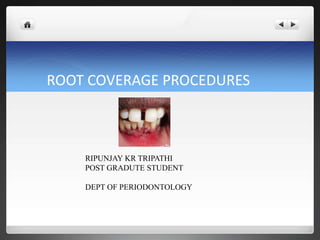
- 1. ROOT COVERAGE PROCEDURES RIPUNJAY KR TRIPATHI POST GRADUTE STUDENT DEPT OF PERIODONTOLOGY
- 2. Contents • Definition • Classifications of gingival recession • Treatment of gingival recession Nonsurgical Surgical • Conclusion • Références
- 3. Definition GINGIVAL RECESSION- Gingival recession is defined as the apical migration of the junctional epithelium with exposure of root surfaces. [Kassab MM, Cohen RE-2003]. Gingival recession is the apical shift of the marginal gingiva from its normal position on the crown of the tooth to levels on the root surface beyond the cemento enamel junction [Loe H-1992].
- 4. Free Gingival Graft (FGG) - A soft tissue graft that is completely detached from one site and transferred to a remote site. No connection with the donor site is maintained
- 5. Sub-epithelial Connective Tissue Graft (CTG) - A detached connective tissue graft that is placed beneath a partial thickness flap. This variation of the free gingival graft provides the tissue graft with a nutrient supply on two surfaces
- 6. Classifications Of Gingival Recession
- 9. Root Coverage Procedures Indications: root sensitivity esthetics protect root surface from caries/abrasions improved hygiene
- 10. Treatment Of Gingival Recession
- 11. NON-SURGICAL
- 12. Non –surgical Treatment Monitoring and prevention Use of de-sensitizing agents, varnishes Composite restoration Removable gingival veneers Orthodontics
- 13. Monitoring and prevention If the recession is not progressing and does not provoke tooth sensitivity or poor aesthetics, then tooth- brushing instructions and regular observation through a strict maintenance program would be the optimal treatment.
- 14. De-sensitizing agents, varnishes Treatment of dentine hypersensitivity is based on blocking the dentinal tubules and preventing nerve stimulation .
- 15. Composite restorations Use of composite resin to mask recession defects and eliminate black triangles caused by recession.
- 17. Orthodontics In some cases surgical intervention and grafting may help to treat the recession defect; however, if orthodontic treatment is an option that the patient is willing to consider then any surgical intervention should be delayed until after orthodontic tooth movement has been completed.
- 18. Indications For Surgical Intervention The need to improve soft tissue aesthetics Reduce hypersensitivity Improve plaque control Prevent further progression of recession defect
- 19. Key factors in the selection of surgical procedures RECIPIENT SITE Gingival recession is limited to one tooth or extends to multiple teeth Degree of gingival recession Amount and thickness of existing keratinized gingiva in the area of recession
- 20. Whether the area of recession protrudes labially from the dental arch Restorative/Prosthodontic treatment after root coverage is necessary
- 21. Donor site Whether area adjacent to gingival recession can be used as a donor site. Amount of Keratinized gingiva Thickness of keratinized gingiva
- 22. Size of adjacent interdental papilla Thickness of the alveolar bone covering the donor tissue Thickness of palatal soft tissue used as donor tissue
- 23. if adequate width is present at the donor site the following procedures can be selected: a. Laterally (horizontally) displaced flap. b. Double-papilla flap. c. Coronally-positioned flap If the donor site is associated with inadequate width: Free soft tissue auto graft Sub epithelial connective tissue grafts are available Depending on the width of the attached gingiva
- 24. Root Coverage Procedures Pedicle soft tissue graft procedures : Rotational flaps Laterally positioned flap Double papilla flap Advanced flaps Coronally positioned flap Semilunar flap
- 25. Free soft tissue grafts Non-submerged graft One stage (free gingival graft) Two stage (free gingival graft + coronally positioned flap)
- 26. Submerged grafts Connective tissue graft + laterally positioned flap Connective tissue graft + double papilla flap Connective tissue graft + coronally positioned flap subepithelial connective tissue graft Envelope techniques
- 27. Additive treatments •Root surface modification agents • Enamel matrix proteins •Guided tissue regeneration •Non-resorbable membrane barriers •Resorbable membrane barriers
- 28. Laterally Positioned flap Advantages a. One surgical site b. Good vascularity of the pedicle flap. c. Ability to cover isolated, denuded roots that have adequate donor tissue laterally.
- 29. Disadvantages a. Limited by the amount of adjacent keratinized attached gingiva. b. Possibility of recession at the donor site. c. Dehiscence or fenestration at the donor site. d. Limited to one or two teeth with gingival recession.
- 30. Indications: a. For covering the isolated denuded root. b. When there is sufficient width of interdental papilla in the adjacent teeth, and Sufficient vestibular depth. Contraindications: a. Presence of deep interproximal pockets. b. Excessive root prominence. c. Deep or extensive root abrasion or erosion.
- 31. Procedure for laterally Positioned flap: Step I : Preparation of the recipient site
- 32. Step 2: Prepare the flap of the donor site. Step 3: Transfer the flap. Step 4: Protect the flap and donor site.
- 33. VARIANTS Satffileno, 1964 partial thickness flap to avoid recession at donor site Grupe,1966 Submarginal incision
- 35. Oblique rotated pedicle flap
- 36. Double papilla flap Indications: 1. When the interproximal papillae adjacent to the mucogingival problem are sufficiently wide. 2. When the attached gingiva on an approximating tooth is insufficient to allow for a Lateral Pedicle Flap. Advantages: 1. The risk of loss of alveolar bone is minimized because the interdental bone is more resistant to loss than is radicular bone. 2. The papillae usually supply a greater width of attached gingiva than from the radicular surface of a tooth.
- 38. Coronally positioned flap Indications: • Esthetic coverage of exposed roots. • For tooth sensitivity owing to gingival recession. Advantages: • Treatment of multiple areas of root exposure. • No need for involvement of adjacent teeth. • High degree of success. • Even if the procedure does not work, it does not increase the existing problem.
- 39. Coronally positioned flap First technique: Step 1: With 2 vertical incisions. Step 2: Root preparation
- 40. Step 3: Return the flap and suture it coronal to the pretreatment position. Step 4: Cover the area with a periodontal dressing.
- 41. Second Technique (Semilunar flap) Indication: Small localized area Advantages: • No vestibular shortening, as occurs with the coronally positioned flap. • No esthetic compromise of interproximal papillae. • No need for sutures.
- 42. Disadvantages: • Inability to treat large areas of gingival recession. • The need for a free gingival graft if there is an underlying dehiscence or fenestration.
- 43. Step 1: Semilunar incision is made and ending about 2 to 3 mm short of the tip of the papillae.
- 44. Step 2: Perform a split-thickness dissection coronally from the incision, and connect it to an intrasulcular incision.
- 45. Step 3: The tissue will collapse coronally, covering the denuded root. then held in its new position for a few minutes with a moist gauze. Many cases do not require either sutures or periodontal dressing.
- 46. Double Lateral sliding bridge flap Multiple gingival recession with or without adequate attached gingiva Coronally advanced flap Vestibular plastic surgery
- 48. Reasons for pedicle flap failure Narrow Flap Tension Bone exposed poor stabilization
- 49. Free Gingival Autograft that consist of epithelium and a thin layer of underlying CT completely detached from one site and transferred to a remote site. Advantages Increase keratinized tissue around teeth, implants or crowns and under removable prostheses. Increase vestibular depth.
- 50. Surgi cal Technique Step 1: Prepare the recipient site. Step 2: Root preparation: Root planing of exposed root to remove cementum and affected dentin. Etch root surface with tetracycline (pH 2.0).
- 51. Step 3: Obtain the graft from the donor site: The ideal thickness of a graft is 1.0 - 1.5 mm.
- 52. Step 4: Graft transferred to recipient site. Step 5: Protect the donor site.
- 53. Sub-epithelial Connective Tissue Graft Indications: • Where esthetics is of prime concern • For covering multiple denuded roots • In the absence of sufficient width of attached gingiva in the adjacent areas. Advantages: • High degree of cosmetic enhancement • Incurs no additional cost for autogenous donor tissue • Minimal palatal trauma • Increased graft vascularity.
- 54. Disadvantages: • High degree of technical skills required. • Complicated suturing
- 55. I. Preparation of recipient site: The initial horizontal right angle incision is made into the adjacent interdental papillae at, or slightly coronal to the cementoenamel junction of the tooth with an exposed root surface. preserve the papillary blood supply A partial thickness flap is raised without vertical incisions SRP Root Conditioning with citric acid pH 1.0 or tetracycline HCl in a concentration of 250 mg mixed in 5 ml of sterile water approximate mesio distal width necessary for the graft is measured with a periodontal probe.
- 57. II. Excision of the donor tissue 1st incision horizontal incision 2-3mm apical to gingival margin 2nd incision parallel to the long axis of the teeth, 1 to 2 mm apical to the first incision raise a full thickness periosteal connective tissue graft
- 58. III. Grafting to the recipient site: With interrupted sutures
- 59. Pouch and Tunnel technique Create “pouch” using full thickness incision and maintain papilla for bilaminar blood supply.
- 60. Extend incision to adjacent teeth and undermine flap beyond MGJ, which allows the coronal positioning of the flap.
- 61. Insertion of CTG and suture.
- 62. Guided Tissue Regeneration Indications • Esthetic demand. • Indicated for single tooth with wide, deep localized recessions. • For areas of root sensitivity where oral hygiene is impaired. • For repair of recessions associated with failing or unesthetic class V restorations.
- 63. Advantages: • Techniques does not require a secondary donor surgical site reducing postoperative discomfort. • New tissue blends evenly with the adjacent tissue, providing highly esthetic results. Disadvantages: • It is sensitive technique. • Insurance of additional cost of barrier membrane.
- 64. Step 1: A full-thickness flap is reflected to MGJ, continuing as a partial- thickness flap 8 mm apical to MGJ. Step 2: Root preparation.
- 65. Step 3: A membrane is placed over the root surface and the adjacent tissue at least 2 mm of marginal periosteum. Step 4: The flap is then positioned coronally and sutured.
- 66. The use of Allografts and Xenografts in management systematic review concluded that these grafts may be useful in situations where 1- A large recession defect needs to be treated . 2- Graft tissue harvested from the palate would provide an insufficient volume of tissue.
- 67. Modifications a. Titanium–reinforced membranes used to create the space below the membrane. b. Resorbable membranes have been used to prevent a second surgery
- 68. Criteria of successful root coverage The gingival margin is on the CEJ in class I, Class II. The depth of gingival sulcus is within 2mm. There is no bleeding on probing , hypersensitivity. Color match with adjacent tissue
- 69. Conclusion The management of gingival recession and its sequelae is based on a thorough assessment of the etiological factors and the degree of involvement of the tissues. The initial part of the management of the patient with gingival recession should be preventive and any pain should be managed and disease should be treated. The degree of gingival recession should be monitored for signs of further progression. When esthetics is the priority and periodontal health is good then surgical root coverage is a potentially useful therapy. Numerous therapeutic solutions for recession defects have been proposed in the periodontal literature and modified with time according to the evolution of clinical knowledge.
- 70. The subepithelial connective tissue graft with a cornonally advanced flap is gold standard grafting procedure . Prognosis (amount of root coverage achieved) will depend on the severity (size )of recession . Careful case selection and surgical management are critical if a successful outcome is to be achieved.
- 71. References Chambrone L, Sukekava F, Araújo MG, Pustiglioni FE, Chambrone LA, Lima LA. Root coverage procedures for the treatment of localised recession-type defects (Review). The Cochrane Library 2009, Issue 2 Umberto Pagliaro, Michele Nieri, Debora Franceschi,Carlo Clauser,and Giovanpaolo Pini-Prato. Evidence-Based Mucogingival Therapy. Part 1: A Critical Review of the Literature on Root Coverage Procedures. J Periodontol May 2003 Paulom. Camargo, Philip R.Melnick & E. Barrie Kenney.The use of free gingival grafts for aesthetic purposes . Periodontology 2000, Vol. 27, 2001,
- 72. Philippe bouchard, Jacquesmalet & Alain borghetti. Decision-making in aesthetics: root coverage revisited - - Periodontology 2000, Vol. 27, 2001 Kassab MM, Badawi H, Dentino AR. Treatment of gingival recession. Dent Clin North Am. 2010;54:129-140. M. Zalkind.Alternative method of conservative esthetic treatment for gingival recession J Prosthet dent 1997 77 561-563
- 73. Carranza’s. Clinical Periodontology. 12th ed.New Delhi:Elsevier ;2015. pp. 628, ch.-63
Editor's Notes
- P.D MILLER CLASSIFICATION
- Class III recession extends to, or beyond MGJ loss of interdental bone or soft tissue apical to CEJ , but coronal to apical extent of marginal tissue recession Class IV recession extends to, or beyond MGJ loss of interdental bone or soft tissue apical to the extent of marginal tissue recession
- I. Shallow-narrow. Ii. Shallow-wide. iii. Deep-narrow. iv. Deep-wide
- Introduced by Grupe and warren 1956
- d. Significant loss of interproximal bone height.
- Advantage Prevent recession at donor site
- Dhalberg, 19689 ADVANTAGES Good tissue blend 2. Usually one surgical site 3. Pedicle to be moved over donor site without tension and releasing incision DISADVANTAGE 1. Possible recession at the donor site
- Introduced by Waienberg in 1964 Modified by Cohen and Ross,1968 3. When periodontal pockets are not present.
- Disadvantage: Technique sensitive—Having to join together the small flap in such a way so that they act as a single flap
- Disadvantage: There is a need for two surgical procedures if the zone of keratinized gingiva is inadequate
- Introduced by norberg Harvey in 1965 used it with FGG
- Tarnow in 1986
- Introduced by margraff, 1985 adv- does not require separate frenectomy increase vestibular depth
- Disadvantages Difficult to achieve root coverage. High esthetic demand. Large, uncomfortable donor site.