Basics of mechanical ventilation
•Download as PPSX, PDF•
138 likes•38,778 views

Ventilator Nursing Care

Recommended
More Related Content
What's hot
What's hot (20)
Similar to Basics of mechanical ventilation
Similar to Basics of mechanical ventilation (20)
More from BP KOIRALA INSTITUTE OF HELATH SCIENCS,, NEPAL
More from BP KOIRALA INSTITUTE OF HELATH SCIENCS,, NEPAL (20)
Recently uploaded
Recently uploaded (20)
Basics of mechanical ventilation
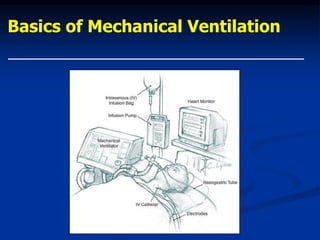
- 1. Basics of Mechanical Ventilation
- 2. Origins of mechanical ventilation •Negative-pressure ventilators (“iron lungs”) • Non-invasive ventilation first used in Boston Children’s Hospital in 1928 • Used extensively during polio outbreaks in 1940s – 1950s •Positive-pressure ventilators • Invasive ventilation first used at Massachusetts General Hospital in 1955 • Now the modern standard of mechanical ventilation The era of intensive care medicine began with positive-pressure ventilation The iron lung created negative pressure in abdomen as well as the chest, decreasing cardiac output. Iron lung polio ward at Rancho Los Amigos Hospital in 1953.
- 3. Outline •Theory • Ventilation vs. Oxygenation • Pressure Cycling vs. Volume Cycling •Modes •Ventilator Settings •Indications to intubate •Indications to extubate •Management algorithim
- 4. Principles (1): Ventilation The goal of ventilation is to facilitate CO2 release and maintain normal PaCO2 •Minute ventilation (VE) • Total amount of gas exhaled/min. • VE = (RR) x (TV) • VE comprised of 2 factors • VA = alveolar ventilation • VD = dead space ventilation • VD/VT = 0.33 • VE regulated by brain stem, responding to pH and PaCO2 •Ventilation in context of ICU • Increased CO2 production • fever, sepsis, injury, overfeeding • Increased VD • atelectasis, lung injury, ARDS, pulmonary embolism • Adjustments: RR and TV V/Q Matching. Zone 1 demonstrates dead-space ventilation (ventilation without perfusion). Zone 2 demonstrates normal perfusion. Zone 3 demonstrates shunting (perfusion without ventilation).
- 5. Principles (2): Oxygenation The primary goal of oxygenation is to maximize O2 delivery to blood (PaO2) •Alveolar-arterial O2 gradient (PAO2 – PaO2) • Equilibrium between oxygen in blood and oxygen in alveoli • A-a gradient measures efficiency of oxygenation • PaO2 partially depends on ventilation but more on V/Q matching •Oxygenation in context of ICU • V/Q mismatching • Patient position (supine) • Airway pressure, pulmonary parenchymal disease, small- airway disease • Adjustments: FiO2 and PEEP V/Q Matching. Zone 1 demonstrates dead-space ventilation (ventilation without perfusion). Zone 2 demonstrates normal perfusion. Zone 3 demonstrates shunting (perfusion without ventilation).
- 6. Non- invasive ventilation Advantages: 1.allows speech 2.ideal for patients with nocturnal hypoventilation 3.complications of intubation –avoided Disadvantages: 1. patient must be NPO 2. lack of airway protection 3. patient should be alert with normal respiratory drive 4. cannot fully sedate patients 5. slower correction of blood gas abnormalities 6. upper airway must be intact 7. cannot provide full 100% ventilatory support 8. apparatus uncomfortable for patients
- 7. Patients likely to benefit from non-invasive ventilation: 1. respiratory failure likely to be reversible within 24-48 hrs acute reversible cardiogenic pulmonary oedema acute exacerbation of COPD post-op respiratory failure laryngeal oedema 2.Chronic use in progressive respiratory failure neuromuscular disease with respiratory muscle weakness lung transplant candidiates awaiting doners 3. Acute respiratory failure in patients not willing intubation terminally ill patients & AIDS patients
- 8. Types of non-invasive ventilation: CPAP & BiPAP 1.CPAP: Patient continuously receives a set air pressure, during both inspiration and expiration. Patient has full control over respiratory rate, inspiratory time , and depth of inspiration. Best suited for: patients with obstructive sleep apnea cardiac patients requiring ventilatory assistance but not requiring immediate intubation Advantages of increase in pressure at end- expiration: increase in FRC decrease afterload to the left ventricle increased patency of alveoli ( esp those atelectatic due to pulmonary edema or poor inspiratory effort) Mode of delivery: nasal vs face mask. Pressure 5-20cm of H2O.
- 9. 2.BiPAP This provides a set inspiratory pressure and a different set expiratory pressure Patient has full control over the respiratory rate, inspiratory time and depth of inspiration The unit senses the beginning and end of inspiration so that the delivery pressure can be changed from the expiratory pressure to the inspiratory pressure and vice versa. In simple way BiPAP=CPAP+Pressure support during inspiration
- 10. BiPAP is indicated only when CPAP alone is insufficient to improve dyspnea or hypoxemia in patients with respiratory muscle fatigue underlying COPD high pCO2. Initial setting: IPAP: 8cm H2O EPAP: 4cm H2O O2 @ 2-5 L/min
- 11. Invasive Ventilation: Pressure ventilation vs. volume ventilation Pressure-cycled modes deliver a fixed pressure at variable volume (neonates) Volume-cycled modes deliver a fixed volume at variable pressure (adults) •Pressure-cycled modes • Pressure Support Ventilation (PSV) • Pressure Control Ventilation (PCV) • CPAP • BiPAP •Volume-cycled modes • Control • Assist • Assist/Control • Intermittent Mandatory Ventilation (IMV) • Synchronous Intermittent Mandatory Ventilation (SIMV) Volume-cycled modes have the inherent risk of volutrauma.
- 12. Pressure Support Ventilation (PSV) Patient determines RR, VE, inspiratory time – a purely spontaneous mode • Parameters • Triggered by pt’s own breath • Limited by pressure • Affects inspiration only • Uses • Complement volume-cycled modes (i.e., SIMV) • Does not augment TV but overcomes resistance created by ventilator tubing • PSV alone • Used alone for recovering intubated pts who are not quite ready for extubation • Augments inflation volumes during spontaneous breaths • BiPAP (CPAP plus PS) PSV is most often used together with other volume-cycled modes. PSV provides sufficient pressure to overcome the resistance of the ventilator tubing, and acts during inspiration only.
- 13. Advantages of PSV Avoids patient-ventilator asynchrony Patient comfortable-having full control over their ventilatory pattern and minute ventilation Avoids breath stacking and auto PEEP Disadvantages Spontaneous mode –can’t be used in heavily sedated, paralyzed or comatose patients Respiratory muscle fatigue can develop if pressure support is set too low
- 14. Pressure Control Ventilation (PCV) Ventilator determines inspiratory time – no patient participation •Parameters •Triggered by time •Limited by pressure •Affects inspiration only •Best reserved for patients with ARDS •Disadvantages •No guaranteed tidal volume thus no guaranteed VE •Requires frequent adjustments to maintain adequate VE •Pt with noncompliant lungs may require alterations in inspiratory times to achieve adequate TV •Air trapping can be a problem •Heavy sedation required
- 15. CPAP and BiPAP CPAP is essentially constant PEEP; BiPAP is CPAP plus PS •Parameters CPAP – PEEP set at 5-10 cm H2O BiPAP – CPAP with Pressure Support (5-20 cm H2O) Shown to reduce need for intubation and mortality in COPD pts Indications When medical therapy fails (tachypnea, hypoxemia, respiratory acidosis) Use in conjunction with bronchodilators, steroids, oral/parenteral steroids, antibiotics to prevent/delay intubation Weaning protocols Obstructive Sleep Apnea
- 16. Volume cycled modes •Control Mode • Pt receives a set number of breaths and cannot breathe between ventilator breaths • Similar to Pressure Control •Assist Mode • Pt initiates all breaths, but ventilator cycles in at initiation to give a preset tidal volume • Pt controls rate but always receives a full machine breath Ventilator delivers a fixed volume
- 17. Assist/Control Mode Assist mode unless pt’s respiratory rate falls below preset value Ventilator then switches to control mode and provides full Vt at a minimum preset rate Advantage provides near complete resting of ventilatory muscles can be used in awake, sedated or paralyzed patients. Disadvantages • Rapidly breathing pts can overventilate and induce severe respiratory alkalosis and hyperinflation (auto-PEEP)
- 18. IMV and SIMV Volume-cycled modes typically augmented with Pressure Support •IMV • Pt receives a set number of ventilator breaths • Different from Control: pt can initiate own (spontaneous) breaths • Different from Assist: spontaneous breaths are not supported by machine with fixed TV • Ventilator always delivers breath, even if pt exhaling
- 19. SIMV Most commonly used mode Ventilator provides set tidal volume at a preset rate If pt has respiratory drive, the mandatory breaths are synchronized with the pt’s inspiratory effort When a ventilator breath is programmed to occur, the ventilator waits a predetermined trigger period; any patient initiated breath during this trigger period results in a programmed ventilator delivered breath
- 20. SIMV contd. The patient can take additional breaths but Vt of these extra breaths is dependent on the patient’s inspiratory effort Advantages of SIMV Theoritically results in improved blood return to the right ventricle owing to intermittent negative pressure (spontaneous) breaths Patient comfort due to the control over their ventilatory pattern and minute ventilation Disadvantage Can result in chronic respiratory fatigue if set rate is too low; characterized by high respiratory rate rising pCO2 Air trapping can occur
- 21. Vent settings to improve <oxygenation> •FIO2 • Simplest maneuver to quickly increase PaO2 • Long-term toxicity at >60% • Free radical damage •Inadequate oxygenation despite 100% FiO2 usually due to pulmonary shunting • Collapse – Atelectasis • Pus-filled alveoli – Pneumonia • Water/Protein – ARDS • Water – CHF • Blood - Hemorrhage PEEP and FiO2 are adjusted in tandem
- 22. Vent settings to improve <oxygenation> •PEEP • Increases FRC • Prevents progressive atelectasis and intrapulmonary shunting • Prevents repetitive opening/closing (injury) • Recruits collapsed alveoli and improves V/Q matching • Resolves intrapulmonary shunting • Improves compliance • Enables maintenance of adequate PaO2 at a safe FiO2 level • Disadvantages • Increases intrathoracic pressure (may require pulmonary a. catheter) • May lead to ARDS • Rupture: PTX, pulmonary edema PEEP and FiO2 are adjusted in tandem Oxygen delivery (DO2), not PaO2, should be used to assess optimal PEEP.
- 23. Vent settings to improve <ventilation> •Respiratory rate • Max RR at 35 breaths/min • Efficiency of ventilation decreases with increasing RR • Decreased time for alveolar emptying •TV • Goal of 10 ml/kg • Risk of volutrauma •Other means to decrease PaCO2 • Reduce muscular activity/seizures • Minimizing exogenous carb load • Controlling hypermetabolic states •Permissive hypercapnea • Preferable to dangerously high RR and TV, as long as pH > 7.15 RR and TV are adjusted to maintain VE and PaCO2 •I:E ratio (IRV) • Increasing inspiration time will increase TV, but may lead to auto-PEEP •PIP • Elevated PIP suggests need for switch from volume-cycled to pressure-cycled mode • Maintained at <45cm H2O to minimize barotrauma •Plateau pressures • Pressure measured at the end of inspiratory phase • Maintained at <30-35cm H2O to minimize barotrauma
- 24. Alternative Modes • I:E inverse ratio ventilation (IRV) • ARDS and severe hypoxemia • Prolonged inspiratory time (3:1) leads to better gas distribution with lower PIP • Elevated pressure improves alveolar recruitment • No statistical advantage over PEEP, and does not prevent repetitive collapse and reinflation • Prone positioning • Addresses dependent atelectasis • Improved recruitment and FRC, relief of diaphragmatic pressure from abdominal viscera, improved drainage of secretions • Logistically difficult • No mortality benefit demonstrated • ECHMO • High-Frequency Oscillatory Ventilation (HFOV) • High-frequency, low amplitude ventilation superimposed over elevated Paw • Avoids repetitive alveolar open and closing that occur with low airway pressures • Avoids overdistension that occurs at high airway pressures • Well tolerated, consistent improvements in oxygenation, but unclear mortality benefits • Disadvantages • Potential hemodynamic compromise • Pneumothorax • Neuromuscular blocking agents
- 25. Airway Pressure Release (APR) Ventilator supplies a low level of CPAP alternating with a relatively high level of CPAP At intermittent times the higher CPAP level is reduced for one or more breaths, permitting rapid exhausting of the gas in the expiratory reserve volume and resulting in a higher tidal volume Advantage: Venous return preserved Barotrauma and overdistension is minimized Permits spontaneous breaths Disadvantage: requires high CPAP, suited only for post op & mildly diseased lungs
- 26. Treatment of respiratory failure •Prevention • Incentive spirometry • Mobilization • Humidified air • Pain control • Turn, cough, deep breathe •Treatment • Medications • B-2 Agonist • Theophylline • Steroids • CPAP, BiPAP • Intubation The critical period before the patient needs to be intubated
- 27. Indications for intubation •Criteria • Clinical deterioration • Tachypnea: RR >35 • Hypoxia: pO2<60mm Hg • Hypercarbia: pCO2 > 55mm Hg • Minute ventilation<10 L/min • Tidal volume <5-10 ml/kg • Negative inspiratory force < 25cm H2O (how strong the pt can suck in) •Initial vent settings • FiO2 = 50% • PEEP = 5cm H2O • RR = 12 – 15 breaths/min • VT = 10 – 12 ml/kg • COPD = 10 ml/kg (prevent overinflation) • ARDS = 8 ml/kg (prevent volutrauma) • Permissive hypercapnea • Pressure Support = 10cm H2O How the values trend should significantly impact clinical decisions
- 28. Indications for extubation •Clinical parameters • Resolution/Stabilization of disease process • Hemodynamically stable • Intact cough/gag reflex • Spontaneous respirations • Acceptable vent settings • FiO2< 50%, PEEP < 8, PaO2 > 75, pH > 7.25 •General approaches • SIMV Weaning • Pressure Support Ventilation (PSV) Weaning • Spontaneous breathing trials and use of T-piece • Demonstrated to be superior No weaning parameter completely accurate when used alone Numerical Parameters Normal Range Weaning Threshold P/F > 400 > 200 Tidal volume 5 - 7 ml/kg 5 ml/kg Respiratory rate 14 - 18 breaths/min < 40 breaths/min Vital capacity 65 - 75 ml/kg 10 ml/kg Minute volume 5 - 7 L/min < 10 L/min Greater Predictive Value Normal Range Weaning Threshold NIF (Negative Inspiratory Force) > - 90 cm H2O > - 25 cm H2O RSBI (Rapid Shallow Breathing Index) (RR/TV) < 50 < 100 Marino P, The ICU Book (2/e). 1998.
- 29. Spontaneous Breathing Trials •Settings • PEEP = 5, PS = 0 – 5, FiO2 < 40% • Breathe independently for 30 – 120 min • ABG obtained at end of SBT •Failed SBT Criteria • RR > 35 for >5 min • SaO2 <90% for >30 sec • HR > 140 • Systolic BP > 180 or < 90mm Hg • Sustained increased work of breathing • Cardiac dysrhythmia • pH < 7.32 SBTs do not guarantee that airway is stable or pt can self-clear secretions Causes of Failed SBTs Treatments Anxiety/Agitation Benzodiazepines or haldol Infection Diagnosis and tx Electrolyte abnormalities (K+, PO4-) Correction Pulmonary edema, cardiac ischemia Diuretics and nitrates Deconditioning, malnutrition Aggressive nutrition Neuromuscular disease Bronchopulmonary hygiene, early consideration of trach Increased intra-abdominal pressure Semirecumbent positioning, NGT Hypothyroidism Thyroid replacement Excessive auto-PEEP (COPD, asthma) Bronchodilator therapy Sena et al, ACS Surgery: Principles and Practice (2005).
- 30. T-piece trial Advantage Simple Provides strenuous exercise to the ventilatory muscles followed by periods of full ventilatory muscle rest; theoretically the best way to condition the inspiratory muscles
- 31. Continued ventilation after successful SBT •Commonly cited factors • Altered mental status and inability to protect airway • Potentially difficult reintubation • Unstable injury to cervical spine • Likelihood of return trips to OR • Need for frequent suctioning Inherent risks of intubation balanced against continued need for intubation
- 32. Need for tracheostomy •Advantages • Issue of airway stability can be separated from issue of readiness for extubation • May quicken decision to extubate • Decreased work of breathing • Avoid continued vocal cord injury • Improved bronchopulmonary hygiene • Improved pt communication •Disadvantages • Long term risk of tracheal stenosis • Procedure-related complication rate (4% - 36%) Prolonged intubation may injure airway and cause airway edema 1 - Vocal cords. 2 - Thyroid cartilage. 3 - Cricoid cartilage. 4 - Tracheal cartilage. 5 - Balloon cuff.
- 33. Ventilator management algorithim Initial intubation • FiO2 = 50% • PEEP = 5 • RR = 12 – 15 • VT = 8 – 10 ml/kg SaO2 < 90% SaO2 > 90% SaO2 > 90% • Adjust RR to maintain PaCO2 = 40 • Reduce FiO2 < 50% as tolerated • Reduce PEEP < 8 as tolerated • Assess criteria for SBT daily SaO2 < 90% • Increase FiO2 (keep SaO2>90%) • Increase PEEP to max 20 • Identify possible acute lung injury • Identify respiratory failure causes Acute lung injury No injury Fail SBT Acute lung injury • Low TV (lung-protective) settings • Reduce TV to 6 ml/kg • Increase RR up to 35 to keep pH > 7.2, PaCO2 < 50 • Adjust PEEP to keep FiO2 < 60% SaO2 < 90% SaO2 > 90% SaO2 < 90% • Dx/Tx associated conditions (PTX, hemothorax, hydrothorax) • Consider adjunct measures (prone positioning, HFOV, IRV) SaO2 > 90% • Continue lung-protective ventilation until: • PaO2/FiO2 > 300 • Criteria met for SBT Persistently fail SBT • Consider tracheostomy • Resume daily SBTs with CPAP or tracheostomy collar Pass SBT Airway stable Extubate Intubated > 2 wks • Consider PSV wean (gradual reduction of pressure support) • Consider gradual increases in SBT duration until endurance improves Prolonged ventilator dependence Pass SBT Pass SBT Airway stable Modified from Sena et al, ACS Surgery: Principles and Practice (2005).
- 34. Afterload afterload = wall tension (T) during contraction where Ptm= transmural pressure, R=radius and H=wall thickness transmural pressure=intraventricular pressure-pleural pressure pleural pressure increased by positive pressure therefore transmural pressure and afterload must be decreased by positive pressure ventilation
- 35. Thank You !
- 37. Advanced Mechanical Ventilation Outline Consequences of Elevated Alveolar Pressure Implications of Heterogeneous Lung Unit Inflation Adjuncts to Ventilation • Prone Positioning • Recruitment Maneuvers Difficult Management Problems • Acute Lung Injury • Severe Airflow Obstruction • An Approach to Withdrawing Ventilator Support Advanced Modes for Implementing Ventilation Case Scenarios
- 38. Consequences of Elevated Alveolar Pressure--1 Mechanical ventilation expands the lungs and chest wall by pressurizing the airway during inflation. The stretched lungs and chest wall develop recoil tension that drives expiration. Positive pressure developed in the pleural space may have adverse effects on venous return, cardiac output and dead space creation. Stretching the lung refreshes the alveolar gas, but excessive stretch subjects the tissue to tensile stresses which may exceed the structural tolerance limits of this delicate membrane. Disrupted alveolar membranes allow gas to seep into the interstitial compartment, where it collects, and migrates toward regions with lower tissue pressures. Interstitial, mediastinal, and subcutaneous emphysema are frequently the consequences. Less commonly, pneumoperitoneum, pneumothorax, and tension cysts may form. Rarely, a communication between the high pressure gas pocket and the pulmonary veins generates systemic gas emboli.
- 39. Partitioning of Alveolar Pressure is a Function of Lung and Chest Wall Compliances Lungs are smaller and pleural pressures are higher when the chest wall is stiff.
- 40. Hemodynamic Effects of Lung Inflation Lung inflation by positive pressure causes • Increased pleural pressure and impeded venous return • Increased pulmonary vascular resistance • Compression of the inferior vena cava • Retardation of heart rate increases These effects are much less obvious in the presence of • Adequate circulating volume • Adequate vascular tone • Spontaneous breathing efforts • Preserved adrenergic responsiveness
- 41. Hemodynamic Effects of Lung Inflation With Low Lung Compliance, High Levels of PEEP are Generally Well Tolerated.
- 42. Effect of lung expansion on pulmonary vasculature. Capillaries that are embedded in the alveolar walls undergo compression even as interstitial vessels dilate. The net result is usually an increase in pulmonary vascular resistance, unless recruitment of collapsed units occurs.
- 43. Conflicting Actions of Higher Airway Pressure Lung Unit Recruitment and Maintenance of Aerated Volume • Gas exchange Improved Oxygenation Distribution of Ventilation • Lung protection Parenchymal Damage Airway trauma Increased Lung Distention • Impaired Hemodynamics • Increased Dead Space • Potential to Increase Tissue Stress …Only If Plateau Pressure Rises
- 45. Diseased Lungs Do Not Fully Collapse, Despite Tension Pneumothorax …and They cannot always be fully “opened” Dimensions of a fully Collapsed Normal Lung
- 46. Tension Cysts
- 47. Tidally Phasic Systemic Gas Embolism End-Expiration End-Inspiration
- 48. Consequences of Elevated Alveolar Pressure--2 In recent years there has been intense interest in another and perhaps common consequence of excessive inflation pressure-- Ventilator-Induced Lung Injury (VILI). VILI appears to develop either as a result of structural breakdown of the tissue by mechanical forces or by mechano-signaling of inflammation due to repeated application of excessive tensile forces. Although still controversial, it is generally agreed that damage can result from overstretching of lung units that are already open or from shearing forces generated at the junction of open and collapsed tissue. Tidally recurrent opening and closure of small airways under high pressure is thought to be important in the generation of VILI. Prevention of VILI is among the highest priorities of the ICU clinician caring for the ventilated patient and is the purpose motivating adoption of “Lung Protective” ventilation strategies. Translocation of inflammatory products, bacteria, and even gas may contribute to remote damage in systemic organs and help explain why lung protective strategies are associated with lower risk for morbidity and death.
- 49. Recognized Mechanisms of Airspace Injury “Stretch” “Shear” Airway Trauma
- 50. Mechanisms of Ventilator-Induced Lung Injury (VILI) High airway pressures may injure the lungs by repeated overstretching of open alveoli, by exposing delicate terminal airways to high pressure, or by generating shearing forces that tear fragile tissues. These latter shearing forces tend to occur as small lung units open and close with each tidal cycle and are amplified when the unit opens only after high pressures are reached. To avoid VILI, end-inspiratory lung pressure (“plateau”) should be kept from rising too high, and when high plateau pressures are required, sufficient PEEP should be applied to keep unstable lung units from opening and closing with each tidal cycle. Independent of opening and closure, tissue strain is dramatically amplified at the junctions of open and closed lung units when high alveolar pressures are reached. Extremely high tissue strains may rip the alveolar gas-blood interface. Repeated application of more moderate strains incite inflammation. Such factors as breath frequency, micro-vascular pressure, temperature, and body position modify VILI expression.
- 51. End-Expiration Tidal Forces (Transpulmonary and Microvascular Pressures) Extreme Stress/Strain Moderate Stress/Strain Mechano signaling via integrins, cytoskeleton, ion channels inflammatory cascade Cellular Infiltration and Inflammation Rupture Signaling Marini / Gattinoni CCM 2004 Pathways to VILI
- 52. Microvascular Fracture in ARDS A Portal for Gas & Bacteria? Hotchkiss et al Crit Care Med 2002; 1
- 53. The Problem of Heterogeneity The heterogeneous nature of regional mechanical properties presents major difficulty for the clinician, who must apply only a single pressure or flow profile to the airway opening. Heterogeneity means that some lung units may be overstretched while others remain airless at the same measured airway pressure. Finding just the right balance of tidal volume and PEEP to keep the lung as open as possible without generating excessive regional tissue stresses is a major goal of modern practice. Prone positioning tends to reduce the regional gradients of pleural and trans- pulmonary pressure.
- 54. Spectrum of Regional Opening Pressures (Supine Position) Superimposed Pressure Inflated 0 Alveolar Collapse (Reabsorption) 20-60 cmH2O Small Airway Collapse 10-20 cmH2O Consolidation (from Gattinoni) Lung Units at Risk for Tidal Opening & Closure = Opening Pressure
- 55. Different lung regions may be overstretched or underinflated, even as measures of total lung mechanics appear within normal limits. Alveolar Pressure LungVolume UPPER LUNG TOTAL LUNG LOWER LUNG
- 56. Recruitment Parallels Volume As A Function of Airway Pressure Recruitment and Inflation (%) Frequency Distribution of Opening Pressures (%) Airway Pressure (cmH2O)
- 57. % Opening and Closing Pressures in ARDS 50 Paw [cmH2O] 0 5 10 15 20 25 30 35 40 45 50 0 10 20 30 40 Opening pressure Closing pressure From Crotti et al AJRCCM 2001. High pressures may be needed to open some lung units, but once open, many units stay open at lower pressure.
- 58. Zone of ↑ Risk
- 59. Dependent to Non-dependent Progression of Injury
- 60. Histopathology of VILI Belperio et al, J Clin Invest Dec 2002; 110(11):1703-1716
- 61. Links Between VILI and MSOF Biotrauma and Mediator De-compartmentalization Slutsky, Chest 116(1):9S-16S
- 62. Airway Orientation in Supine Position
- 64. Prone Positioning Evens The Distribution of Pleural & Transpulmonary Pressures
- 65. Prone Positioning Relieves Lung Compression by the Heart Supine Prone
- 66. Proning May Benefit the Most Seriously Ill ARDS Subset SAPS II MortalityRate > 49 40- 49 31- 40 0 - 31 0.0 0.1 0.2 0.3 0.4 0.5 Supine * p<0.05 vs Supine Prone * Quartiles of SAPS II
- 67. Proning Helped Most in High VT Subgroup At Risk For VILI VT/Kg < 8.2 8.2- 9.7 9.7- 12 > 12 0.0 0.1 0.2 0.3 0.4 0.5 SupineMortalityRate Quartiles of VT /Predicted body weight * p<0.05 vs Supine Prone *
- 68. How Much Collapse Is Dangerous Depends on the Plateau R = 100% 20 60 100 Pressure [cmH2O] 20 40 60 TotalLungCapacity[%] R = 22% R = 81% R = 93% 0 0 R = 0% R = 59% From Pelosi et al AJRCCM 2001 Some potentially recruitable units open only at high pressure More Extensive Collapse But Lower PPLAT Less Extensive Collapse But Greater PPLAT
- 69. Recruiting Maneuvers in ARDS The purpose of a recruiting maneuver is to open collapsed lung tissue so it can remain open during tidal ventilation with lower pressures and PEEP, thereby improving gas exchange and helping to eliminate high stress interfaces. Although applying high pressure is fundamental to recruitment, sustaining high pressure is also important. Methods of performing a recruiting maneuver include single sustained inflations and ventilation with high PEEP .
- 70. Theoretical Effect of Sustained Inflation on Tidal Cycling Rimensberger ICM 2000 VOLUME (% TLC) Benefit from a recruiting maneuver is usually transient if PEEP remains unchanged afterward.
- 71. Three Types of Recruitment Maneuvers S-C Lim, et al Crit Care Med 2004
- 72. How is the Injured Lung Best Recruited? Prone positioning Adequate PEEP Adequate tidal volume (and/or intermittent ‘sighs’?) Recruiting maneuvers Minimize edema (?) Lowest acceptable FiO2 (?) Spontaneous breathing efforts (?)
- 73. Severe Airflow Obstruction A major objective of ventilating patients with severe airflow obstruction is to relieve the work of breathing and to minimize auto-PEEP. Reducing minute ventilation requirements will help impressively in reducing gas trapping. When auto-PEEP is present, it has important consequences for hemodynamics, triggering effort and work of breathing. In patients whose expiratory flows are flow limited during tidal breathing, offsetting auto-PEEP with external PEEP may even the gas distribution and reduce breathing effort.
- 74. Auto-PEEP Adds To the Breathing Workload The pressure-volume areas correspond to the inspiratory mechanical workloads of auto-PEEP (AP) flow resistance and tidal elastance.
- 75. Gas Trapping in Severe Airflow Obstruction Disadvantages the respiratory muscles and increases the work of tidal breathing Often causes hemodynamic compromise, especially during passive inflation Raises plateau and mean airway pressures, predisposing to barotrauma Varies with body position and from site to site within the lung
- 77. Volume Losses in Recumbent Positions Note that COPD patients lose much less lung volume than normals do, due to gas trapping and need to keep the lungs more inflated to minimize the severity of obstruction. Orthopnea may result.
- 78. PEEP in Airflow Obstruction Effects Depend on Type and Severity of Airflow Obstruction Generally Helpful if PEEP Original Auto-PEEP Potential Benefits • Decreased Work of Breathing • Increased VT During PSV or PCV • (?) Improved Distribution of Ventilation • (?) Decreased Dyspnea
- 79. Inhalation Lung Scans in the Lateral Decubitus Position for a Normal Subject and COPD Patient Normal COPD No PEEP PEEP10 Addition of 10 cm H2O PEEP re-opens dependent airways in COPD
- 81. Adding PEEP that approximates auto-PEEP may reduce the difference in pressure between alveolus (Palv) and airway opening, thereby lowering the negative pleural (Pes) pressure needed to begin inspiration and trigger ventilation.
- 82. Adding PEEP Lessens the Heterogeneity of End-expiratory Alveolar Pressures and Even the Distribution of Subsequent Inspiratory Flow.
- 83. PEEP may offset (COPD) or add to auto-PEEP (Asthma), depending on flow limitation. Note that adding 8 cmH2O PEEP to 10 cmH2O of intrinsic PEEP may either reduce effort (Pes, solid arrow) or cause further hyper- inflation (dashed arrow). Ranieri et al, Clinics in Chest Medicine 1996; 17(3):379-94 ASTHM A COP D
- 84. Conventional Modes of Ventilatory Support The traditional modes of mechanical ventilation—Flow-regulated volume Assist Control (“Volume Control”, AMV, AC)) or Pressure-Targeted Assist Control (“Pressure Control”), Synchronized Intermittent Mandatory Ventilation (SIMV)—with flow or pressure targeted mandatory cycles), Continuously Positive Airway Pressure (CPAP) and Pressure Support can be used to manage virtually any patient when accompanied by adequate sedation and settings well adjusted for the patient’s needs. Their properties are discussed in the “Basic Mechanical Ventilation” unit of this series.
- 85. Positive Airway Pressure Can Be Either Pressure or Flow Controlled—But Not Both Simultaneously Dependent Variable Dependent Variable Set Variable Set Variable
- 86. Decelerating flow profile is an option in flow controlled ventilation but a dependent variable in pressure control. Decelerating Flow Pressure Control Peak pressure is a function of flow; plateau pressure is not
- 87. Patient-Ventilator Interactions Coordination of the patient’s needs for flow, power, and cycle timing with outputs of the machine determine how well the ventilator simulates an auxiliary muscle under the patient’s control. Flow controlled, volume cycled ventilator modes offer almost unlimited power but specify the cycle timing and flow profile. Traditional pressure targeted modes (Pressure Control (PCV) and Pressure support (PSV)) provide no greater power amplitude than that set by the clinician but do not limit flow. PCV has a cycle time fixed by the clinician, and in that sense is as inflexible as VCV. Pressure Support (PSV) allows the patient to determine both flow and, within certain limits, cycle length as well. Because flow is determined by respiratory mechanics as well as by effort, adjustments may be needed to the inspiratory flow offswitch criterion of PSV so as to coordinate with the patient’s needs. Modification of the rate at which the pressure target is reached at the beginning of inspiration (the ramp or ‘attack’ rate) may be needed to help ensure comfort
- 88. Pressure Support ‘off-switch’ is a set flow value or a set % of peak inspiratory flow. The patient with airflow obstruction may need to put on the brake with muscular effort to slow flow quickly enough to satisfy his intrinsic neural timing.
- 89. Tapered inspiratory ‘attack’ rate and a higher percentage of peak flow off switch criterion are often more appropriate in airflow obstruction than are the default values in PSV. Airflow Obstruction
- 90. Although early flows are adequate, mid- cycle efforts may not be matched by Decelerating Flow Control (VCV). Pressure Controlled breaths (PCV) do not restrict flow. Since the flow demands of severely obstructed patients may be nearly unchanging in severe airflow obstruction , decelerating VCV may not be the best choice.
- 91. Interactions Between Pressure Controlled Ventilation and Lung Mechanics The gradient of pressure that determines tidal volume is the difference between end-inspiratory (Plateau) and end-expiratory alveolar pressure (total PEEP). In PCV, tidal volume can be reduced if the inspiratory time or the expiratory time is too brief to allow the airway and alveolar pressures to equilibrate. The development of auto-PEEP reduces the gradient for inspiratory flow and curtails the potential tidal volume available with any given set inspiratory airway pressure. These conditions are most likely to arise in the setting of severe airflow obstruction. Therefore, modifications of the breathing frequency and inspiratory cycle period (‘duty cycle’) may powerfully impact ventilation efficiency.
- 94. Deformed Airway Pressure Waveforms Paw Reflects Effort and Dys-Synchrony During Constant Flow Ventilation
- 95. High Pressure Alarm Variable Tidal Volume or Pressure Limit Alarm During Pressure Contro
- 96. What to Do When the Patent and Ventilator are “Out of Synch”? Frequent pressure alarms during Volume Assist Control or Tidal Volume alarms during Pressure Control both mean that the patient is not receiving the desired tidal volume. If not explained by an important underlying change in the circuit properties (e.g., disconnection or plug) or in the impedance of the respiratory system, this often arises from a timing “collision” between the patient and ventilator’s cycling rhythms. To quickly regain smooth control without deep sedation, the patient must be given back some degree of control over cycle timing. This timing control is conferred by introducing flow off-switched cycles in the form of high level pressure support. Pressure Support alone, or SIMV at a relatively low mandated rate together with PSV, are the logical choices to provide adequate power, achieve flow synchrony and yield cycle timing to the patient. Pressure Controlled SIMV, where the inspiratory time of the PCV cycle is set to be a bit shorter than the observed PSV cycle, is a good strategy for this purpose.
- 97. Advanced Interactive Modes of Mechanical Ventilation Airway pressure release/BiPAP/Bi-Level Combination or “Dual control” modes Proportional assist ventilation Adaptive support ventilation Automatic ET tube compensation
- 98. Combination “Dual Control” Modes Combination or “dual control” modes combine features of pressure and volume targeting to accomplish ventilatory objectives which might remain unmet by either used independently. Combination modes are pressure targeted Partial support is generally provided by pressure support Full support is provided by Pressure Control
- 99. Combination “Dual Control” Modes Volume Assured Pressure Support (Pressure Augmentation) Volume Support (Variable Pressure Support) Pressure Regulated Volume Control (Variable Pressure Control, or Autoflow) Airway Pressure Release (Bi-Level, Bi-PAP)
- 100. Pressure Regulated Volume Control Characteristics Guaranteed tidal volume using “pressure control” waveform Pressure target is adjusted to least value that satisfies the targeted tidal volume minimum Settings: Minimum VE Minimum Frequency Inspiratory Time per Cycle
- 101. Compliance Changes During Pressure Controlled Ventilation
- 102. PRVC Automatically Adjusts To Compliance Changes
- 103. Several modes allow the physician to allow for variability in patient efforts while achieving a targeted goal. Volume support monitors minute ventilation and tidal volume , changing the level of pressure support to achieve a volume target. Volume assured pressure support allows the patient to breathe with pressure support, supplementing the breath with constant flow when needed to achieve the targeted tidal volume within an allocated time. Proportional assist (see later) varies pressure output in direct relation to patient effort.
- 104. Airway Pressure Release and Bi-Level Airway Pressure Over the past 20 years, modes of ventilation that move the airway pressure baseline around which spontaneous breaths occur between two levels have been employed in a variety of clinical settings. Although the machine’s contribution to ventilation may resemble inverse ratio ventilation, patient breathing cycles occur through an open (as opposed to closed) circuit, so that airway pressure never exceeds that value desired by the clinician. Transpulmonary pressure—the pressure across the lung—can approach dangerous levels, however, and mean airway pressure is relatively high.
- 105. Inverse Ratio Airway Pressure Release (APRV), and Bi-Level (Bi-PAP)
- 106. Bi-Pap &Airway Pressure Release Characteristics Allow spontaneous breaths superimposed on a set number of “pressure controlled” ventilator cycles Reduce peak airway pressures “Open” circuit / enhanced synchrony between patient effort and machine response Settings: Pinsp and Pexp (Phigh and Plow) Thigh and Tlow
- 107. Unlike PCV, BiPAP Allows Spontaneous Breathing During Both Phases of Machine’s Cycle
- 108. Bi-Level Ventilation
- 109. Bi-Level Ventilation With Pressure Support
- 110. Modes That Vary Their Output to Maintain Appropriate Physiology Proportional Assist Ventilation (Proportional Pressure Support) - Support pressure parallels patient effort Adaptive Support Ventilation - Adjusts Pinsp and PC-SIMV rate to meet “optimum” breathing pattern target Neurally Adjusted Ventilatory Assist - Ventilator output is keyed to neural signal
- 111. Proportional Assist Ventilation (PAV) Proportional assist ventilation attempts to regulate the pressure output of the ventilator moment by moment in accord with the patient’s demands for flow and volume. Thus, when the patient wants more, (s)he gets more help; when less, (s)he gets less. The timing and power synchrony are therefore nearly optimal—at least in concept. To regulate pressure, PAV uses the monitored flow and a calculated assessment of the mechanical properties of the respiratory system as inputs into the equation of motion of the respiratory system. More than with any other currently available mode, PAV acts as an auxiliary muscle whose strength is regulated by the proportionality constant determined by the clinician. At present, PAV cannot account for auto-PEEP, and there is a small chance for inappropriate support to be applied.
- 112. Proportional Assist Amplifies Muscular Effort Muscular effort (Pmus) and airway pressure assistance (Paw) are better matched for Proportional Assist (PAV) than for Pressure Support (PSV).
- 113. Goals of Adaptive Support Ventilation Reduce Dead-space Ventilation Avoid Auto-PEEP Discourage Rapid Shallow Breathing Mirror Changing Patient Activity
- 114. Adaptive Lung Ventilation Synchronized Intermittent Pressure Control Rate Inspiratory Pressure PSV cycling characteristics Physician Settings Ideal body weight % Minute Volume Maximal Allowed Pressure
- 116. Neural Control of Ventilatory Assist (NAVA) Although still in its development phase, the possibility of controlling ventilator output by sensing the neural traffic flowing to the diaphragm promises to further enhance synchrony. NAVA senses the desired assist using an array of esophageal EMG electrodes positioned to detect the diaphragm’s contraction signal. The reliability of neurally-controlled ventilator assistance needs to be determined before its deployment in the clinical setting.
- 117. Neural Control of Ventilatory Assist (NAVA) Neuro-VentilatoryCoupling Central Nervous System Phrenic Nerve Diaphragm Excitation Diaphragm Contraction Chest Wall and Lung Expansion Airway Pressure, Flow and Volume New Technology Ideal Technology Current Technology Ventilator Unit
- 118. Electrode Array in Neurally Adjusted Ventilatory Assist (NAVA) Sinderby et al, Nature Medicine; 5(12):1433-1436
- 119. NAVA Provides Flexible Response to Effort Volume PAW DGM EMG Sinderby et al, Nature Medicine; 5(12):1433- 1436
- 120. Automatic Tube Compensation The endotracheal tube offers resistance to ventilation both on inspiration and on expiration. A low level of pressure support can help overcome this pressure cost, but its effect varies with flow rate. Automatic tube compensation (ATC) adjusts its pressure output in accordance with flow, theoretically giving an appropriate amount of pressure support as needed as the cycle proceeds and flow demands vary within and between subsequent breaths. Some variants of ATC drop airway pressure in the early portion of expiration to help speed expiration. Supplemental pressure support can be provided to assist in tidal breath delivery.
- 121. External and Tracheal Pressures Differ Because of Tube Resistance ATC offsets a fraction of tube resistance
- 122. Valve Control Maintains Tracheal Pressure During ATC Pressure Support Pressure Support ATC ATC Fabry et al, ICM 1997;23:545-552
- 123. ATC Adjusts Inspiratory Pressure to Need Postop Critically Ill
- 124. Discontinuation of Mechanical Ventilation To discontinue mechanical ventilation requires: Patient preparation Assessment of readiness For independent breathing For extubation A brief trial of minimally assisted breathing An assessment of probable upper airway patency after extubation Either abrupt or gradual withdrawal of positive pressure, depending on the patient’s readiness
- 125. Preparation: Factors Affecting Ventilatory Demand
- 127. The frequency to tidal volume ratio (or rapid shallow breathing index, RSBI) is a simple and useful integrative indicator of the balance between power supply and power demand. A rapid shallow breathing index < 100 generally indicates adequate power reserve. In this instance, the RSBI indicated that spontaneous breathing without pressure support was not tolerable, likely due in part to the development of gas trapping. Even when the mechanical requirements of the respiratory system can be met by adequate ventilation reserve, congestive heart failure, arrhythmia or ischemia may cause failure of spontaneous breathing.
- 128. Integrative Indices Predicting Success
- 129. Measured Indices Must Be Combined With Clinical Observations
- 130. Three Methods for Gradually Withdrawing Ventilator Support Although the majority of patients do not require gradual withdrawal of ventilation, those that do tend to do better with graded pressure supported weaning than with abrupt transitions from Assist/Control to CPAP or with SIMV used with only minimal pressure support.
- 131. Extubation Criteria Ability to protect upper airway Effective cough Alertness Improving clinical condition Adequate lumen of trachea and larynx “Leak test” during airway pressurization with the cuff deflated
- 132. Drager Savina Modes and Waveforms
- 133. Terms Vt Set volume ∆P PIP Vt dependent upon C/Raw f Set Spontaneous MV = Vt x f PEEP Ti Set Patient regulated
- 134. April 17 Advanced Core Effects of PIP and PEEP on Vt
- 135. Mean Airway Pressure (Paw, MAP)
- 136. Reminders Ventilation relates to CO2 Managed by f and Vt (MV) Oxygenation Function of MAP and FiO2
- 137. Preset volume is delivered to patient Inspiration ends once volume is delivered Volume constant, pressure variable Ensures desired amount of air is delivered to lungs regardless of lung condition May generate undesirable(high) airway pressures Volume –Targeted Ventilation
- 138. Preset inspiratory pressure is delivered to patient Pressure constant, volume variable Clinician determines ventilating pressures Volumes may increase or decrease in response to changing lung conditions Pressure-Targeted Ventilation
- 139. Breath Types Two basic breath types Mandatory breaths – delivered according to set ventilator parameters Demand breaths (spontaneous) – triggered by the patient
- 140. Phase Variables A Trigger: START Patient (Assisted)- flow or pressure Machine (Time controlled) B Limit: TARGET Volume Pressure C Cycle: STOP Time Flow AB B B A C With permission: Rob Diblasi, RRT-NPS, Clinical Specialist, Viasys
- 141. Continuous Mandatory Ventilation CMV Controlled Ventilation Can be volume oriented or pressure oriented Intended for patients without spontaneous breathing Inspiration is initiated by timing device. Machine controlled breath Set: Vt (tidal volume) or PIP f (frequency/rate) (Vt x f = MV) Ti (inspiratory time) PEEP
- 142. Press ure Flow Volume (L/min) (cm H2O) (ml) Time (sec) Time-Cycled Set PC level From CD “Esssentials of Ventilator Graphics”. ©2000 RespiMedu. With permission Control Mode (Pressure-Targeted)
- 144. CMVAssist Synchronized mandatory breaths with spontaneous breathing Breaths triggered by patient unless rate falls below set level Actual rate may be > set f (due to patient triggering vent Each breaths pressure of volume is pre set
- 145. Assist-Control Mode (Pressure-Targeted Ventilation) Pressure Flow Volume (L/min) (cm H2O) (ml) Set PC level Time (sec) Time-Cycled Patient Triggered, Pressure Limited, Time Cycled Ventilation From CD “Esssentials of Ventilator Graphics”. ©2000 RespiMedu. With permission
- 146. Assist-Control Mode (Volume-Targeted Ventilation) Time (sec) Flow (L/m) Pressure (cm H2O) Volume (mL) Preset VT Volume Cycling Patient triggered, Flow limited, Volume cycled Ventilation From CD “Esssentials of Ventilator Graphics”. ©2000 RespiMedu. With permission
- 147. Pressure Assist Control (Assisted) Paw Paw Paw With permission: Rob Diblasi, RRT-NPS, Clinical Specialist, Viasys
- 148. SIMV Synchronized Intermittent Mandatory Ventilation Fixed pattern and rate Synchronized with patient’s spontaneous breathing Patient’s may breathe between ventilator breaths, but breaths are not supported Used for weaning Apnea back up rate
- 149. SIMV (Volume-Targeted Ventilation) Spontaneous Breaths Flow (L/m) Pressure (cm H2O) Volume (mL)
- 150. Pressure Support Decelerating, variable inspiratory flow (varies depending upon patient needs) No set rate. Patient initiates all breaths Flow triggered Set pressure above PEEP (Delta P) Pressure limited Patient initiates exhalation Flow cycled Set PEEP All breaths supported Apnea back up available Commonly used as a “weaning mode”
- 151. Pressure Support (PS) Augmented pressure for insufficient spontaneous breathing Triggered by flow Or Inspired vol > 25 ml Terminated by insp flow of 0 during phase I Or Insp flow phase II <25% peak insp flow of previous breath Or 4 sec
- 152. Pressure Support Ventilation Paw CPAP Paw PS With permission: Rob Diblasi, RRT-NPS, Clinical Specialist, Viasys
- 153. Flow Synchronized Ventilation Aka “flow cycle”- allows patient to determine their own I- time by terminating the breath once a certain percentage of the peak inspiratory flow is met May improve preload and eliminate V/Q mismatching Improves patient/ventilator dsy-synchrony May improve oxygenation and ventilation in spontaneously breathing patients
- 154. Synchronized IMV with PS (SIMV/PS) Constant inspiratory pressure Decelerating, variable inspiratory flow rate SIMV breaths Set minimum rate (Mandatory breaths): Synchronized with patients spontaneous efforts Set pressure above PEEP (Delta P) Pressure limited Time cycled or Flow cycled PS breaths Spontaneous efforts are supported with lower ∆P Flow cycled with Set Max Ti Set PEEP (same for SIMV and PS breaths) Used for “acute” phase or as a “weaning” mode Weaning commonly consists of weaning IMV rate and then IMV ∆P
- 155. SIMV + PS PS adds to SIMV Provides augmentation of unsupported spontaneous breath Each breath is a pt triggered breath Vol. delivered during mechanical breaths is preset Vol delivered by PS breath is dependent on Level of PS Pt’s lung compliance Airway resistance Pt’s inspiratory effort Flow-time curve flow pattern of spontaneous breaths is decelerating instead of rounded-shape as in unsupported spontaneous breaths Pressure-time curve indicates the level of set pressure support PS level will be maintained throughout inspiration. Essentials of Ventilator Graphics ©2000 RespiMedu SIMV+PS (Volume-Targeted Ventilation) SIMV+PS (Volume-Targeted Ventilation) Time (sec)Time (sec) FlowFlow PressurePressure VolumeVolume (L/min)(L/min) (cm H(cm H22O)O) (ml)(ml) Set PSlevelSet PSlevel
- 156. Press ure Flow Volume (L/min) (cm H2O) (ml) SIMV + PS (Pressure-Targeted Ventilation) PS Breath Set PS level Set PC level Time (sec) Time-Cycled Flow-Cycled
- 157. Paw Paw SIMV/PS Mandatory Breaths SIMV: Mandatory (patient or machine init Spontaneous breaths With permission: Rob Diblasi, RRT-NPS, Clinical Specialist, Viasys
- 158. SIMV/PS Spontaneous Breaths With permission: Rob Diblasi, RRT-NPS, Clinical Specialist, Viasys
- 159. Preset pressure is maintained in the airway Patient must breathe spontaneously - no mechanical breaths delivered “breathing at an elevated baseline” Increases lung volumes, improves oxygenation. Continuous Positive Airway Pressure (CPAP)
- 160. CPAP Flow Pressure Volume (L/min) (cm H2O) (ml) CPAP level Time (sec) From CD “Esssentials of Ventilator Graphics”. ©2000 RespiMedu. With permission
- 161. PCV+ (BIPAP) A pressure controlled /time cycled mode Patient can breath spontaneously any time So, a time cycled alternation between two CPAP levels Time cycled change of pressure produces controlled vent similar to PCV Non-invasive ventilation Set IPAP to obtain level of pressure support Improve ventilation Set EPAP to obtain level of CPAP Improve oxygenation
- 162. Consequences of Asynchrony “Fighting” the ventilator Inconsistent Vt delivery Increased work of breathing Inefficient gas exchange Increased caloric expenditure Increased oxygen demand Barotrauma Disturbances in cerebral perfusion
- 163. Trigger Sensitivity- Inappropriate Flow Trigger e Sensitivity level Time Flow Time Patient Effort With permission: Rob Diblasi, RRT-NPS, Clinical Specialist, Viasys
- 164. Trigger Sensitivity- Appropriate Flow Trigger With permission: Rob Diblasi, RRT-NPS, Clinical Specialist, Viasys
- 165. Air leak - Related to ETT or Circuit Volume Pressure Flow Volume Time With permission: Rob Diblasi, RRT-NPS, Clinical Specialist, Viasys
- 166. Air leak- Related to ETT or Circuit Volume Flow Volume Time With permission: Rob Diblasi, RRT-NPS, Clinical Specialist, Viasys
- 167. Autocycling- Secondary to Leak With permission: Rob Diblasi, RRT-NPS, Clinical Specialist, Viasys
- 168. Resolved Autocycle- Flow Trigger Increased With permission: Rob Diblasi, RRT-NPS, Clinical Specialist, Viasys
- 169. Excessive Inspiratory Time Created when Inspiratory time exceeds the time constants of the lung or when active exhalation occurs May increase WOB and “Fighting” of the ventilator May increase intra-thoracic pressure compromising cardiovascular status May result in an insufficient expiratory time and gas trapping May cause hypercarbia
- 170. Excessive Inspiratory Time Inspiratory Time= .5 secs With permission: Rob Diblasi, RRT-NPS, Clinical Specialist, Viasys
- 171. Excessive Inspiratory Time With permission: Rob Diblasi, RRT-NPS, Clinical Specialist, Viasys
- 172. Insufficient Expiratory Time Expiratory flow is unable to return to baseline prior to the initiation of the next mechanical breath Incomplete exhalation causes: gas trapping dynamic hyper-expansion development of intrinsic PEEP Can be fixed by decreasing I-time
- 173. Gas Trapping with Inappropriate Inspiratory Time Inspiratory Time 1.2 s Inspiratory Time 0.8 s With permission: Rob Diblasi, RRT-NPS, Clinical Specialist, Viasys
- 174. Air Trapping Inspiration Expiration Normal Patient Time (sec) Flow(L/min) Air Trapping Auto-PEEP }
- 175. Before and After Flow Cycle Added With permission: Rob Diblasi, RRT-NPS, Clinical Specialist, Viasys Before After
- 176. Increased Expiratory Resistance Prolonged expiratory flow indicates an obstruction to exhalation Possible causes include: Obstruction of a large airway Bronchospasm Secretions Bias flow too high
- 177. Increased Expiratory Resistance Normal Resistance Increased Resistance With permission: Rob Diblasi, RRT-NPS, Clinical Specialist, Viasys
- 178. Response to Bronchodilator Before Time (sec) Flow(L/min) PEFR After Long TE Higher PEFR Shorter TE
- 179. Airway Obstruction- Secretions With permission: Rob Diblasi, RRT-NPS, Clinical Specialist, Viasys
- 180. Airway Obstruction- Secretions BEFORE SXN AFTER SXN With permission: Rob Diblasi, RRT-NPS, Clinical Specialist, Viasys
- 181. Insufficient PSV Negative hysteresis With permission: Rob Diblasi, RRT-NPS, Clinical Specialist, Viasys
- 182. Rise Time
- 183. Rise Time- Slow With permission: Rob Diblasi, RRT-NPS, Clinical Specialist, Viasys
- 184. Rise Time- Fast With permission: Rob Diblasi, RRT-NPS, Clinical Specialist, Viasys
- 185. Lung Compliance Changes and the P-V Loop Volume (mL) Preset PIP VTlevels Paw (cm H2O) COMPLIANCE Increased Normal Decreased PressureTargetedVentilation
- 187. Airway Secretions/ Water in the Circuit Inspiration Expiration Volume (ml) Flow (L/min) Normal Abnormal
- 188. Air Trapping Inspiration Expiration Volume (ml) Flow (L/min) Does not return to baseline Normal Abnormal
- 189. Air Leak Inspiration Expiration Volume (ml) Flow (L/min) Air Leak in mL Normal Abnormal
- 190. References Sunil K Sinha, Steven M Donn Weaning from assisted ventilation: art or science? Arch Dis Child Fetal neonatal Ed 2000;83:F64-F70 Steven M Donn, Sunil K Sinha Newer modes of mechanical ventilation for the neonate. Pediatrics 2001, 13:99-103 Martin Keszler Pressure support ventilation and other approaches to overcome imposed work of breathing. Neo Reviews Vol. 7 No. 5 May 2006 March C. Mammel Editorial: New modes of neonatal ventilation: Let there be light. Journal of Perinatology (2005) 25, 624-625 Amal Jubran Critical illness and mechanical ventilation: Effects on the diaphragm. Respiratory Care Sept. 2006 Vol 51 No 9 Senaida C. Reyes, Nelson Claure, Markus Tauscher, Carmen D’Ugard, Silvia Vanbuskirk, Eduardo Bancalari Randomized, controlled trial comparing synchronized intermittent mandatory ventilation and synchronized intermittent mandatory ventilation plus pressure support in preterm infants. Pediatrics 2006:118:1409-1417 Waldo Osorio, Nelson Claure, Carmen D’Ugard, Lamlesh Athavale, Eduardo Bancalari Effects of pressure support during and acute reduction of synchronized intermittent mandatory ventilation in preterm infants. Journal of Perinatology 2005: 25:412-416
- 191. References Yoshitsugu Yamada MD and Hong-Lin Du MD Effects of Different Pressure Support Termination on Patient-Ventilator Synchrony. Respir Care 1998;43(12):1048-1057 Jan Klimiek, Colin Morley, Rosalind lau, Peter Davis Does measuring respiratory function improve neonatal ventilation? Journal of Paediatrics and Child Health 42 (2006) 140-142 Steven M Donn, Sunil Sinha Invasive and Noninvasive neonatal mechanical ventilation. Respiratory Care April 2003 Vol 48 No 4 Martin Keszler Volume targeted ventilation Neo Reviews Vol. 7 No. 5 May 2006 Irfan U Cheema, Jagjit S.Ahluwalia Feasibility of tidal volume guided ventilation in newborn infants: a randomized, crossover trial using the volume guarantee modality. Pediatrics 2001; 107;1323-1328 Viasys Clinical Monograph Series, Avea Modes Viasys Clinical Monograph Series, Ventilator Management of neonates and infants using respiratory mechanics Viasys Clinical Monograph Series, New approaches to mechanical ventilatory support
- 192. THANK YOU!
- 193. Pressure Limiting
- 194. Sigh
- 195. Auto Flow Settings menu Insp flow automatically adjusted to changes in C, R and to patient demands Always set high Paw and Vt alarms
- 196. Notes on Setting Pressure A/C Monitor the I:E ratio to ensure it doesn’t inverse If it does, ensure Trigger and Ti are appropriately set If your “adjustable Ti” is not sufficient to provide a good I:E ratio, notify the MD. He/she may “order” a Flow Cycle Monitor Vte to ensure the Ti is long enough to provide adequate Vt
- 197. Pressure Control Ventilation Constant inspiratory pressure Decelerating, variable inspiratory flow rate Set pressure above PEEP ((Delta P) for ALL breaths Pressure limited Set minimum rate (Mandatory breaths): Time cycled (Set Ti) or Flow cycled Patients spontaneous breaths (Demand breaths): Time cycled (Set Ti) or Flow cycled At SAME settings as Mandatory Breaths Set PEEP (Same for both Mandatory and Demand breaths) All breaths (set + demand) look exactly the same
- 198. Pressure Control Variable flow capability based on patient demand Reduced patient inspiratory muscle workload Adjustable inspiratory time (Time or Flow) Uniform Vt delivery Commonly used for “acute” phase of illness Rate commonly set below infants “spontaneous” rate Beware I:E ratio and risk of breath stacking, inadvertent PEEP if Ti not set appropriately Weaning from this mode is generally accomplished by lowering pressure above PEEP (Delta P)
- 199. SIMV + PS + PEEP In this graph, flow-time and volume-time curves are similar to the SIMV + PS Pressure-time waveform shows elevated baseline of pressure. This indicates the level of applied PEEP
- 200. Notes on Setting SIMV/PS SIMV Inspiratory Rise Time to match PS Rise Time SIMV Ti: Flow Term (if used) to match PS Flow Term PS Rise Time and Flow Term to match SIMV setting Max Ti to match SIMV set Ti Monitor Vte to ensure Ti is not set too short Monitor flow/pressure waveforms to ensure Ti is not too long
- 201. Pressure Control Ventilation C = VT / PC Flow Pressure Volume Cl Cl Set PC level Time (sec) (L/min) (cm H2O) (ml)
- 202. Controlled Mode Volume Targeted Flow Pressure Volume Time (sec) (L/min) (cm H2O) (ml)
- 203. Controlled Mode (Pressure- Targeted Ventilation) Press ure Flow Volume (L/min) (cm H2O) (ml) Time (sec) Time-Cycled Set PC level Time Triggered, Pressure Limited, Time Cycled Ventilation
- 204. Autocycling? Things To Try! Ensure there is NO leak in circuit Perform EST (from Set-Up menu) Ensure flow sensor is not wet Increase flow trigger sensitivity Increase Bias flow 1.5 ml > Trigger Sensitivity Set Pressure Trigger Remove flow trigger (From patient wye & vent) Notify MD of % leak – May choose to ∆ ETT
- 205. With permission: Rob Diblasi, RRT-NPS, Clinical Specialist, Viasys Flow Cycle
- 206. Begin Inspiration Begin Expiration Paw(cmH2O) Time (sec) Distending (Alveolar) Pressure Expiration Inflation Hold (seconds) Begin Expiration Paw(cmH2O) Time (sec) Begin Inspiration PIP Pplateau (Palveolar Transairway Pressure (PTA) } Exhalation Valve Opens Expiration PIP
- 207. CPAP + PSV Set PS level CPAP level Time (sec) Flow (L/m) Pressure (cm H2O) Volume (mL) Flow Cycling
- 208. PIP vs Pplat Normal High Raw High Flow Low Compliance Time (sec) Paw(cmH2O) PIP PPlat PIP PIP PIP PPlat PPlat PPlat Interpretation of Ventilator Graphics v.1 ©2000 RespiMedu